




Introduction
Recent progress in dynamic microfluidics has supported efforts to create in vitro embryo production (IVEP) environments that better reflect the physiological conditions of the reproductive tract (Kim et al., Reference Kim, Bae, Wee, Han and Park2009; Heo et al., Reference Heo, Cabrera, Bormann, Shah, Takayama and Smith2010; Ferraz et al., Reference Ferraz, Rho, Hemerich, Henning, van Tol, Hölker, Besenfelder, Mokry, Vos, Stout, Le Gac and Gadella2018; Chen et al., Reference Chen, Lo, Huang, Li, Wang, Yao, Hsu and Liu2021). In contrast, the static nature of conventional culture systems, such as Petri dishes, differs significantly from the biomechanical and biochemical conditions experienced by embryos in vivo within the oviduct and uterus. Microfluidic culture platforms offer a promising alternative, enabling the incorporation of key physiological features, including: (1) mechanical stimuli that mimic embryo transport and oviductal peristalsis (Kim et al., Reference Kim, Bae, Wee, Han and Park2009; Heo et al., Reference Heo, Cabrera, Bormann, Shah, Takayama and Smith2010; Ferraz et al., Reference Ferraz, Rho, Hemerich, Henning, van Tol, Hölker, Besenfelder, Mokry, Vos, Stout, Le Gac and Gadella2018); (2) the generation of molecular gradients reflective of oviductal and uterine fluid secretions (Heo et al., Reference Heo, Cabrera, Bormann, Shah, Takayama and Smith2010; Chen et al., Reference Chen, Lo, Huang, Li, Wang, Yao, Hsu and Liu2021); and (3) the temporal modulation of signals that more closely resemble the dynamic progression of the reproductive environment (Heo et al., Reference Heo, Cabrera, Bormann, Shah, Takayama and Smith2010; Chen et al., Reference Chen, Lo, Huang, Li, Wang, Yao, Hsu and Liu2021).
The growing application of dynamic microfluidic systems has been facilitated by advances in affordable, high-resolution three-dimensional (3D) printing technologies (Bhattacharjee et al., Reference Bhattacharjee, Urrios, Kang and Folch2016; de Almeida Monteiro Melo Ferraz et al., Reference de Almeida Monteiro Melo Ferraz, Nagashima, Venzac, Le Gac and Songsasen2020). Combined with soft lithography, 3D printing enables the rapid and cost-effective fabrication of customized microfluidic devices. In this approach, a 3D-printed negative mould is created to define the desired microarchitecture. An elastomeric prepolymer, typically polydimethylsiloxane (PDMS), is poured into the mould and allowed to cure, forming a solidified microstructure replica. Once cured, the elastomeric device is demoulded and ready for use. The reusable nature of the 3D-printed mould supports high-throughput production and rapid prototyping of microfluidic systems tailored to specific biological applications.
The prepolymer PDMS offers several advantages, including ease of use, gas permeability, optical transparency, and general biocompatibility (Berthier et al., Reference Berthier, Young and Beebe2012). However, one of its key limitations is the leaching of uncrosslinked compounds, particularly when devices are fabricated using 3D-printed resin moulds (de Almeida Monteiro Melo Ferraz et al., Reference de Almeida Monteiro Melo Ferraz, Nagashima, Venzac, Le Gac and Songsasen2020). To mitigate the potential toxicity of leachates, the choice of mould material is critical, and implementing an appropriate cleaning protocol is essential for the mould, the PDMS device, or both. Given the limited control over the specific composition of the commercial resins used in our 3D printing process, we hypothesized that PDMS devices fabricated from such moulds could be rendered biocompatible through an optimized cleaning protocol. Accordingly, we evaluated a range of post-fabrication treatments, including sequential washes, sonication, and extended incubation in washing solutions, to identify an effective method. Our results demonstrate that the optimized protocol enables bovine embryo development within PDMS devices at rates equivalent to standard conditions, with no increase in apoptosis or oxidative stress, thereby validating its suitability for IVEP applications.
Material and methods
Chemicals, reagents, and incubation conditions
Unless stated otherwise, all chemicals were obtained from Sigma Chemical Co. (St. Louis, MO) at the highest available purity, and incubations were performed in a highly humidified atmosphere, 5% CO2, at 38.5°C.
Experimental design
Microfluidic devices were produced by the soft lithography technique, in which the mould was fabricated using a stereolithography 3D printer, and the PDMS was cast into the mould (Figure 1a). The PDMS devices were submitted to different cleaning protocols to evaluate their biocompatibility with bovine IVEP (Figure 1b). In experiment 1 (Figure 1c-i), PDMS devices were incubated in an antibiotic and antifungal solution for 30 minutes, followed by washes with in vitro culture (IVC) media. In experiment 2 (Figure 1c-ii), PDMS devices were incubated in an antibiotic and antifungal solution for 48 hours, followed by sonication and washes with IVC media. In experiments 1 and 2, embryos were cultured in conditioned IVC media collected from inside the PDMS devices. Last, in experiment 3 (Figure 1c-iii), embryos were cultured in contact with cleaned PDMS prepared in the 3D-printed mould (PDMS Mould) or not (PDMS Ctrl). Experimental conditions are described in detail below.
Experimental design. (a) Microfluidic devices production: devices were produced by the soft lithography technique, in which the mould was fabricated using a 3D printer, and the PDMS was used as the polymer ink. Once solidified in the oven, the cured PDMS device was unmoulded, autoclaved, and submitted to one of the three experiments. (b) In vitro embryo production. On day 5, the cleavage rate was assessed, and all groups were transferred to identical standard IVC medium. On day 8, the blastocyst rate was assessed, and blastocysts were submitted to apoptosis rate and reactive oxygen species (ROS) detection, and Hoechst as counterstaining. Unless otherwise mentioned, all the incubations were performed inside the cell incubator. (c) Experimental Design, (c-i) Experiment 1 – Sequential washes of the PDMS device. (c-ii) Experiment 2 – Sonication of the PDMS device. (c-iii) Experiment 3 – In vitro embryo culture with PDMS substrate. 3D: three-dimensional, D: day, IVC: in vitro culture, IVEP: in vitro embryo production, PDMS: poly(dimethylsiloxane).
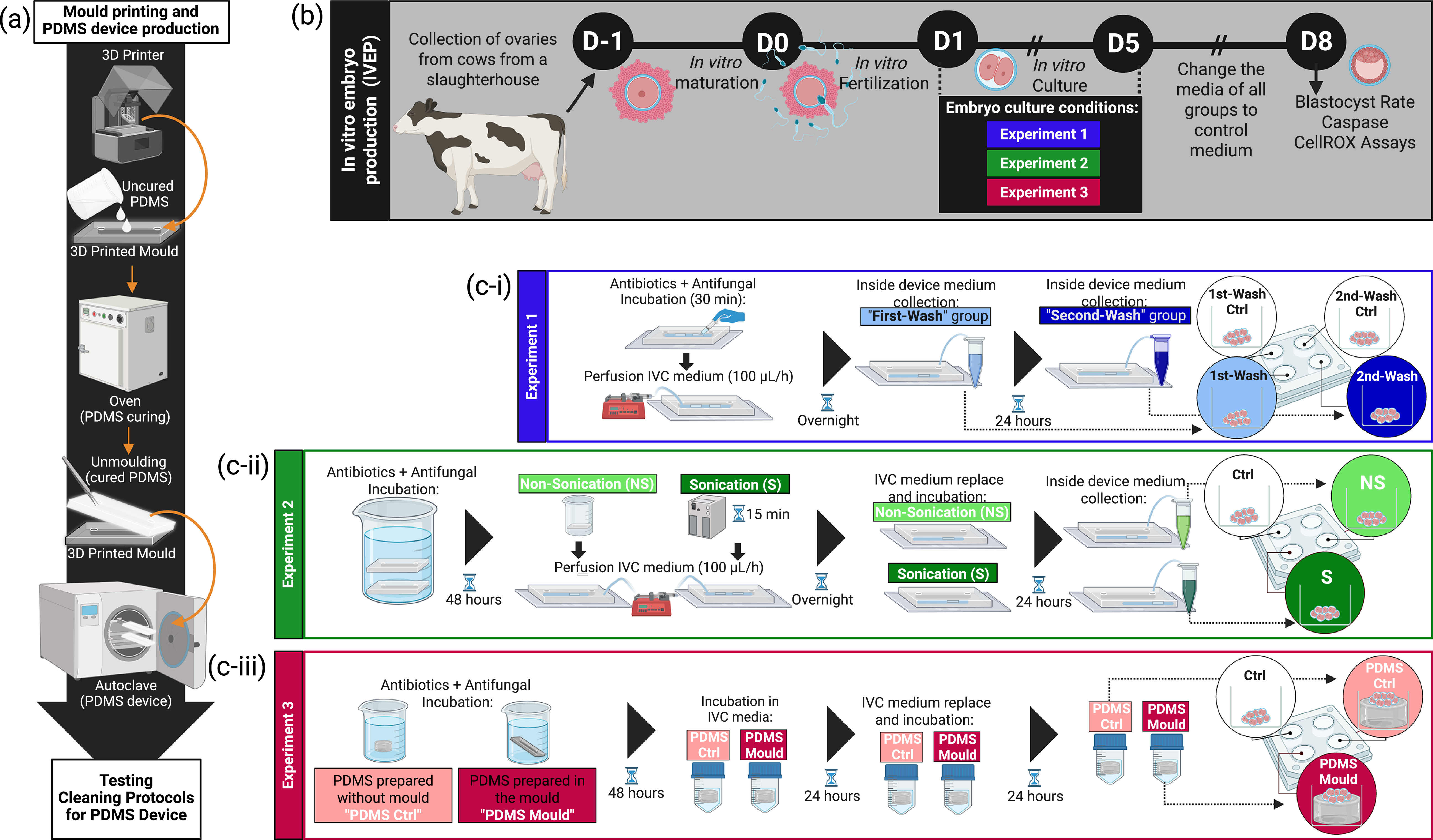
Figure 1. Long description
Panel A: A flowchart depicting the production of microfluidic devices using the soft lithography technique. It shows the steps of 3D printing the mold, using PDMS as the polymer ink, curing the PDMS in an oven, unmolding the cured PDMS device, and autoclaving it before testing cleaning protocols. Panel B: A timeline diagram outlining the in vitro embryo production process. It includes the collection of ovaries from cows, in vitro maturation, in vitro fertilization, embryo culture with different experiments, changing the media, and assessing the blastocyst rate and caspase cell assays. Panel C: Three sub-experiments detailing the cleaning protocols for the PDMS device. Experiment 1 involves sequential washes of the PDMS device. Experiment 2 involves sonication of the PDMS device. Experiment 3 involves in vitro embryo culture with the PDMS substrate.
Experiment 1
In the first experiment, after being rinsed with Wash-PBS solution (PBS supplemented with 100 IU/mL penicillin and 100 μg/mL streptomycin, and 0.25 μg/mL amphotericin B) for 30 minutes, the device was filled with IVC medium and submitted to perfusion (overnight at a flow rate of 100 μL/h in the incubator) using a programmable syringe pump (AL-1000, World Precision Instruments, USA). On D0 of IVEP, the medium inside the device was collected. Embryos cultured in this conditioned medium were designated the ‘First-Wash’ experimental group. Simultaneously, IVC medium without device contact was collected, and embryos cultured in this unconditioned medium were designated the ‘First-Wash-Ctrl’ experimental group. Subsequently, the device was filled again with newly prepared IVC medium, followed by 24 hours of incubation in static conditions inside the incubator. On D1, the conditioned medium inside the device was collected, and embryos cultured in this conditioned medium were designated the ‘Second-Wash’ experimental group. For comparison, embryos were also cultured in medium that stayed incubated for another 24 hours without device contact and were designated the ‘Second-Wash-Ctrl’ experimental group (Figure 1c-i).
Experiment 2
In the second experiment, the PDMS device was subjected to sonication to test improvement in the cleaning process. After being submerged in Wash-PBS solution (48 hours at 4°C), PDMS devices were either subjected to bath sonication (70% v/v ethanol, 45 kHz, at 30°C for 15 minutes, followed by distilled water, 45 kHz, at 30°C for 15 minutes), or left untreated (control). Then, sonicated and non-sonicated devices were perfused with IVC medium (overnight). Last, the devices were filled with newly prepared IVC medium and incubated for 24 hours in static conditions. On the following day, conditioned medium was collected from inside the devices; embryos cultured in medium from sonicated devices were designated the ‘Sonicated’ experimental group, and those from non-sonicated devices the ‘Non-Sonicated’ experimental group. In addition, IVC medium that had no contact with the PDMS device was used for the ‘Ctrl’ experimental group (Figure 1c-ii).
Experiment 3
In the third experiment, embryos were cultured under three conditions: with a PDMS fabricated using the 3D-printed resin mould (hereafter ‘PDMS-Mould’) which may contain leachable compounds from the resin; or with PDMS piece prepared in a petri-dish well (i.e., not having contact with the 3D-printed mould), referred to as ‘PDMS-Ctrl’, and without PDMS, referred to as ‘Ctrl’ experimental groups. The PDMS pieces from both groups were 1.9 cm2 in area and 0.2 cm in thickness. For cleaning, the PDMS pieces underwent the same ‘Non-Sonicated’ protocol used in Experiment 2, except that all the incubations with IVC medium were under static conditions. On D1, the PDMS pieces were placed in a four-well plate for the duration of embryo culture days (D1–D5; Figure 1c-iii).
Embryos from all the experimental groups were cultured for four days under the conditions described above. On the fifth day of culture, embryos from all groups were transferred to a similar standard culture medium, and the culture medium was renewed (Santos et al., Reference Santos, Fonseca Junior, Lima, Ispada, Silva and Milazzotto2021). The total number of oocytes in this study was n = 1,775, distributed for each experiment as follows: experiment 1, n = 864 (First-Wash-Ctrl, n = 215; First-Wash, n = 200; Second-Wash-Ctrl, n = 225; Second-Wash, n = 224), experiment 2, n = 495 (Ctrl, n = 161; Sonicated, n = 168; Non-Sonicated, n = 166), and experiment 3, n = 416 (Ctrl, n = 140; PDMS-Mould, n = 136; PDMS-Ctrl, n = 140). Six devices were produced for experiment 1, each constituting one replicate, and six for experiment 2 (3 replicates, n = 3 devices non-sonicated and n = 3 devices sonicated). For experiment 3, 3 replicates were carried out.
Mould 3D printing
The moulds for the PDMS device consisted of the negative shape of the device, designed as two mould parts to obtain a single-channel device (670 mm long, 1.2 mm wide, and 1.2 mm high). The moulds were printed and cured as previously described (Franko and de Almeida Monteiro Melo Ferraz, Reference Franko and de Almeida Monteiro Melo Ferraz2025). Briefly, a 3D Printer (Elegoo Mars Pro 2, Elegoo) and translucent 3D Printing UV Sensitive Basic Resin (Shenzhen Anycubic Technology Co.) were used for the mould preparation, which was washed [70% v/v Isopropanol (IPA), Elegoo Mercury Plus (Elegoo)], completely dried, cured (405 nm UV-light at 14 mW/cm2 for 5 minutes), and submitted to sonication (Transsonic, TI-H-5, Elma Schmidbauer GmbH, Germany) to avoid PDMS curing inhibition.
PDMS device fabrication
The devices were fabricated as previously described (Franko and de Almeida Monteiro Melo Ferraz, Reference Franko and de Almeida Monteiro Melo Ferraz2025). Briefly, the soft-lithography technique was used with the 3D-printed mould. PDMS was prepared as a 12:1 mixture of pre-polymer to curing agent (Sylgard™ 184 Silicone Elastomer Kit, Dow Europe GmbH, Germany). After complete curing, vertical inlets and outlets were created in the devices using a 3-mm biopsy punch (PFM Medical, Germany), and silicone tubes (Tygon®, Darwin Microfluidics, France) were attached. New devices were prepared for each experiment (first use). Before all the experiments, all the devices were rinsed with Milli-Q water, followed by 70% ethanol. Considering that the final volume inside the PDMS channel was measured as approximately 640 μL, each channel was rinsed at least seven times with each solution. Last, the devices were autoclaved (20 minutes, 121°C) and stored at room temperature until use.
IVEP and embryonic apoptosis rate and ROS detection
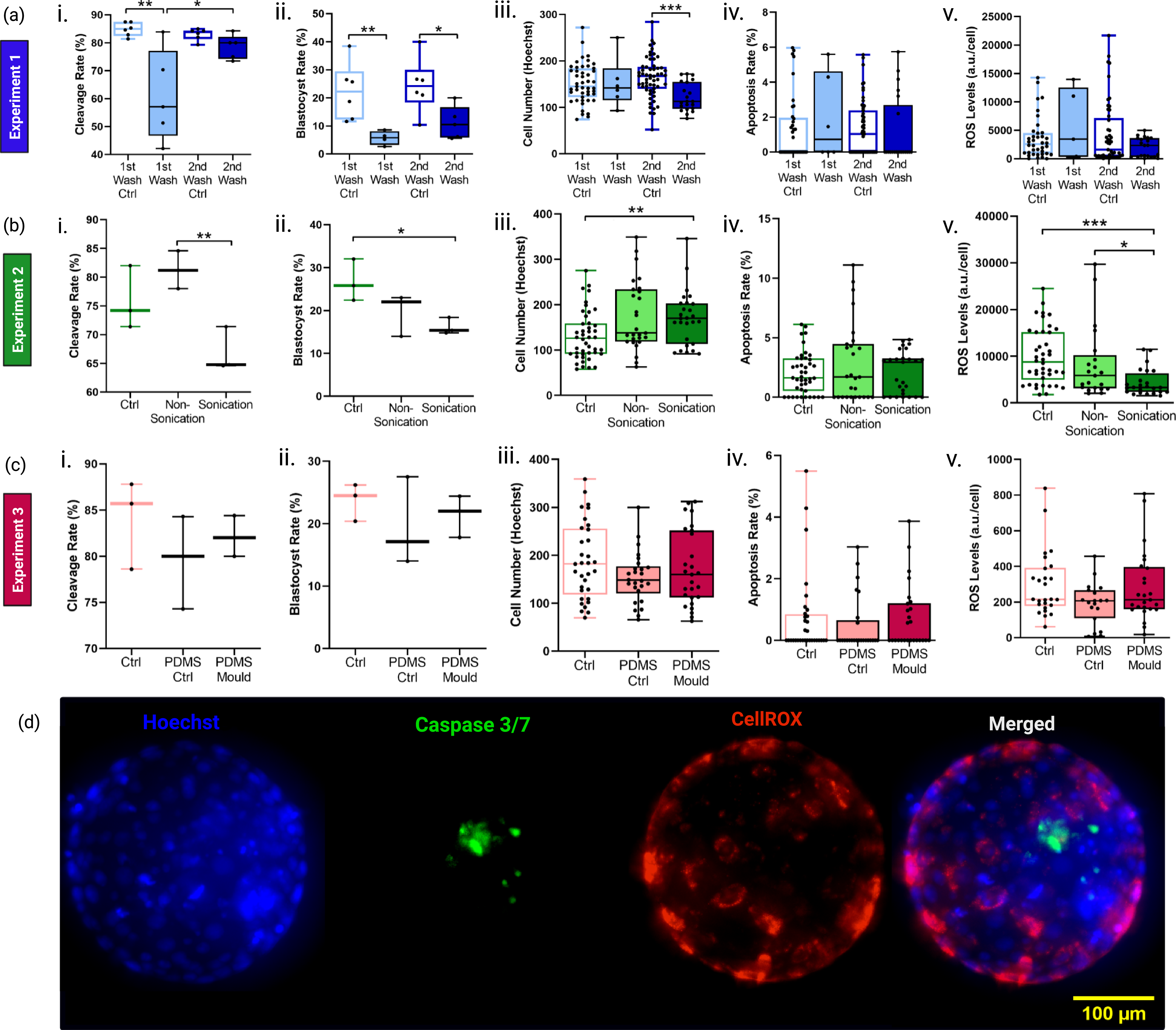
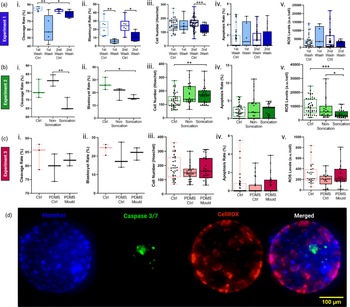
IVEP was performed as previously described (Fontes et al., Reference Fontes, Milazzotto and Ferraz2025). On Day 5, the cleavage rate was assessed, and the medium was refreshed. On Day 8 after fertilization, the blastocyst rate was assessed. Embryos were submitted to apoptosis rate analysis and reactive oxygen species (ROS) detection, with CellEvent™ Caspase-3/7 Green ReadyProbes™ Reagent (Invitrogen™, ThermoFisher, UK) and CellROX™ Deep Red Reagent (Invitrogen™, ThermoFisher, UK), respectively. DNA was counterstained with Hoechst 33342 (NucBlue™ Live ReadyProbes™ Reagent, Invitrogen™, ThermoFisher, UK). All the procedures were performed as previously described (Fontes et al., Reference Fontes, Milazzotto and Ferraz2025). Representative images of fluorescence assays are presented in Figure 2d.
Biocompatibility of cleaning protocol for PDMS devices on in vitro embryo production yield (cleavage and blastocyst rates) and quality (total cell number, apoptosis rate, and oxidative stress level). (a) Results of experiment 1, (b) Results of experiment 2, and (c) Results of experiment 3 for: i. Cleavage rate (%), ii. Blastocyst rate (%), iii. Total blastocyst cell number (nuclei counted by Hoechst staining), iv. Caspase-positive cell rate (%), and v. ROS levels (arbitrary unit/cell). Graphs are presented as medians with bars for the minimum and maximum values, and black circles represent the individual values of independent replicates. (d) Representative fluorescent image of blastocyst analysis (D8) for Hoechst for nuclei (blue), Caspase-3/7 for apoptosis (green), CellROX for ROS level (red), and merged image (maximum projections). Statistical difference: *p < 0.05; **p < 0.01; ***p < 0.0001.
Data and statistical analysis
All the data are expressed as medians with minimum and maximum values, including the individual values. All statistical analyses were performed using Prism 8.0 GraphPad software (GraphPad Software Inc., USA). Data normality was assessed using the D’Agostino-Pearson test. Normally distributed data were analysed by one-way ANOVA followed by Tukey’s post hoc test; non-normally distributed data were analysed by the Kruskal–Wallis H test. Data were considered significantly different when p < 0.05.
Results and discussion
The cleaning methods evaluated in the first experiment were inefficient in producing bovine embryos, as evidenced by the detrimental effect on cleavage rate (Figure 2a-i), blastocyst yield (Figure 2a-ii), and the total blastocyst cell number (Figure 2a-iii). Therefore, for the second experiment, devices were subjected to extended PBS incubation, with or without an additional sonication step (Figure 1c-ii). The sonication process has been used by other studies as a general cleaning step (McKenna, Reference McKenna2016) and stencil remover (Hawkins et al., Reference Hawkins, Miao, Cui and Sun2022). However, in our study, the sonication process had a detrimental effect on the cleavage (Figure 2b-i) and blastocyst rates (Figure 2b-ii). Surprisingly, the sonication process resulted in the production of embryos with higher cell number compared to the Ctrl group (Figure 2b-iii) and lower ROS levels compared to other experimental groups (Figure 2b-v). However, this result should be carefully interpreted. Although higher blastocyst cell number and lower ROS levels might indicate better embryo quality, we should not assume that embryos cultured in conditioned medium from sonicated PDMS devices improve embryo quality. Such a result is probably caused by a selection of embryos that developed until the blastocyst stage, since the blastocyst rate was much lower in this experimental group. On the other hand, the Non-Sonication condition seems to support IVEP. Therefore, to confirm the efficiency of the Non-Sonication sterilization method, in experiment 3, the IVC was performed not only in PDMS-conditioned medium, but in the presence of the PDMS. As a result, the embryos produced in the PDMS-Mould group presented similar cleavage (Figure 2c-i) and blastocyst (Figure 2c-ii) rates and similar quality to the Ctrl and PDMS-Ctrl groups, as shown by the markers of total blastocyst cell number (Figure 2c-iii), apoptosis rate (Figure 2c-iv), and ROS levels (Figure 2c-v).
Previous studies are consistent in using culture media to wash PDMS devices before use. However, the definition of the incubation period required for proper safety cleaning varies greatly between studies. Reported incubation periods ranged from 1 hour (Ferraz et al., Reference Ferraz, Henning, Costa, Malda, Melchels, Wubbolts, Stout, Vos and Gadella2017) to overnight (de Almeida Monteiro Melo Ferraz et al., Reference de Almeida Monteiro Melo Ferraz, Nagashima, Venzac, Le Gac and Songsasen2020) and 24 hours (Belda-Perez et al., Reference Belda-Perez, Heras, Cimini, Romero-Aguirregomezcorta, Valbonetti, Colosimo, Colosimo, Santoni, Barboni, Bernabò and Coy2023). Such variation is mainly linked to the cell type used to test the toxicity of the PDMS device. For instance, HeLa cells and feline ovarian tissues showed normal physiological growth and survival rates once PDMS devices were submitted to 24 hours of perfusion of culture medium at a 2 μL/min flow rate (de Almeida Monteiro Melo Ferraz et al., Reference de Almeida Monteiro Melo Ferraz, Nagashima, Venzac, Le Gac and Songsasen2020). However, it is known that somatic cells are more resistant to certain 3D print substrates than gametes and embryos (de Almeida Monteiro Melo Ferraz et al., Reference de Almeida Monteiro Melo Ferraz, Henning, Ferreira da Costa, Malda, Le Gac, Bray, van Duursen, Brouwers, van de Lest, Bertijn, Kraneburg, Vos, Stout and Gadella2018; Ferraz et al., Reference Ferraz, Rho, Hemerich, Henning, van Tol, Hölker, Besenfelder, Mokry, Vos, Stout, Le Gac and Gadella2018; Belda-Perez et al., Reference Belda-Perez, Heras, Cimini, Romero-Aguirregomezcorta, Valbonetti, Colosimo, Colosimo, Santoni, Barboni, Bernabò and Coy2023). As such, a 24-hour rinse resulted in impaired embryo development (Belda-Perez et al., Reference Belda-Perez, Heras, Cimini, Romero-Aguirregomezcorta, Valbonetti, Colosimo, Colosimo, Santoni, Barboni, Bernabò and Coy2023), even with a significant reduction in the detection of compounds leached from PDMS devices cast from 3D-printed resin moulds (de Almeida Monteiro Melo Ferraz et al., Reference de Almeida Monteiro Melo Ferraz, Nagashima, Venzac, Le Gac and Songsasen2020). As an alternative to the cleaning procedure, another strategy to improve the biocompatibility of PDMS devices is to use surface coating protocols (Heo et al., Reference Heo, Cabrera, Song, Futai, Tung, Smith and Takayama2007; Yao et al., Reference Yao, Guan, Park, Choi, Kim and Park2021; Hawkins et al., Reference Hawkins, Miao, Cui and Sun2022); however, simplicity and practicality are key considerations for applications of microfluidic devices in IVEP. Therefore, the development of tailored cleaning steps to ensure material safety remains an important priority. In our study, we demonstrated that a straightforward treatment involving long-term incubation in PBS (supplemented with antibiotics and antifungal agents), followed by 48 hours of incubation with IVC medium, was necessary to support embryo development. Based on previous studies, the extended PBS treatment may facilitate the removal of leachable compounds from the PDMS surface (de Almeida Monteiro Melo Ferraz et al., Reference de Almeida Monteiro Melo Ferraz, Nagashima, Venzac, Le Gac and Songsasen2020), thereby improving biocompatibility. However, the specific leachate compounds responsible for the embryotoxicity were not identified within the scope of this study. Future investigations, ideally involving medium composition analysis via mass spectrometry, are warranted to directly quantify leachates and validate this proposed mechanism.
Conclusion
This study establishes an effective cleaning protocol for PDMS microfluidic devices fabricated using 3D-printed resin moulds, enabling their safe application in future bovine IVEP procedures. The successful integration of PDMS devices into embryo culture systems is a necessary step towards enabling dynamic in vitro culture platforms, including organ-on-a-chip applications designed to mimic the in vivo environment of the bovine reproductive tract. It is important to note that in the present study, embryos were not cultured directly inside the devices; instead, biocompatibility was assessed using conditioned medium and PDMS substrate contact as proxies. Direct embryo culture within the microfluidic channel represents the logical next step to fully validate the proposed cleaning protocol under dynamic flow conditions.
Data availability statement
The data that support the findings of this study are available from the authors upon request.
Author contributions
Patricia Kubo Fontes: Conceptualization-Equal, Formal analysis-Equal, Investigation-Equal, Methodology-Equal, Writing – original draft-Equal, Writing – review & editing-Equal; Roksan Franko: Formal analysis-Equal, Investigation-Equal, Methodology-Equal, Writing – review & editing-Equal; Giuliana de Avila Ferronato: Formal analysis-Equal, Investigation-Equal, Methodology-Equal, Writing – review & editing-Equal; Marcella Milazzotto: Funding acquisition-Equal, Supervision-Equal, Writing – review & editing-Equal; Marcia de Almeida Monteiro Melo Ferraz: Conceptualization-Equal, Formal analysis-Equal, Funding acquisition-Equal, Methodology-Equal, Supervision-Equal, Writing – review & editing-Equal.
Funding information
This work was supported by the Alexander von Humboldt Foundation in the framework of the Sofja Kovalevskaja Award endowed by the German Federal Ministry of Education and Research and the São Paulo Research Foundation (FAPESP, grant 2022/12169-9).
Competing interests
The authors have no competing interests to declare that are relevant to the content of this article.
Ethical standards
Ethics declaration: not applicable.

 Open access

