

Secondary intracranial hypertension is a rare complication of Guillain-Barré syndrome (GBS). We describe a case with clinical, and neurophysiological evidence of GBS with headache and progressive bilateral vision loss 3 weeks after the onset of weakness. Fundoscopic examination showed bilateral papilledema with retinal hemorrhages. She had features of secondary intracranial hypertension, and required ventriculoperitoneal (VP) shunt. Our case emphasized that headache and vision changes in the setting of GBS should require detailed ophthalmology assessment, adequate monitoring, and treatment as indicated.
A 33-year-old severely obese female without any past medical history presented with a 5-day history of progressive ascending weakness involving all limbs. Her weakness was preceded by an upper respiratory infection 2 weeks before presentation. There was no evidence of dysautonomia. She did not report vision changes on presentation. Cranial nerve examination revealed normal fundi, but dysarthric speech, along with bilateral lower motor neuron facial weakness, weak gag reflex, and weak tongue movements. Motor examination revealed flaccid tone in all limbs. Muscle strength was 3/5 proximally and 1-2/5 distally of all limbs using the Medical Research Council scale. Deep tendon reflexes were absent and plantar response was flexor bilaterally. Sensory examination revealed symmetrical glove/stocking sensory loss to wrist and knee level, respectively. CSF analysis revealed cytoalbuminologic dissociation with white blood cell (WBC) of 3×10⁶/L, and protein at 132 mg/dl. Human immunodeficiency virus test, and hepatitis B antigen were negative, and thyroid-stimulating hormones and T3/T4, antinuclear antibody, and extractable nuclear antigen antibodies were normal. Serum electrophoresis was unremarkable. She was immunized for rubella and measles in childhood. She did not have recent immunizations. Nerve conduction studies showed acquired demyelinating features with prolonged distal motor latencies at both upper and lower extremities, conduction blocks, absent F waves, and absent sensory nerve action potentials at median, ulnar, peroneal, and sural nerves. She was diagnosed with acute inflammatory demyelinating polyradiculoneuropathy, and started with intravenous immunoglobulin (IVIg) at the dose of 0.4g/kg daily for 5 days. After 2 weeks in hospital, she started to complain of increasing headache and vision changes. Examination revealed no meningsmus, but bilateral upgaze restriction and limited abduction with extraocular movements. Fundoscopic examination showed grade IV papilledema bilaterally with hemorrhages. Head CT scan and brain MRI with MR venography (MRV) were normal. Repeated lumbar puncture (LP) showed a high opening pressure greater than 500 mm H2O, protein of 143 mg/dl, and WBC of 2×10⁶/l. A total of 30 ml of CSF was removed. She reported symptomatic improvement. Idiopathic intracranial hypertension (IIH) was considered as differential diagnosis given her age, gender, and body mass index. Other secondary conditions that can cause headache were also considered, including sleep apnea, chronic hypertension, and endocrine causes. She, however, did not have previous headache history. Given the temporal relationship with the motor symptoms, increased CSF protein, lack of apnea history and symptoms, normal blood pressure, and normal endocrine profile, the diagnosis of secondary intracranial hypertension was made. She was treated with acetazolamide up to 500 mg twice a day. However, she remained symptomatic with progressive visual scotoma in the following 5 days post-LP. She required Codman intracranial pressure (ICP) monitoring. The ICP was consistently high (between 300 and 500 mm H2O) with intermittent spikes up to 900 mm H2O. After a week, eventually, VP shunt was inserted to prevent permanent optical injury. She reported significant improvement of her headache and visual symptoms within 24 hours post-procedure.
Over the following 3 weeks in hospital, her ICP remained in the normal range (between 80 and 120 mm H2O), and she no longer reported headache or vision change. She did not require further IVIg doses, and started to show substantial improvement in her motor symptoms while working with the rehabilitation team. She was eventually discharged with the VP shunt remaining in situ, and was able to walk using a four-wheel walker. She continued to regain function and currently only requires minimal assistance, and has not reported any symptomatic/functional deterioration, or VP shunt complications for the course of 2 years since discharge.
Secondary intracranial hypertension has been reported in association with GBS in a few case reports (Table 1). Papilledema has been reported as a rare complication of GBS, and it has been associated with elevated CSF protein in most of the reports. Taylor and McDonaldReference Taylor and McDonald 1 were the first group to report the occurrence of blurred optical discs associated with GBS. However, the first report of definite papilledema was by Gilpin et al.Reference Gilpin, Moersch and Kernohan 2 As then a number of case reports and a case series summarized by Morley et al.Reference Morley and Reynolds 3 have been available. The main differentiating features from the IIHReference Morley and Reynolds 3 are high CSF protein and developing symptoms related to increased ICP in association with onset of GBS. Headache can be a frequent adverse effect from IVIg treatment. In our case, however, the signs and symptoms related to increased ICP and the remarkable response to the LP and VP shunt would argue against the possibility of having aseptic meningitis from IVIg use.
Secondary intracranial hypertension in Guillain-Barré syndrome (GBS)
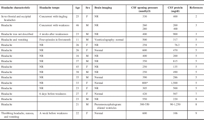
NR=not recorded.
This table shows previous case reports with secondary intracranial hypertension in the sitting of GBS.
* Measured in sitting position.
The pathophysiologic basis of headache in such cases remains uncertain, with two main competing theories. The most frequently cited cause is that increased CSF protein concentration slows the reabsorption in the arachnoid granulations, and subsequently leads to increased ICP.Reference Denny-Brown 4 However, there are cases in the literature of disc edema with normal range CSF protein concentration. This remained unchallenged until JoyntReference Joynt 5 suggested that the basis for papilledema was cerebral edema, rather than the impaired absorption of CSF. To support this, historical brain biopsy specimens have shown marked swelling of nerve cells.Reference Joynt 5 The increased intracranial pressure may, like the IIH, arise from intrinsic cerebral edema rather than impaired CSF reabsorption. Almost all the cases in the literature as well as this case are young-onset. Whether elderly patients with certain degree of brain atrophy who can compensate better for increased ICP, and thus are less symptomatic remain unclear.
Brain imaging with MRI/MRV brain are indicated to rule out structural causes for increased pressure. This condition is extremely rare, and updated literature on this topic is lacking. Thus, probably general guidelines on intracranial hypertension management should be applied in such cases. However, some patients from previous case reports showed spontaneous improvement as the motor symptoms resolve.Reference Farmakidis, Inan, Milstein and Herskovitz 6 Optic nerve fenestration, repeated LPs, VP shunts, and lumbar drainsReference Pyati, Razis and Desai 7 are indicated in cases of impending vision loss. Although intracranial hypertension is a rare complication of GBS, one should be cautious that visual complaints, and especially papilledema should require monitoring of ICP, and adequate treatment to prevent permanent visual loss.
Acknowledgments
The authors acknowledge Douglas Zochodne and George Elleker for detailed nerve conductance study, and Matt Wheatley for surgical intervention with ICP monitor and VP shunt. The authors also acknowledge S. Nizam Ahmed and Rajive Jassal who were involved in the care of the patient.
Disclosure
AA and FB have nothing to disclose.
Conflicts of Interest
None.
Financial Support
None.

