


A 15-year-old male patient presented with persistent headache, refractory to analgesia, and panhypopituitarism. Brain magnetic resonance imaging (MRI) revealed a well-defined suprasellar cystic lesion, with internal fat-fluid level, presenting hyperintense signal in its upper aspect, due to fat, low signal in its lower portion, due to fluid, and a floating round structure with isointense signal in the central region, on T1-weighted imaging, without gadolinium enhancement (Figure 1). The lesion had the appearance of the “Poké Ball sign,” compatible with suprasellar mature cystic teratoma/dermoid cyst. The patient was submitted to craniotomy for lesion resection and histopathological analysis, which confirmed the diagnosis.
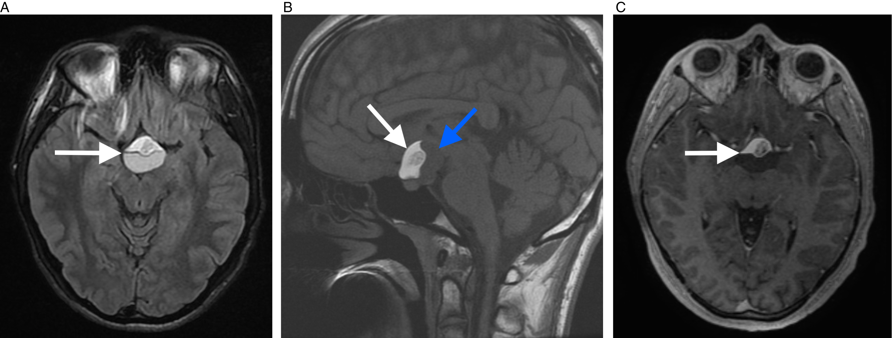
Suprasellar mature teratoma/dermoid cyst, with the “Poké Ball sign.” Brain MRI demonstrated a well-defined suprasellar cystic lesion, with internal fat-fluid level on FLAIR (arrow in A). On T1-weighted imaging, the lesion presented with hyperintense signal in its upper aspect, which represented the fat portion (white arrow in B), and hypointense signal in its dependent portion, which represented the fluid portion (blue arrow in B). The lesion also had a floating round structure, with isointense signal, configuring the “Poké Ball sign.” There was no gadolinium enhancement (arrow in C). Histopathologic examination revealed a mature teratoma/dermoid cyst.
Although some authors consider dermoid cyst and mature cystic teratoma to be synonymous, Reference Outwater, Siegelman and Hunt1 others consider that mature cystic teratomas are congenital inclusion cysts composed of well-differentiated cells from at least two of the three germ cell layers (ectoderm, mesoderm, and endoderm), whereas dermoid cysts occur when the ectodermal component predominates in the cyst. Reference Chen, Ruiz, Killeen, Coté, Wu and Correa2 Generally, they contain sebaceous glands and desquamated epithelium. They are also extremely rare, accounting for 0.5% of all primary intracranial tumors and are slightly more common in females. Reference Giambelluca, Caruana and Cannella3
Dermoid cyst in the central nervous system is usually identified by the demonstration of internal fat on imaging exams. On MRI, this lesion has a high signal on T1-weighted imaging, associated with signal suppression on fat-suppressed sequences, confirming the fat content. On computed tomography, fat can be characterized by its low density. Reference Giambelluca, Caruana and Cannella3,Reference Kuyumcu and Jhaveri4
Originally, Poké Ball is a round compartment, used to store pokémons, from the Japanese cartoon series Pokémon (Figure 2). The “Poké Ball sign” is a radiological finding widely described in mature ovarian cystic teratomas, characterized by a cyst with a fat-fluid level, associated with a globular structure floating in its central portion. Histopathologically, the cyst is represented by oily fluid in its dependent portion and fat in its superior portion. The central floating round component usually is composed of multiple spherules of fibrin, keratin, fat, sebum, and hair. Reference Giambelluca, Caruana and Cannella3 To the best of our knowledge, this sign has not yet been described in intracranial mature teratomas/dermoid cysts, although it is well described in the ovaries. Although some authors reported a sensitivity of 25% for this sign, Reference Şahin, Akdoğan, Ayaz, Karadeniz and Sancı5 it can be considered pathognomonic of teratoma. Reference Giambelluca, Caruana and Cannella3,Reference Şahin, Akdoğan, Ayaz, Karadeniz and Sancı5 We consider that the “Poké ball sign” can also be of great value for the diagnosis of intracranial dermoid cyst/teratoma.
Drawing of the original Poké Ball, from the Pokémon series. The red portion represents the fat, which is less dense and stays in the upper portion of the cyst. The white portion represents the fluid, which is in denser, and stays in the dependent portion of the cyst. The central round portion represents the floating nodule in the fat-fluid interface, composed of multiple spherules of fibrin, keratin, fat, sebum, and hair.

Imaging exams are fundamental for both diagnosis and surgical planning. Resection is the only effective treatment for dermoid cysts. MRI accurately evaluates the topography, size, structures involved by the lesion and estimates the extent of resection, allowing an appropriate surgical approach and early diagnosis. Reference Vaz-Guimaraes, Koutourousiou and de Almeida6 This is important because one possible complication of a dermoid cyst is rupture, in which fat droplets spread out into the subarachnoid space, causing chemical meningitis. Reference Osborn and Preece7 Thus, an early diagnosis may prevent this complication and assist in the proper surgical planning, reducing the risk of intraoperative rupture.
Therefore, the “Poké Ball sign” is very illustrative of the importance of imaging in diagnosis and surgical planning in these cases, by directly suggesting a histological diagnosis.
Disclosures
The authors do not have anything to disclose.
Statement of Authorship
ARSdS undertook conception, drafted the article, and had final approval of the last version.
DAdA collected the patient data, edited the article, and had final approval of the last version.
AA collected the patient data and had final approval of the last version
DGC edited the article, provided direct supervision, and had final approval of the last version.


