


Introduction
Wild beasts were used in Roman animal spectacles (venationes), providing public entertainment. Descriptions of these spectacles appear in numerous Roman texts, many of which refer to the largest and most monumental amphitheatre, the Colosseum in Rome (Jennison Reference Jennison2005), where a variety of exotic and indigenous domestic and wild animals were mustered for the games. Among these, the brown bear (Ursus arctos) features prominently in written accounts and iconography, deployed as performing animals, combatants for gladiators or other animals and executioners for convicts (Friedlaender Reference Friedlaender1979; Jennison Reference Jennison2005). Provincial amphitheatres are believed to have primarily featured animals that could be sourced locally. This included the brown bear, but also wild boars and cervids, and domestic animals, predominantly bulls (Bomgardner Reference Bomgardner1992; Jennison Reference Jennison2005). Skeletal remains of brown bears have been discovered at and around amphitheatres, prompting speculation regarding their role in spectacles (e.g. De Grossi Mazzorin et al. Reference De Grossi Mazzorin, Minniti, Rossella, Malerba and Visentini2005; Gostenčnik Reference Gostenčnik2008; Vuković Reference Vuković2012). Recent research, including stable isotope analysis of bear remains from Augusta Raurica in present-day Switzerland, has explored the possibility that these animals were held in captivity (Gerling Reference Gerling2023), but definitive evidence, such as skeletal trauma consistent with fights, has yet to be identified.
This study provides osteological evidence of the participation of captive brown bears in Roman spectacles. Excavations of Viminacium amphitheatre (in present-day Serbia) in 2016 uncovered the fragmented cranium of a brown bear in square B/7, in a building situated 13.5m south-west of the western entrance to the stone-and-wood amphitheatre. The skull was found during the excavation of the walls, at an absolute height between 75.7 and 76m above the level of the Adriatic Sea. The building was constructed at a higher elevation than the amphitheatre, which was set into a natural slope, with the arena sitting 4–4.5m lower. A part of the skeleton (left scapula, humerus, ulna and radius) of a leopard was also discovered in the same structure (Bogdanović et al. Reference Bogdanović, Rogić, Vuković-Bogdanović, Korać and Pop-Lazić2018: 47, cat. 1), while the remains of other wild animals, including other brown bears (Vuković Reference Vuković2012, Reference Vuković2015; Bogdanović et al. Reference Bogdanović, Rogić, Vuković-Bogdanović, Korać and Pop-Lazić2018), were found in the broader area of the amphitheatre. The bear skull is not directly dated, but another bone from the same feature yielded a radiocarbon date of cal AD 240–350 (95.4% probability, calibrated using IntCal20 (Reimer et al. Reference Reimer2020); lab no. Dea-42615; uncal. 1761±16 BP) (Figures 1, 2 & 3; see also online supplementary material (OSM)).
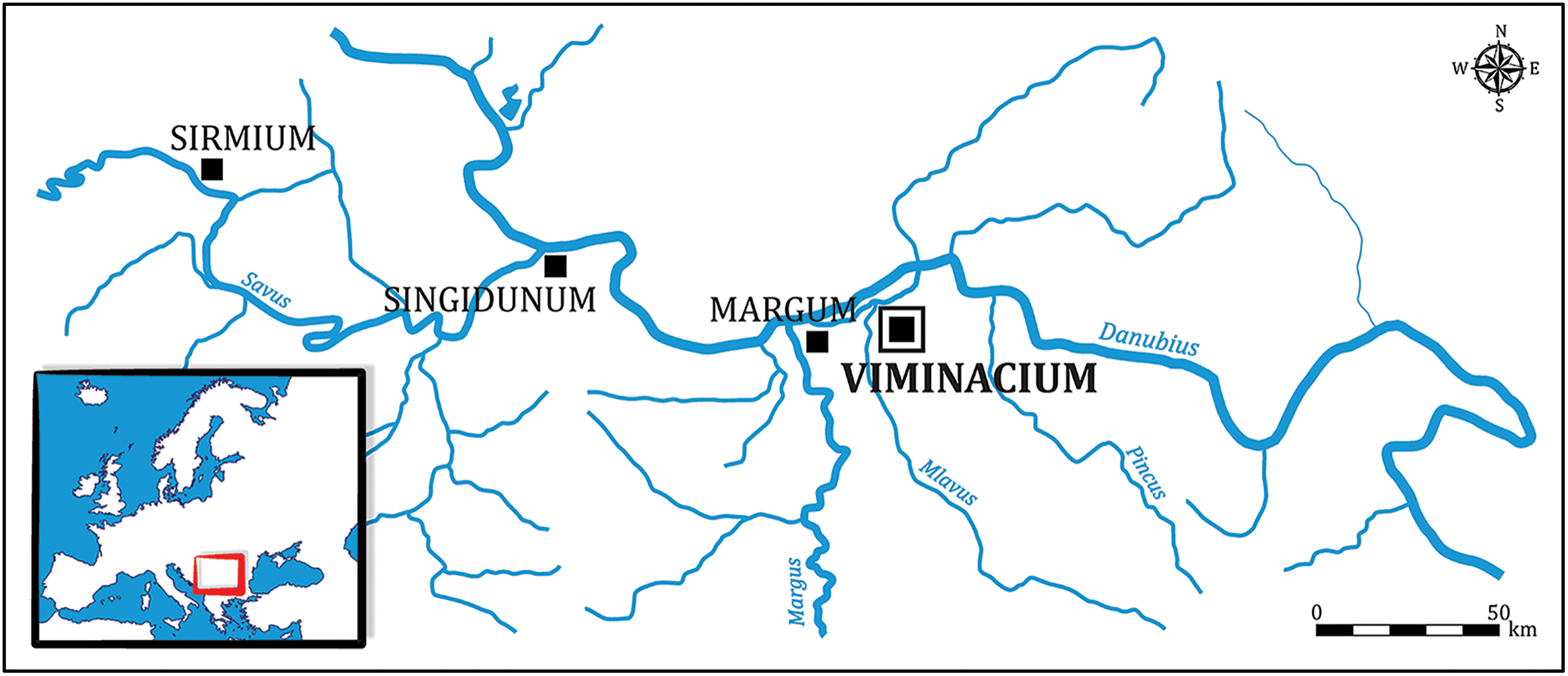
Location of Viminacium in relation to other larger Roman towns on the Danube Limes (figure by authors).

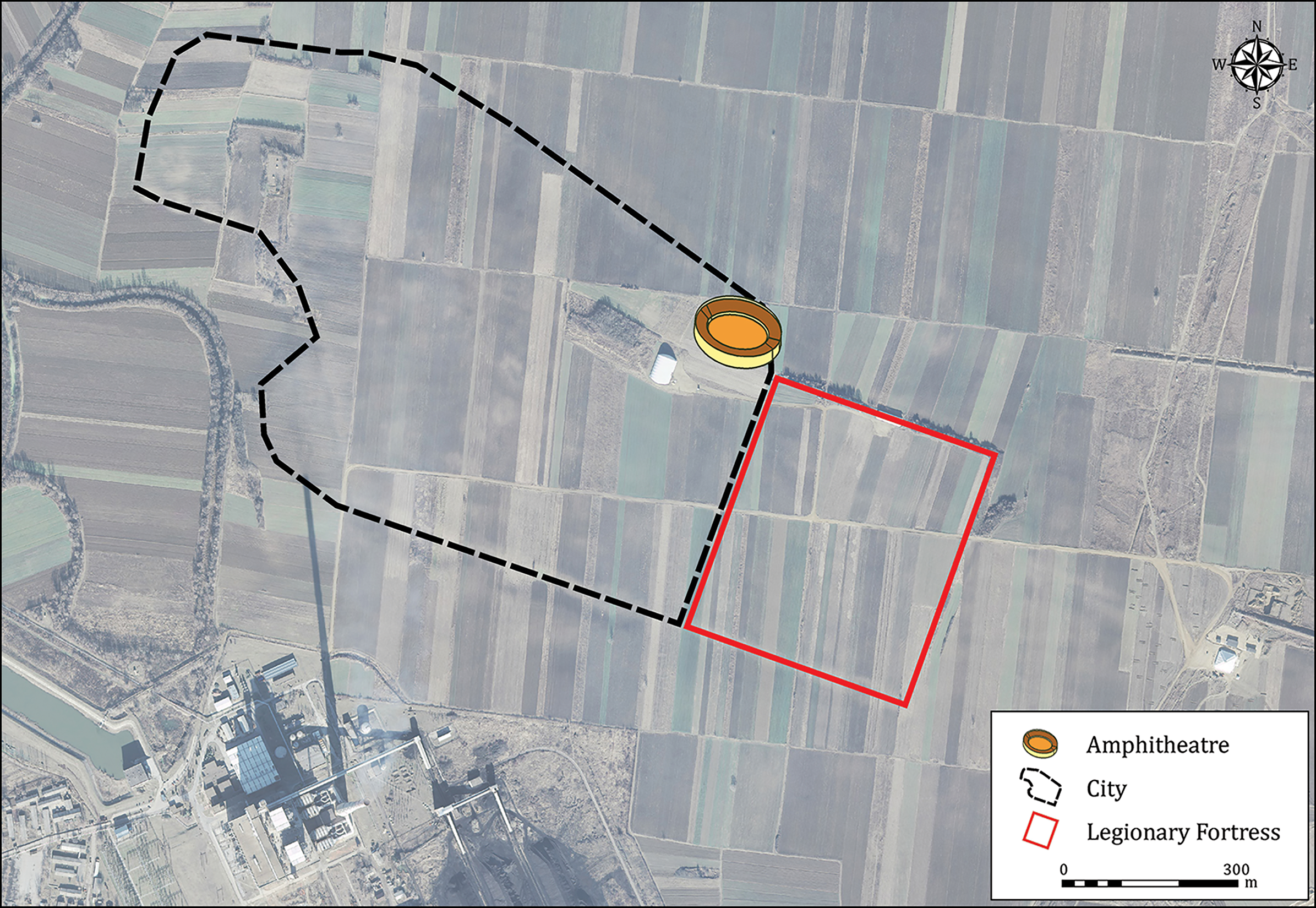
An aerial photograph of the current landscape at Viminacium, highlighting the extent of the city and location of the amphitheatre (figure by authors).

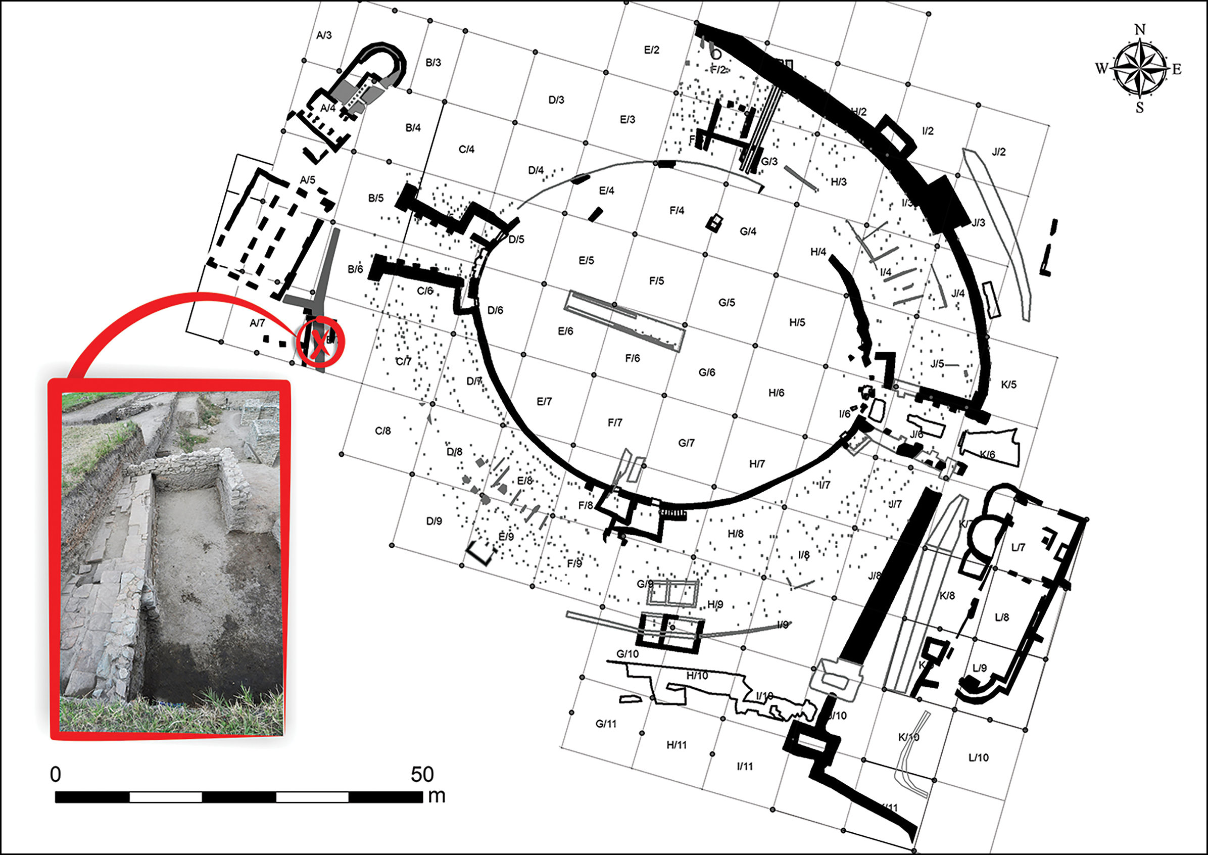
Plan of the Viminacium amphitheatre with inset image of the area where the fragmented brown bear cranium was found (figure by authors).

Viminacium was an important military base at the Roman frontier, located at the confluence of the Mlava and the Danube rivers. Initially it was a legionary fortress, established in the 80s of the first century AD, and it was home to the Legio VII Claudia during most of its history. Next to the fortress, a settlement grew to become the capital of the province of Moesia Superior (Mirković Reference Mirković1968; Bogdanović et al. Reference Bogdanović, Stojić, Jevtović, Vitezović, Radišić and Obradović2024). Following the administrative reforms in the last decades of the third century, it became the capital of a new province, Moesia Prima, established in the northern part of the older province of Moesia Superior (Ferjančić Reference Ferjančić, Popović and Borić-Brešković2013).
The brown bear skull fragments were studied by using a combination of zooarchaeological, palaeopathological, palaeoradiological, cementochronological and ancient DNA (aDNA) analyses. This multiproxy approach aims to provide a comprehensive evaluation of the specimen from Viminacium amphitheatre and, by further contextualisation with other osteological finds and written and iconographic evidence, to explore the role and significance of brown bears in amphitheatre spectacles of the Roman world.
Materials and methods
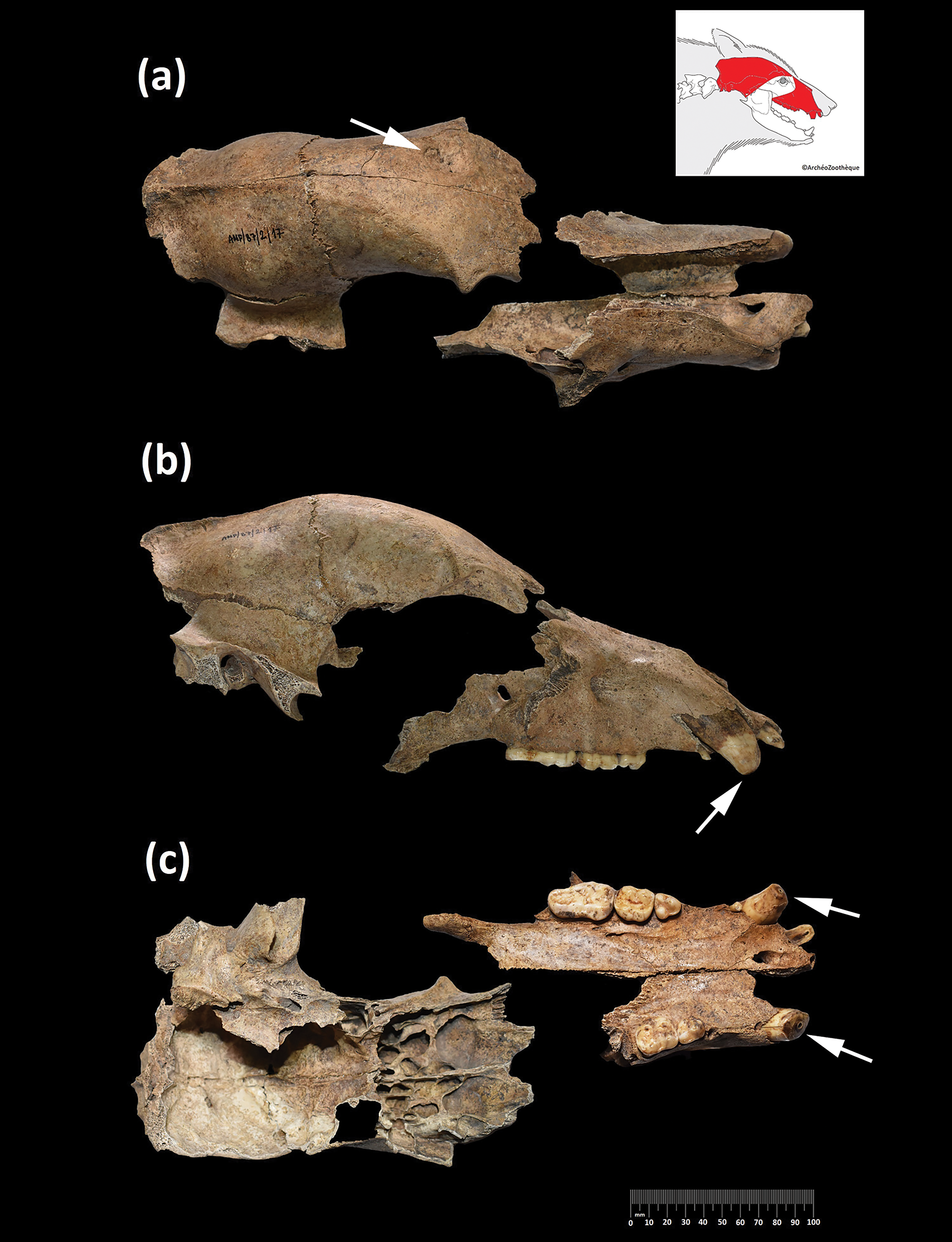
Large parts of the cranial vault are preserved (Figure 4), including most of the frontal bone, the left and the right parietal bones (with sagittal crest), a fragment of the occipital bone and a large part of the right temporal bone (with a fragment of the zygomatic process and the entire tympanic petrous portions). A fragment of the left maxilla preserves the in situ permanent canine, fourth premolar and first molar teeth, with damaged alveolus for the first and third premolars and the second molar. Almost the entire right maxilla is preserved, containing the canine, first and fourth premolars, and first and second molars, with an alveolus for the third premolar. There is also a fragment of the right premaxilla with damaged alveoli for the first and second incisors, and an in situ third incisor with a damaged crown. The second premolar is bilaterally absent in this specimen. Although the dental formula indicates a complete set of premolars, research has shown that brown bears exhibit variation in premolar presence (Strömquist et al. Reference Strömquist, Fahlman, Arnemo and Pettersson2009).
The fragmented brown bear cranium from the Viminacium amphitheatre: a) dorsal view, showing pathological changes on the left part of the frontal bone; b) right lateral view, showing a shortened canine; c) ventral view, showing shortening of both upper canines (brown bear skull template by ©ArchéoZoo.org; figure by authors).

Determination of biological sex was first achieved by correlating metric data from the upper canines with corresponding data from modern brown bear populations (Kurtén Reference Kurtén1955), then tested through aDNA analysis. Phylogenetic analysis of mitochondrial DNA was also performed to determine which brown bear population (clade) the individual from Viminacium belonged to and thus its geographical origin. A sample from the right petrous bone was submitted for aDNA analysis, which was conducted in accordance with established protocols (see OSM for details; Table S2).
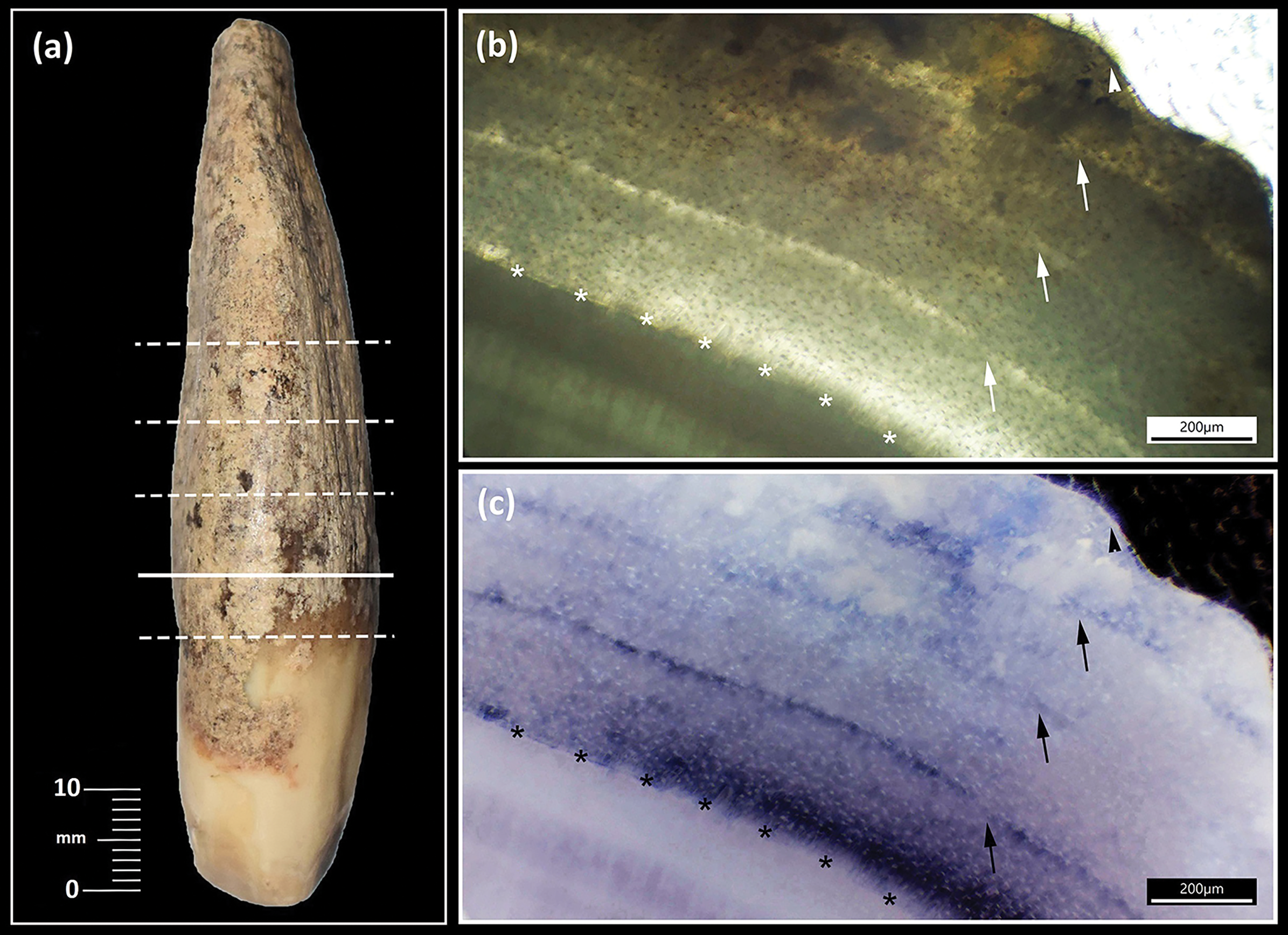
Analysis of the incremental growth of dental cementum (cementochronology) in the root of the upper right canine allowed estimation of the age of the bear and the season in which it died (Figure 5a; see OSM). Dental wear on the crowns of the canines and pathological changes to bone surfaces were observed by using a Dino-Lite digital microscope equipped with polarised light (model AM73115MZT, 220× magnification). Pathological changes on the left part of the frontal bone were further subjected to x-ray and computed tomography (CT) imaging for reliable differential diagnosis. Radiographic analysis was conducted with a portable Ecotron EPX-F5000 x-ray apparatus set at 60kV and 4mAs. CT imaging was performed by using SOREDEX Scanora 3D, with settings of 90kV and 30mAs and capturing continuous slices of 0.25mm thickness. Post-processing of the scans was carried out in the Carestream Vue PACS software (v. 12.2.0.0314), designed for professional medical examination reading in DICOM format. Differential diagnosis of pathological changes is based on reference texts on palaeopathology (Rothschild & Martin Reference Rothschild and Martin2006; Bartosiewicz & Gál Reference Bartosiewicz and Gál2013; Rothschild et al. Reference Rothschild, Surmik and Pellegrini2023) and veterinary medicine (Craig et al. Reference Craig, Dittmer, Thompson and Grant2016).
Cementochronology of the upper right canine: a) mesial view of the tooth, transverse sections were taken along each of the lines shown, with the solid line indicating the position of the section displayed in b and c; b) photomicrograph of transverse section in plane-polarised light—asterisks show the dentin-cementum junction line, arrows indicate lines of arrested growth and the arrowhead shows the tooth margin; c) photomicrograph of the same section with colours inverted to more clearly show the lines of arrested growth (figure by authors).

Results
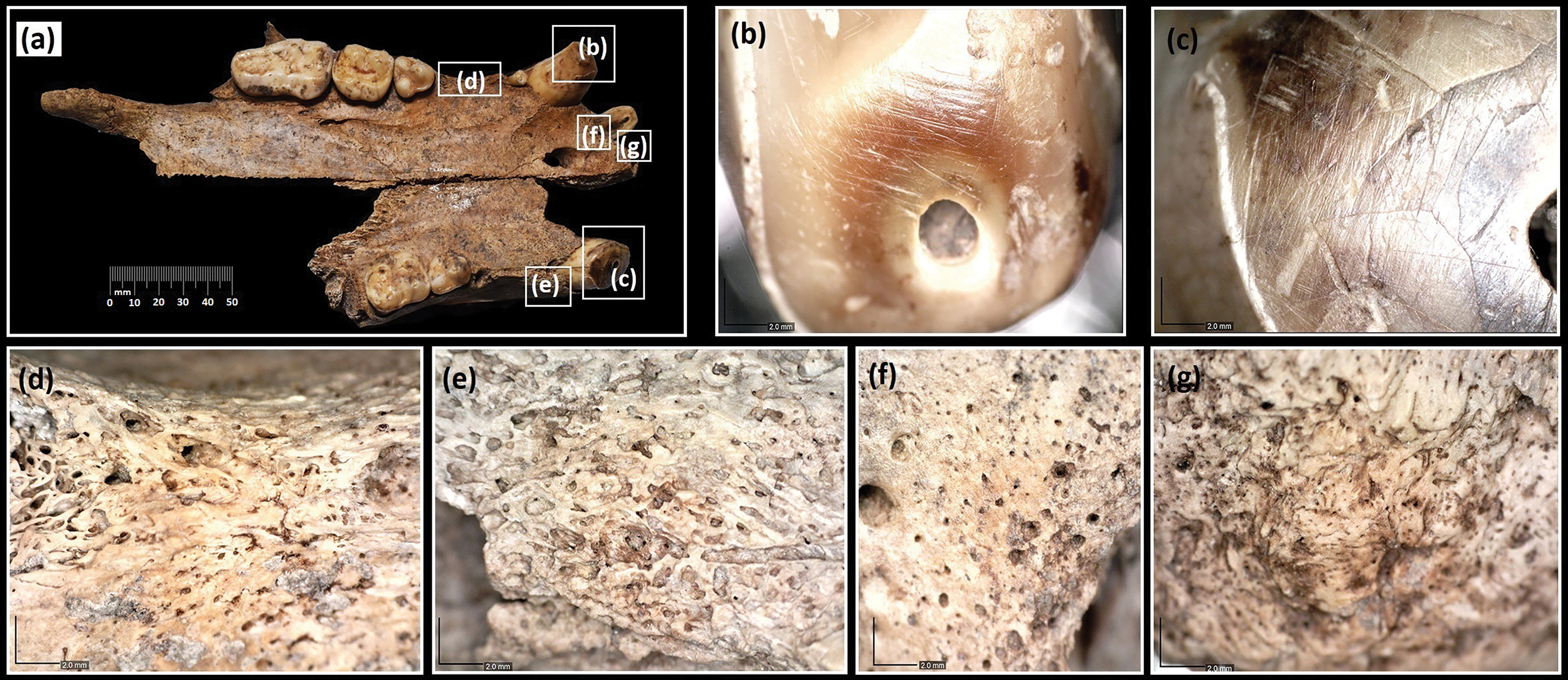
Despite the degree of fragmentation, the bone surface is well preserved, with no evidence of gnawing, weathering or butchery. The tips of both canines are heavily worn, with visible damage, in the form of transverse and oblique micro-scratch marks, observed under higher magnification and polarised light. The crowns of the left and right canines are almost equal in height, with approximately 5mm difference. The pulp canal is exposed in both teeth, and additional damage is noted on the enamel on the lingual and labial/mesial edges of the right canine (Figures 4b–c & 6b–c). The other permanent teeth show only slight wear (Table S1).
a) Ventral view of the fragmented maxilla; b & c) tips of the canines under polarised light revealing microwear; high magnification of pathological changes to the occlusal (d–f) and rostral (g) surfaces of the palate caused by periodontal disease (figure by authors).

Sex is determined to be male based on upper canine breadth (13.90mm), which aligns with the available average measurements (13.70±0.27mm) for modern populations of brown bear in Scandinavia (Kurtén Reference Kurtén1955). This was confirmed by molecular sex determination based on an X-to-autosomal chromosome coverage ratio of 0.52 (see OSM). An almost complete (more than 97%) sequence of the mitochondrial genome of the specimen from Viminacium amphitheatre (ID_AW084) was obtained (Table S2). The sequence falls within clade 1b and clusters with modern bears from Greece, belonging more broadly to the local Balkan population of brown bears (Figure S3).
Cementochronological analysis of the upper right canine indicates that the individual was approximately six years old at the time of death. Three lines of arrested growth are observed in the cementum. Due to the wide growth zone observed between the first annulus/line of arrested growth and the dentine-cementum junction line, a correction factor of 2–2.5 years was applied to the number of lines of arrested growth, resulting in the estimated age-at-death of six years. The formation of a new growth zone, which is nearly fully developed, is noted at the edge of the cementum. This observation suggests that the death occurred at the end of a warm season or the beginning of a cold season (Figure 5b–c).
Pathological changes are identified on the right premaxilla, both maxillae and the frontal bone. Macroscopic and microscopic examinations reveal periosteal reactions on the diastema of both maxillae, specifically at the edge of the alveolus of the left canine and the right third premolar (Figure 6d–e). Additionally, traces of increased vascularisation are observed on the premaxilla in the area around the alveoli of the right first and second incisors (Figure 6f–g). These changes are characteristic of early alveolar osteomyelitis, a chronic periodontal disease (Bartosiewicz & Gál Reference Bartosiewicz and Gál2013; Craig et al. Reference Craig, Dittmer, Thompson and Grant2016).
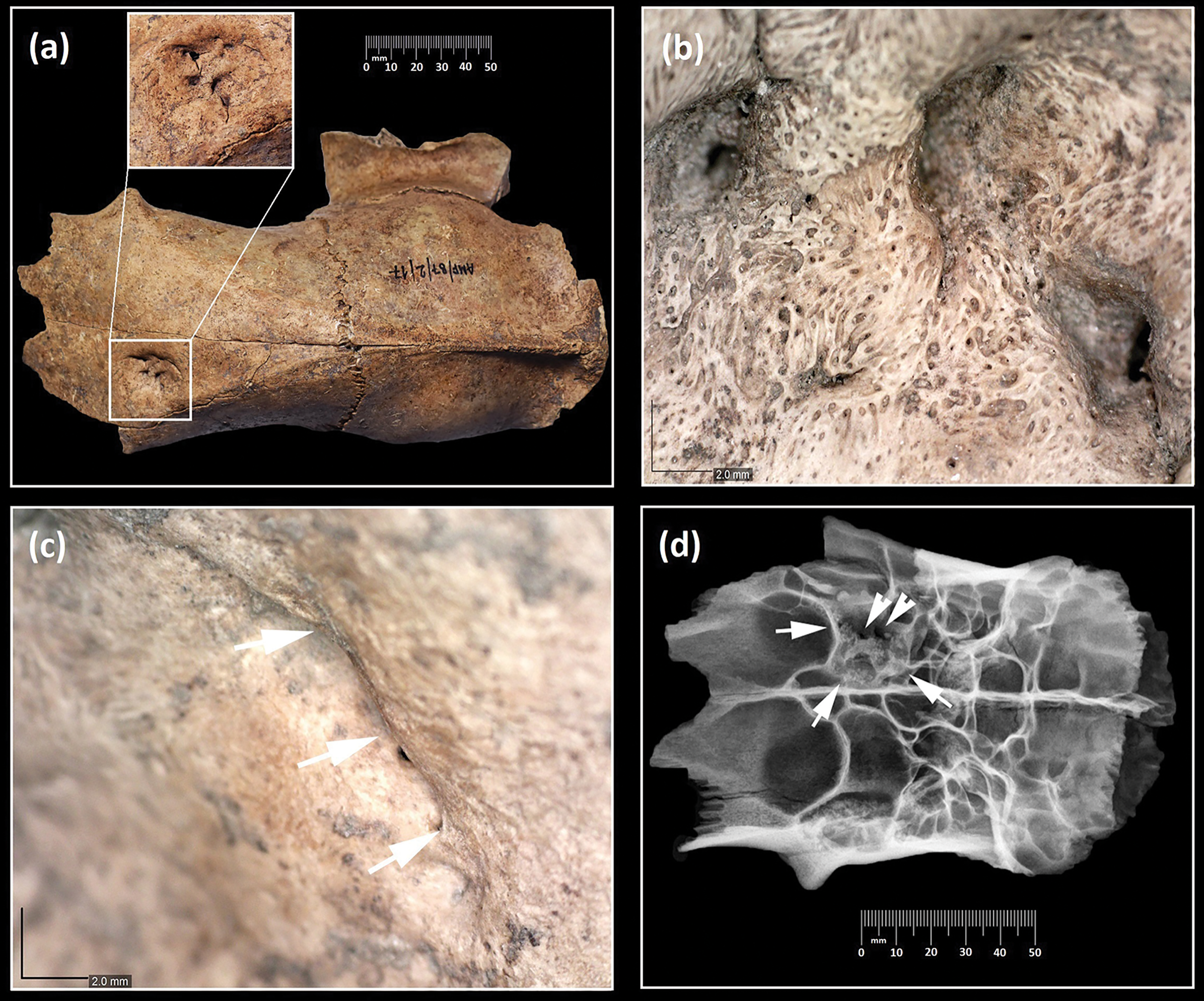
A sharply defined oval depression is located on the left part of the frontal bone, approximately 5–7mm from the frontal suture. It measures 23 × 27mm in maximum diameter, covering an area of 373mm2, with a maximum depth of 10mm and an estimated volume of 80mm3 (±0.4mm3). At the upper aspect of the depression, two short straight linear areas (fracture lines) are visible; they are not connected, instead separated by normal-looking bone. The depression slopes convexly towards the sinus spaces, causing partial disruption of sinus morphology compared to the contralateral side. Bone at the base of the depression is up to 8mm thick, exhibiting a pronounced relief and containing three large and two small fistulous channels. The three larger channels communicate with the sinus cavities; the largest, with a diameter of 3mm, is situated on the medial wall of the depression, while the channels on the lateral and anterior walls measure up to 1mm in diameter. The edges of the fistulous canals and the entire depressions appear rounded. Under higher magnification, the lesion shows a well-defined structure with a semi-lunar superior border and irregular divots, giving way to a less defined basicranial border. A periosteal reaction in the form of porous new bone formation is noted within the defect along the right border. Two remodelled, laterally oriented cracks are observed: the lower one extends from a deeper defect associated with remodelled bone, while the upper one shows less remodelling (Figure 7a–c).
a) Pathological changes on the left part of frontal bone with higher magnification of lesions (inset) revealing three fistulae of different shapes and sizes; b) central part of the lesions showing filigree-bone reaction and fistulae; c) the marginal zone of lesions (outlined with arrows); d) x-ray image in dorsoventral projection: the arrows show the area affected by pathological changes and the arrowheads show two fistulae (figure by authors).

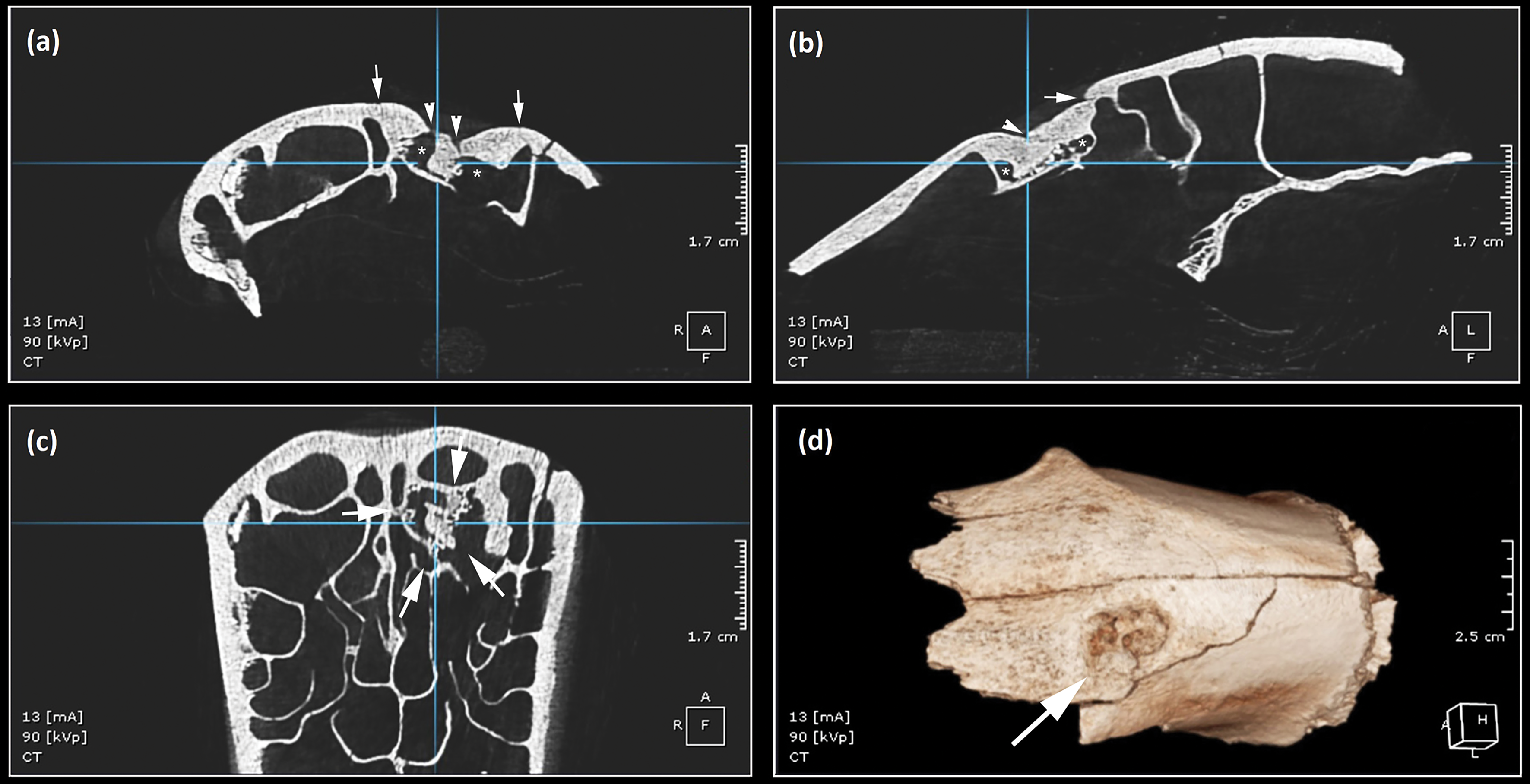
Radiographs of the frontal bone, taken in the dorsoventral projection (Figure 7d), reveal an osteosclerotic (radiodense), irregular semilunar structure in the area of the lesion. Within this structure, three calcific, rice grain-like densities forming a straight line are observed, accompanied by a curvilinear pattern of similar densities. An irregular orbital rim shows reduced thickness, with associated loss of continuity and signs of intracortical resorption. Adjacent major trabeculae appear destroyed, and two fistulous channels are evident in the central area of the lesion (Figure 7d: arrowheads). CT scans reveal a mass with new bone formation visible on the anterior and lateral projections. Fracture lines delineate the new bone formation. A focal thickening of the superficial area encompasses a linear cystic defect containing rice grain-like calcifications. Loss of continuity is associated with trabecular resorption, as well as destruction of surrounding trabeculae in the form of fistula and cystic areas (Figure 8a–b). An irregular orbital trabecular rim of reduced thickness, along with new bone with multiple rice grain-like structures is also observed in ventral projection (Figure 8c–d).
CT scan of the frontal bone: anterior (a) and lateral (b) views showing fracture lines (arrows), fistulae (arrowheads) and cystic areas (asterisks); ventral view (c) showing the area affected by pathological changes; and a three-dimensional reconstruction (d) of the frontal bone and the pathological changes (figure by authors).

The lesions observed on the frontal bone are consistent with an impact fracture that shows signs of healing but which subsequently became infected, leading to osteomyelitis (inflammation of the bone) (Rothschild & Martin Reference Rothschild and Martin2006; Bartosiewicz & Gál Reference Bartosiewicz and Gál2013; Craig et al. Reference Craig, Dittmer, Thompson and Grant2016).
Discussion
Animal spectacles were held in circuses during the early Principate (30 BC–AD 37), but were later moved to amphitheatres alongside gladiatorial contests (Futrell Reference Futrell2006). The spectacles, which took place in the mornings, included animal fights, combat between animal fighters (venators) and beasts, as well as animal hunts and displays. Wild animals were also used for the execution of convicts (damnatio ad bestias) during midday shows (ludi meridiani) (Futrell Reference Futrell2006: 89–95). The height of the arena wall at the Viminacium amphitheatre, as well as the small chambers (carceres) next to the main entrances to the arena (Nikolić & Bogdanović Reference Nikolić, Bogdanović, Vagalinski and Sharankov2015) and the huge stone chaining block in the central part of the arena (Bogdanović et al. Reference Bogdanović, Rogić, Vuković-Bogdanović, Korać and Pop-Lazić2018: 47, fig. 4), indicate that animal spectacles were enacted in the amphitheatre.
Ancient texts (e.g. Augenti Reference Augenti2001; Jennison Reference Jennison2005; Futrell Reference Futrell2006) and iconography from across the Empire (e.g. Dunbabin Reference Dunbabin1978, Reference Dunbabin1999; Nossov Reference Nossov2009; Sparreboom Reference Sparreboom2016) indicate the significant presence of bears in different types of games—ranging from combat and execution to performances featuring trained and tamed animals. Bears also appear in animal combat scenes on various artefacts across the Balkans, including a stone relief from Sofia that shows bears attacking venators (Vagalinski Reference Vagalinski2009). In Serbia, imported relief terra sigillata wares found at Viminacium and Singidunum depict bears in convict executions (Bjelajac Reference Bjelajac1990; Vuković Reference Vuković2012). Ancient texts also show that bears were transported from regions such as Lucania, Caledonia, North Africa and the Balkans (Bomgardner 2000; Jennison Reference Jennison2005) to participate in games in Rome. Phylogenetic analysis of mitochondrial DNA from the bear cranium found at Viminacium indicates that this individual most likely originated from the local Balkan brown bear population.
The presence of the cranium in archaeological strata associated with the Viminacium amphitheatre strongly suggests that events involving brown bears were hosted there. Previous research based on zooarchaeological evidence (Vuković Reference Vuković2012) and historical data (Kyle Reference Kyle2001: 187–90) suggested that animals from the arena were butchered near the amphitheatre, with the meat likely distributed to local inhabitants and skeletal remains discarded nearby (Vuković Reference Vuković2015). The lack of brown bear remains at other contemporaneous Roman period sites in the region (Vuković Reference Vuković, Marković and Bulatović2020) adds to the significance of the finds from the Viminacium amphitheatre, particularly as the remains of brown bears have been discovered in large numbers only in the archaeological deposits related to the Colloseum in Rome (De Grossi Mazzorin et al. Reference De Grossi Mazzorin, Minniti, Rossella, Malerba and Visentini2005) and the amphitheatre in Virunum, Austria (Galik Reference Galik, Jernej and Gugl2004). Brown bear bones have also been found in smaller numbers in amphitheatres at Augusta Raurica (Grädel Reference Grädel1989), London (Liddle Reference Liddle, Bateman, Cowan and Wroe-Brown2008) and Serdica, Bulgaria (Velichkov Reference Velichkov and Wilmott2009). Identification of projectile trauma on a red deer tibia also excavated at Viminacium may further support the performance of venationes at the amphitheatre (Marković et al. Reference Marković, Savić and Bogdanovć2023). The presence of the legionary fortress at Viminacium and the evidence for specialised bear-hunters (ursarii) in numerous military inscriptions across the Empire (Epplett Reference Epplett2001) suggest the possibility that this bear may have been captured by soldiers. Though it remains possible that civilians, professional hunters and even venators were involved in capturing beasts for games (MacKinnon Reference MacKinnon, Futrell and Scanlon2021).
High-ranking and wealthy individuals organised spectacles in the provinces, often during religious festivals, after major building activities, triumphs or imperial visits (Mirković Reference Mirković1968). Ancient sources providing accounts of shows in monumental arenas, mostly in Italy and big urban centres, suggest that games occurred throughout the year (Cooley & Cooley Reference Cooley and Cooley2004; Futrell Reference Futrell2006). Inscriptions from Macedonia and Moesia Inferior dated to the first half of the third century indicate that entertainments took place in winter (Vagalinski Reference Vagalinski2009: cat. 35, 42, 56; Babamova Reference Babamova and Blaževska2018), but the cold, windy and snowy winters of the Danubian region likely deterred the hosting of events during this period (Stojić & Jacanović Reference Stojić and Jacanović2008). This aligns with the estimated season-of-death of the six-year-old male bear at Viminacium, which occurred either at the end of the warm season or at the beginning of the cold season.
Evidence of periodontal disease observed in the premaxilla and maxilla of the brown bear indicates chronic alveolar osteomyelitis in its initial stage. Chronic alveolar osteomyelitis, an aggressive form of infection, can severely impair an animal’s ability to eat (Hoefs & Bunch Reference Hoefs and Bunch2001; Fagan et al. Reference Fagan, Oosterhuis and Benirschke2005). As with other periodontal diseases, chronic alveolar osteomyelitis typically arises from a localised bacterial infection that evolves into long-term issues, starting with damage to the gum margins caused by food (Bartosiewicz & Gál Reference Bartosiewicz and Gál2013). A comparative analysis of brown bear skulls from the Bern bear pit (Switzerland) and skulls from wild populations reveals that captive bears experienced greater canine wear, likely due to chewing on cage bars (Wenker et al. Reference Wenker, Stich, Müller and Lussi1999). Stereotypical behaviours in bears in modern zoos, such as cage chewing, lead to dental and alveolar issues similar to those observed in the specimen from Viminacium, which are compounded by a nutritionally inappropriate diet and the lack of natural tooth-cleaning opportunities (Wenker et al. Reference Wenker, Stich, Müller and Lussi1999; O’Regan & Kitchener Reference O’Regan and Kitchener2005). Similarly, skeletal remains of baboons from the ancient Egyptian site of Hierakonpolis provide evidence of excessive canine wear potentially resulting from captivity-induced stereotypic behaviours—like the bar-biting observed in primates kept in zoos—or from malocclusion, possibly as a compensatory effect caused by the absence of an incisor in the lower jaw (Van Neer et al. Reference Van Neer, Udrescu, Linseele, De Cupere and Friedman2017). The oral pathology and abnormal canine wear in the six-year-old bear from Viminacium therefore suggest an extended period of captivity. Abnormal canine wear was also observed in a specimen from the Virunum amphitheatre (Gostenčnik Reference Gostenčnik2008), while saw marks on the canines of a bear found in the Roman city of Augusta Raurica indicate that the teeth were deliberately shortened (Mraz Reference Mraz, Golubović and Mrđić2018). Thus, anthropogenic action on the canines of the Viminacium specimen to reduce their sharpness cannot be completely ruled out; possible traces of grinding or sawing on the canines may have been worn away with further use of the teeth. However, chewing on parts of the cage and inadequate nutrition in captivity seems a more plausible scenario for the abnormal wear to the canines.
The basicranial curvature associated with the depth of the depression on the frontal bone and its more superficial proximal component indicate blunt force trauma. Depictions in paintings, mosaics, reliefs and other monuments and finds widely distributed across the Roman Empire suggest that venators fighting bears were typically equipped with spears—probably specialised long hunting spears (venabulum)—and were lightly dressed, occasionally using also swords, shields and helmets (Nossov Reference Nossov2009; Epplett 2014). Transverse bars were often set behind spearheads to prevent the weapons from remaining stuck in animals. Although a spear injury is therefore most likely, other weapons or tools used in the arena could also have caused the injury (Junkelmann Reference Junkelmann2008; Mattesini Reference Mattesini2009; Nossov Reference Nossov2009). A less probable scenario is that the bear was injured while being captured, before being brought to the arena. According to the second-century writer Oppian, bears were caught with nets, tied to wooden planks and transported in cages made of oak and pine (Cynegetica, Book 3; Mair Reference Mair1928: 191–95).
The presence of fracture lines, fistulae, and granulations in the base of the depression suggests that the injury was also subject to a secondary infection. The palaeoradiological examination revealed a surface depression with compacted bone and internal cortical disruption. Subjacent trabecular disruption was irregular and associated with new bone formation, including calcific ‘granules’. Actinomycetes infections often follow trauma, with Actinomyces israelii being the most common infecting pathogen (Clarridge & Zhang Reference Clarridge and Zhang2002; Gomes-Silva et al. Reference Gomes-Silva, Pewreira, Fregnani, Almeida, Armada and Pires2020). Calcified rice-like granules of Actinomyces are known in infections of humans (e.g. Hata & Irei Reference Hata and Irei2011; Henry & Hinze Reference Henry and Hinze2014), while mandibular actinomycosis, also known as lumpy jaw and characterised by swelling of the jaw and slow-growing abscesses with discharging sinuses, is most common in other animals, particularly ruminants and pigs (Bartosiewicz & Gál Reference Bartosiewicz and Gál2013). Actinomycetes infection has been reported in modern black bears (Ursus americanus; Lisowski et al. Reference Lisowski, Chinnici and Huffman2014), but it is not yet documented in brown bears.
Sulfa granules (clumps of invasive and immune cells that can calcify) have also been observed in infections by Nocardia brasiliensis (Minero et al. Reference Minero, Marín, Cercenado, Rabadán, Bouza and Muñoz2009; Gooptu et al. Reference Gooptu, Ali, Singh and Mishra2013) and other bacteria in the botryomycosi group, which result in chronic suppurative granulomatous infections—producing large amounts of pus and bacterial masses or granules (Bonifaz & Carrasco Reference Bonifaz and Carrasco1996; Devi et al. Reference Devi, Behera, Dash, Puhan, Pattnaik and Pagtro2013). Actinomyces, Nocardia and pseudomycoses are potential agents for the secondary infection observed in the cranium of the bear from Viminacium amphitheatre. Differential diagnoses should also consider the calcification of larval stages of Habronema microstoma, H. muscae and Draschia megastoma that cause equine habronemiasis (Barlaam et al. Reference Barlaam, Traversa, Papini and Giangaspero2020), although these nematodes do not typically infect bears. Another nematode, Gongylonema pulchrum, infects humans, but it does not produce the calcific granules relevant to this discussion (Kramar et al. Reference Kramar, Skvarč, Logar, Islamovič, Kolenc and Šoba2019).
Conclusion
Roman spectacles, particularly those involving animals such as brown bears, offer a fascinating insight into ancient entertainment and the relationship between humans and wild animals. By employing a multiproxy approach in the analysis of a fragmented brown bear cranium associated with the Viminacium amphitheatre, this study provides the first direct osteological evidence for the participation of brown bears in Roman spectacles, offering a glimpse of the significance of brown bears in spectacles across the wider Empire. The bear, a six-year-old male, suffered an impact fracture that shows signs of healing but that subsequently became infected. Abnormal tooth wear to both upper canines may be a consequence of long periods spent in captivity, chewing on enclosing metal bars. Thus, the osteological evidence and the archaeological context of the find indicate that this brown bear, probably captured in the local Balkan area, featured repeatedly in the Roman spectacles of Viminacium amphitheatre.
Acknowledgements
We thank Doctor of Veterinary Medicine Ivan Lukić from the veterinary clinic ‘Doggy Dog’, Mladenovac (Serbia) for providing us with the x-ray images and Milutin Mičić from the Dental clinic ‘RO-DENT’, Belgrade, Serbia for providing us with the CT images.
Funding statement
This research is a part of the IDEAS project ‘The Holocene History of Human-Wildlife Conflict and Coexistence: Archaeozoological, Archaeobotanical, Isotopic, Ancient DNA, Iconographic and Written Evidence from the Central Balkans – ARCHAEOWILD’, no. 7750265, funded by the Science Fund of the Republic of Serbia and by the Ministry of Science, Technological Development and Innovation of the Republic of Serbia.
Online supplementary material (OSM)
To view supplementary material for this article, please visit https://doi.org/10.15184/aqy.2025.10173 and select the supplementary materials tab.


 Open access