

INTRODUCTION
Developmental dyslexia is a neurobiological disorder characterized by a deficit in the phonological component of language (speech sounds) and poor reading skills. These deficits occur in the relative absence of other cognitive deficits and neurological disorders (Lyon et al., Reference Lyon, Shaywitz and Shaywitz2003; Shaywitz et al., Reference Shaywitz, Morris and Shaywitz2008). Developmental dyslexia is also associated with deficient memory skills and specific deficits in auditory working memory (AWM) have been reported (Swanson & Siegel, Reference Swanson and Siegel2001; Torgesen, Reference Torgesen1985). AWM is the active maintenance and manipulation of a finite amount of verbal information for a behavioral purpose (Baddeley, Reference Baddeley2003). In adult readers with developmental dyslexia, deficient AWM skills may contribute to their reading and phonological deficits (Berninger et al., Reference Berninger, Abbott, Thomson, Wagner, Swanson, Wijsman and Raskind2006). However, AWM deficits in children diagnosed with dyslexia are also evident during tasks with complex comparisons of non-linguistic (pure tones) stimuli (Banai & Ahissar, Reference Banai and Ahissar2006) and the same may be true for adults diagnosed with dyslexia (Chait et al., Reference Chait, Eden, Poeppel, Simon, Hill and Flowers2007). Banai & Ahissar explored the relationship between behavioral task complexity (identification, discrimination, or AWM) and auditory stimuli complexity (tones, phonemes, or pseudowords) in female adolescents with developmental dyslexia. Overall, behavioral performance differences were most evident only when significant working memory demands (identifying-discriminating a stimuli pair from a set of 3 stimuli) were present for pure tone or pseudoword stimuli. Given the evidence of linguistic and non-linguistic auditory working memory deficits in developmental dyslexia, it has been suggested that impaired auditory working memory skills may be one feature of this disorder (Banai & Ahissar, Reference Banai and Ahissar2006; Berninger et al., Reference Berninger, Abbott, Thomson, Wagner, Swanson, Wijsman and Raskind2006; Kibby et al., Reference Kibby, Marks, Morgan and Long2004).
Post-mortem and structural MRI studies of adults diagnosed with dyslexia identify specific cortical regions that may contribute to deficits in auditory working memory. Post-mortem neuroanatomic investigations of adults with developmental dyslexia report perisylvian regions with aberrant neuronal structures (Galaburda et al., Reference Galaburda, LoTurco, Ramus, Fitch and Rosen2006; Galaburda et al., Reference Galaburda, Sherman, Rosen, Aboitiz and Geschwind1985). Recent evidence indicates these neuronal anomalies may be neurodevelopmental in nature, originating in utero (Galaburda et al., Reference Galaburda, LoTurco, Ramus, Fitch and Rosen2006; Ramus, Reference Ramus2004). The presence of developmental cortical anomalies in perisylvian language cortices of adults with developmental dyslexia suggests that atypical neural activity may be evident in these regions during neuroimaging investigations of AWM.
Neuroimaging studies provide additional evidence about possible relationships between cortical structure, function, and behavioral abilities relative to AWM. Auditory detection and discrimination tasks that had minimal AWM demands (auditory rhyme, phoneme or syllable detection, and phoneme discrimination) have been explored with neuroimaging. When comparing normal readers to adults diagnosed with dyslexia during such tasks, greater left temporoparietal activity in normal readers has been found (Rumsey et al., Reference Rumsey, Andreason, Zametkin, Aquino, King, Hamburger, Pikus, Rapoport and Cohen1992). In contrast, adults diagnosed with dyslexia have shown higher brain metabolism in the medial temporal lobe during syllable identification than normal readers (Hagman et al., Reference Hagman, Wood, Buchsbaum, Tallal, Flowers and Katz1992). For normal readers, neuroimaging studies during auditory phonological tasks with only minimal working memory demands report activity in left fusiform gyrus and Broca's region (Demonet et al., Reference Demonet, Chollet, Ramsay, Cardebat, Nespoulous, Wise, Rascol and Frackowiak1992), left superior temporal gyrus and bilateral operculum (Fiez et al., Reference Fiez, Raichle, Miezin and Petersen1995), and Broca's area, occipital cortex, and fusiform gyrus (Zatorre et al., Reference Zatorre, Evans, Meyer and Gjedde1992; Zatorre et al., Reference Zatorre, Meyer, Gjedde and Evans1996). Reportedly, the frontal activity reflects executive control (Hickok & Poeppel, Reference Hickok and Poeppel2007; Zatorre et al., Reference Zatorre, Meyer, Gjedde and Evans1996), rather than storage or manipulation. Other reports contend that AWM tasks can be separated into monitoring and manipulation demands with respective regions of cortical activity. Mid-dorsolateral prefrontal cortex activity may represent monitoring of manipulated stimuli during AWM tasks, whereas manipulation of stimuli may be represented by posterior parietal cortex activity (Berninger et al., Reference Berninger, Abbott, Thomson, Wagner, Swanson, Wijsman and Raskind2006; Champod & Petrides, Reference Champod and Petrides2007) (see (Wager & Smith, Reference Wager and Smith2003) for a review of neuroanatomic, behavioral, and neuroimaging findings in frontal, temporal (excluding superior temporal), and parietal lobes relative to working memory deficits in children and adults diagnosed with dyslexia). Therefore, neuroimaging studies imply that temporal and parietal regions may demonstrate anomalous activity in adults diagnosed with dyslexia.
It is unknown whether normal readers and adults diagnosed with dyslexia will show differential neural activity during AWM tasks with phonological (linguistic) and pure tone (non-linguistic) stimuli. Prior neuroimaging studies of normal readers report overlapping regions in the superior temporal cortex for linguistic and non-linguistic auditory perception (Binder et al., Reference Binder, Frost, Hammeke, Bellgowan, Springer, Kaufman and Possing2000; Liebenthal et al., Reference Liebenthal, Binder, Spitzer, Possing and Medler2005). Because differential auditory perceptual skills exist between adults diagnosed with dyslexia and normal readers (Tallal, Reference Tallal1980), differential AWM skills may follow and differences between normal readers and those diagnosed with dyslexia during AWM tasks may be observed in superior temporal regions. Unlike neuroimaging during visual reading tasks, which report decreased activity in left inferior temporoparietal regions for adults diagnosed with dyslexia compared to controls (Shaywitz et al., Reference Shaywitz, Morris and Shaywitz2008), no between groups differences in left inferior temporoparietal activity have been reported during AWM tasks for adults diagnosed with dyslexia. Overall, differences in neural activity during minimal load AWM tasks may depend on task demands. However, given the temporal cortex's role in primary auditory processing of linguistic and non-linguistic stimuli, the reported differences in neural activity in this region may be particularly relevant to understanding AWM deficits for linguistic and non-linguistic stimuli in adults diagnosed with dyslexia.
This study used fMRI during linguistic (pseudoword) and non-linguistic (pure tone) AWM tasks to identify patterns of neural activity between adults with and without developmental dyslexia. The study's primary aim was to identify if neural substrates of auditory working memory varied between these groups for one or both types of stimuli, pseudowords and pure tones. If between groups differences in neural activity were found, then a secondary aim was to determine if these differences were related to task accuracy, participants' behavioral strategies, degree of behavioral effort, or other factors.
METHODS
Participants
Twenty-four monolingual (native English speaking) men (12 normal readers, 12 readers diagnosed with dyslexia) were matched for age, education, general intelligence (IQ), right-handedness (Edinburgh Handedness Inventory) (Oldfield, Reference Oldfield1971), and socio-economic status (SES) (Hollingshead, Reference Hollingshead1975). Also, no group differences existed for parental years of education, state anxiety immediately prior to fMRI, and trait anxiety (Spielberger, Reference Spielberger1983). Table 1 lists neuropsychological testing that was administered to all participants. Normal readers had average or above skills on all neuropsychological tests. The mean intellectual ability for the adults diagnosed with dyslexia and normal readers was superior, with standard scores ranging from 97 to 143 and 120 to 140, respectively. Adults diagnosed with dyslexia reported current difficulty with reading and a childhood history of a learning disability. Ten of the adults diagnosed with dyslexia demonstrated reading skills that were at least 1.5 standard deviations below their IQ (meeting an IQ-achievement discrepancy definition of dyslexia), and the eleventh participant's discrepancy was 1.3 standard deviations. The eleventh participant's pseudoword reading was more than three standard deviations below estimated IQ (consistent with a processing deficit definition of dyslexia). Both of these diagnostic approaches identify developmental dyslexia with similar core deficits in phonological abilities (Fletcher & Shaywitz, Reference Fletcher and Shaywitz1994; Stanovich & Siegel, Reference Stanovich and Siegel1994), which persists into adulthood for the current group (Bruck, Reference Bruck1993).
Table 1. Participant demographics and behavioral assessment results

Note
n.s. = not significant. WAIS-III = Wechsler Adult Intelligence Scale 3rd Ed. (Wechsler, Reference Wechsler1997). WRMT-R = Woodcock Reading Mastery Test-Revised (Woodcock, Reference Woodcock1987). GORT-III = Gray Oral Reading Test 3rd Ed. (Wiederholt & Bryant, Reference Wiederholt and Bryant1992). LAC Test (Lindamood, Reference Lindamood1985). LAC-MAC = Lindamood Auditory Conceptualization-Multisyllable Auditory Conceptualization Test-Research Version. CTOPP = Comprehensive Test of Phonological Processing (Wagner et al., Reference Wagner, Torgesen and Rashotte1999).
a WAIS-III Block Design and Vocabulary two-subtest estimated IQ.
b (Oldfield, Reference Oldfield1971).
c Socio-economic Status (Hollingshead, Reference Hollingshead1975); both groups scored in the medium business, minor professional, technical social strata classification, which may be high for most individuals diagnosed with dyslexia.
d Weighted raw score. ePercent correct.
f Standard scores with M = 100 and SD = 15. gScaled scores with M = 10 and SD = 3.
Overall, adults diagnosed with dyslexia demonstrated moderate to severe deficits in oral reading of real words, pseudowords, paragraph reading accuracy, paragraph reading rate, passage comprehension, phonological awareness (CTOPP phonological subtests (Wagner et al., Reference Wagner, Torgesen and Rashotte1999)), and auditory working memory (LAC) (Lindamood, Reference Lindamood1985). On another measure of AWM, Auditory Consonant Trigrams (ACT) (Stuss et al., Reference Stuss, Stethem and Pelchat1988), adults diagnosed with dyslexia showed significant AWM deficits following no delay and 3, 9, and 18 second delays. However, no differences existed between groups on measures of rapid automatized naming (CTOPP; naming subtests) (Wagner et al., Reference Wagner, Torgesen and Rashotte1999). Thus, relative to reading abilities, the adults diagnosed with dyslexia exhibited phonological reading and phonological processing deficits consistent with a diagnosis of developmental phonological dyslexia. The adults diagnosed with dyslexia did not meet criteria for “double-deficit” dyslexia (Wolf & Bowers, Reference Wolf and Bowers1999), which is characterized by impaired rapid automatized naming and impaired phonological reading or processing skills.
One typical reader was withdrawn after MRI revealed a left temporal lobe mass and one participant diagnosed with dyslexia was withdrawn after a questionable history of Asperger's syndrome surfaced. Group matching was unaffected by participant removals. No participants reported a speech disorder, head injury, epilepsy, substance abuse, neurological or psychiatric disorder, visual or auditory acuity impairment, or more than two years of musical training. Participants were recruited by flyers, word of mouth, and printed announcements at two local dyslexia clinics. All participants who contacted the investigator agreed to participate in the study. Informed consent was obtained pursuant to a protocol approved by the University of Florida's Health Science Center Institutional Review Board.
Paradigm Design
During fMRI, participants maintained visual fixation on a cross viewed through a head coil mirror while pairs of pseudowords, pairs of tone series, or white noise were presented binaurally (Fig. 1). Blocks of stimuli were counterbalanced and pseudo randomly alternated with 12.5, 15, and 17.5 second blocks of white noise. Each stimuli block was fixed at 30 seconds by varying the response time after stimulus pairs from 2.4 to 3.7 seconds; response durations were derived from a pilot study with normal readers and adults diagnosed with dyslexia. Each run began with 17.5 seconds of silence, allowing adequate estimation of baseline fMRI signal, and lasted 4 minutes and 36 seconds. Participants completed four fMRI runs per stimuli type, totaling 12 blocks and 17 minutes and 44 seconds.

Fig. 1. Mixed block design with pseudowords, tones and white noise blocks pseudo randomized per run.
During pseudowords comparison, participants silently segmented each pseudoword into constituent phonemes and silently counted the total number of phonemes per pseudoword. Two pseudowords with the same number of phonemes (2, 3, 4, or 5) were trained as target stimuli, indicated by a button press with the thumb of the dominant right-hand. Two pseudowords with an unequal number of phonemes required no response.
In the tones comparison task, participants silently segmented and counted each tone in a series. Two series of tones with the same number of tones (2, 3, 4, or 5) were trained as target stimuli. Participants responded to target stimuli by a button press with the thumb of the dominant hand. Two tone series with an unequal number of tones required no response.
Whereas sensorimotor, attention, and cognitive parameters were equal between the two tasks, the pseudowords comparison task required perception and segmentation of pseudowords instead of non-linguistic (pure tones) stimuli. Participants' button-press responses were recorded via observation of a light-box in the console room. The signal in voxels of cortical tissue, active during task performance, was expected to rise and fall with the alternation of task performance and white noise presentation. Because adults diagnosed with dyslexia show slower response rates to some auditory stimuli our paradigm utilized a block design that was insensitive to response rate and therefore it was not measured (Chait et al., Reference Chait, Eden, Poeppel, Simon, Hill and Flowers2007).
All pseudowords were monosyllable, digitally sampled at 44.1 kHz, normalized, and compressed to an equal amplitude (Cool Edit 2000, Syntrillium Software, 2000). A male speaker with Midwestern dialect of North American English produced all pseudowords with equivalent prosody. Monosyllable pseudowords contained two to five phonemes and followed English phonotactic conventions. Two pseudowords in a pair were presented binaurally and sequentially, separated by 1 second of silence. To prevent masking by white noise blocks, 250 milliseconds of silence preceded each pseudowords block. The average pseudoword frequency of occurrence value per run (Roberts, Reference Roberts1965) was equivalent across all runs. All four pseudowords comparison runs were equated on number of targets, number of targets per ordinal position, and on the average number of phonemes per pseudoword per run, 3.25 phonemes. Equating runs on average pseudoword frequency of occurrence values and average number of phonemes per run controlled for differential cortical activity because of variability in pseudoword novelty, complexity, or unequal word duration across the runs. For example, target pseudoword pairs with the same number of phonemes included /ravz/ & /gluj/, /glo/ & /igz/, /minz/ & /mogz/, and /bo/ & /ni/. Sample foil pairs, different number of phonemes per word, included /grom/ & /daj/, /uj/ & /ezd/, /najd/ & /ovz/, /lud/ & /glabd/.
For tones comparison, each series included combinations of 500, 750, and 1000 Hz pure tones. Each tone was 200 milliseconds in duration, digitally sampled at 44.1 kHz, with equal maximum amplitude, and their amplitudes were equivalent to pseudoword stimuli. Tones in a series were separated by 250 milliseconds of silence and two series of tones comprised a pair. Five-pairs of tone series occurred per block (see Fig. 1). All four runs had equivalent number of targets, targets per ordinal position in the pairs within a block, tones per Hz, and target pair combinations of tones. Also, 3.25 tones occurred on average per series across all four runs. To prevent masking by white noise blocks, tone comparison blocks were preceded by 250 milliseconds of silence.
Experimental Protocol
Prior to fMRI, participants completed the State-Trait Anxiety Inventory (STAI) (Spielberger, Reference Spielberger1983) to measure situational and typical anxiety levels. All participants received detailed instructions and practice on both fMRI tasks. During the pseudoword comparison task, phoneme counting was defined as counting only heard phonemes, with initial consonant blends, (e.g., bl, pl, tr…) and final consonant blends (e.g., nd, gd, bd…) considered as two phonemes. Unlimited practice occurred until participants demonstrated accurate task performance and reported confidence in their performance. During fMRI, task directions were reviewed prior to each run. At the end of a scanning session, each participant completed a debriefing questionnaire to assess strategy use during fMRI, measuring adherence to the fMRI task instructions.
Sufficient sound delivery of stimuli was ensured with audiological testing. Prior to participant scanning, an audiologist measured peak scanner noise (Bruel & Kjer 2237 Controller) during an fMRI spiral sequence, 99.4 decibels. Binaural sound output from the MR compatible sound delivery system was verified with a measuring amplifier (Bruel & Kjer type 2609) and an artificial ear (Bruel & Kjer type 4152), 95 decibels. Foam ear inserts attenuated scanner noise by 30 decibels. Based on these measures, audiologist's professional opinion, and participants' post-scanning self-report, the binaural sound delivery provided sufficient stimuli clarity. After the last participant's fMRI, repeat measurement of sound levels found all levels equivalent to initial measurements.
fMRI Data Acquisition
fMRI data were acquired using a two-shot spiral gradient echo sequence (Noll, Cohen, Meyer, & Schneider, Reference Noll, Cohen, Meyer and Schneider1995) on a 3T GE Signa LX scanner (TE = 18 ms; TR = 1250 ms; FA = 70°, FOV = 20 cm; 128 × 128 matrix, 24 contiguous sagittal 5.5 mm thick slices). The time required for acquisition of each whole-brain volume was 2500 ms. A 124-slice high-resolution T1 weighted 3D spoiled GRASS image set (TE = 8 ms, TR = 23 ms, flip angle = 25°, slice thickness = 1.3 mm, FOV = 24 cm, 256 × 256,) provided anatomic reference for functional data.
fMRI Data Analysis
For all functional runs, head motion detection and correction, image registration, spatial normalization and filtering, and conversion to a standard anatomic reference (Talairach & Tournoux, Reference Talairach and Tournoux1998) were performed with the Analysis of Functional Neuroimages (AFNI) software (Cox, Reference Cox1996). All fMRI runs were detrended of low frequency drifts and concatenated per task. Voxels with a standard deviation of signal change exceeding 5% of mean signal intensity were masked to remove artifacts caused by excessive motion, large vessel effects and other nuisance artifacts. Deconvolution analysis was used to estimate the impulse response function for each condition (Cox, Reference Cox1996) with a maximum lag of 16 images (40 seconds) to allow return to baseline of hemodynamic response function (HRF). Area under the curve (AUC) for estimated HRF was calculated voxel by voxel for each task. AUC values quantified BOLD signal change for each block. Individual structural and functional images were resampled at 1 mm3 voxel-resolution, warped to standard Talairach space (Talairach & Tournoux, Reference Talairach and Tournoux1998). The Talairach Method of Piecewise Linear Scaling (TMPLS), adapted in AFNI, was used to locate anatomical landmarks and perform the necessary rotation and linear scaling to transform anatomic and functional datasets into Talairach space. Images were smoothed with a 5 mm full-width half-maximum isotropic Gaussian filter to compensate for inter-participant variability in structural and functional anatomy.
For within-group analyses, voxel-wise repeated measures t-tests were conducted, comparing each experimental task (normalized AUC) to baseline (white noise); AUC was divided by the mean intensity of the entire time series to produce a normalized AUC. For between-groups analyses (adults diagnosed with dyslexia vs. normal readers), voxel-wise unpaired t-tests were conducted comparing the normalized AUC values between two groups across each stimulus type or a contrast of the two stimulus types (tones vs. pseudowords). To guard against errors in specificity, these group t-score statistical parametric maps were thresholded at t ≥ 3.58 (p-values of ≤0.005) and clustered with connectivity distance 1.8 mm and minimum volume threshold of 150 mL (Forman et al., Reference Forman, Cohen, Fitzgerald, Eddy, Mintun and Noll1995). Neuroanatomical localization of clusters was based on clusters' 3-plane borders and voxel of maximum intensity coordinates.
RESULTS
Behavioral Performance
Reliable and greater than chance task accuracy during fMRI of both tones comparison and pseudowords comparison was present for normal readers and adults diagnosed with dyslexia. On the tones comparison task normal readers and adults diagnosed with dyslexia demonstrated a high percent of average correct responses (M = 99.1, SD = 2.0, M = 91.4, SD = 17.3, respectively); however, no significant differences in performance accuracy existed between the two groups (p < .172). As expected, there was a greater frequency of response to target stimuli than foils (p < .0001 and p < .0001, respectively). On pseudowords comparison, normal readers and adults diagnosed with dyslexia had a low average percent correct (M = 64.6, SD = 10.4; M = 56.8, SD = 11.5, respectively); however, no significant differences in performance accuracy existed between the two groups (p < .158). As expected, both normal readers and adults diagnosed with dyslexia demonstrated a frequency of response to pseudoword targets that was significantly greater than to foils (p < .0003 and p < .01, respectively). Participants' responses to post-scanning strategy debriefing questionnaires indicated a consistent use of the trained auditory segmentation and counting strategy for both scanning tasks.
Imaging Results
Table 2 and Figure 2A (left panel) show distinct patterns of neural activity in normal readers during tones comparison versus baseline white noise. Normal readers showed bilateral activity in primary and secondary auditory cortex (BA 41, 42, 21, 22). Left hemisphere activity occurred in SMG (BA 40). Right hemisphere activity appeared in temporal pole (BA 38; not visible on Fig. 2) and superior frontal gyrus (BA 6). Both participant groups showed bilateral frontal and posterior perisylvian activity (Fig. 2A and 2B left panel).
Table 2. Normal readers' activity during tones comparison versus white noise

Note
L = left. R = right. Ins = insula. MTG = middle temporal gyrus. SFG = superior frontal gyrus. SMG = supramarginal gyrus. STG = superior temporal gyrus. TTG = transverse temporal gyrus. Regions listed from largest to smallest cluster volume.
a Clusters' minimum volume of activity ≥150 μL
b Not visible in Figure 2 because of opacity of image.
* p ≤ .005.

Fig. 2. Activity for normal readers (A), adults diagnosed with dyslexia (B) and significant between group differences (C) during auditory working memory for tones (left panel) and auditory working memory for pseudowords (right pane)
Table 3 and Figure 2A (right panel) show unique patterns of neural activity for normal readers during pseudowords comparison versus baseline white noise. Normal readers exhibited bilateral activity in primary and secondary auditory cortex (BA 41, 42, 21, 22) and pre-SMA. Right hemisphere activity occurred in anterior cingulate gyrus (BA 32) and middle frontal gyrus (BA 8). Decreased activity existed in left superior parietal lobe (BA 7) and post-central gyrus (BA 31). Both participant groups showed bilateral posterior perisylvian activity (Fig. 2A and 2B right panel).
Table 3. Normal readers' activity during pseudowords comparison versus white noise

Note
L = left. R = right. MeFG = medial frontal gyrus. MFG = middle frontal gyrus. MPL = medial parietal lobe. MTG = middle temporal gyrus. PoCG = posterior cingulate gyrus. Pre-SMA = pre-supplementary motor area. SFG = superior frontal gyrus, STG = superior temporal gyrus. TTG = transverse temporal gyrus. Regions listed from largest to smallest cluster volume.
a Clusters' minimum volume of activity ≥150 μL
* p ≤ .005.
Table 4 shows results of contrasting pure tones (non-speech) versus pseudowords (speech stimuli) tasks for normal readers. Two clusters demonstrated greater activity during tones comparison than pseudowords for normal readers. One cluster occurred in left STG (BA 22) and another in right cingulate gyrus (BA 32). No clusters showed greater activity for pseudowords than tones.
Table 4. Normal readers' greater activity during tones versus pseudowords comparison
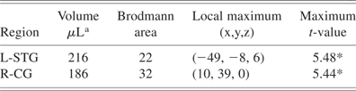
Note
L = left. R = right. STG = superior temporal gyrus. CG = cingulate gyrus. Regions listed from largest to smallest cluster volume.
a Clusters' minimum volume of activity ≥150 μL
* p ≤ .005.
Table 5 shows differences between groups identified by comparing neural activity for adults diagnosed with dyslexia and normal readers during tones comparison, pseudowords comparison or a contrast of tones comparison versus pseudowords comparison. During tones comparison, two clusters in left STG and inferior parietal lobe (BA 22 and 40, 389 μL; BA 42, 349 μL) showed significantly more activity in adults diagnosed with dyslexia than normal readers (Fig. 2C, left panel). During pseudowords comparison, a cluster in left STG (BA 22, extending into BA 40, 295 μL) exhibited more activity in adults diagnosed with dyslexia than normal readers (Fig. 2C, right panel). Between groups differences on a tones versus pseudowords contrast revealed one cluster with greater activity in adults diagnosed with dyslexia than normal readers (left hemisphere STG, BA 42, tones comparison > pseudowords comparison).
Table 5. Adults diagnosed with dyslexia exhibit greater activity than normal readers

Note
L = left. STG = superior temporal gyrus.
a Clusters' minimum volume of activity ≥150 μL
b Cluster represents greater activity for tones than pseudowords.
* p ≤ .005.
DISCUSSION
We used fMRI to identify neural activity associated with linguistic and nonlinguistic auditory working memory (AWM) comparison tasks in adults with normal reading and normal AWM skills versus adults with developmental phonological dyslexia and impaired AWM skills (per Auditory Consonant Trigrams Test (Stuss et al., Reference Stuss, Stethem and Pelchat1988) and Lindamood Auditory Conceptualization Test (Lindamood, Reference Lindamood1985) performances). For this study, the salient cognitive components needed for the AWM comparison task with pseudowords were speech perception, phonological awareness, AWM, comparison of phoneme sequences, and motoric response. To determine if adults diagnosed with dyslexia demonstrated a speech-specific deficit in AWM, we also measured neural activity during AWM of non-linguistic stimuli (pure tones). The salient cognitive components for the AWM comparison task for tones included auditory perception, AWM, comparison of tone sequences and motoric response. Therefore, the primary difference between tasks was the linguistic versus non-linguistic stimuli used for AWM.
Behavioral testing provided evidence of differential cognitive abilities between the adults diagnosed with dyslexia, and those with normal reading skills in the fMRI tasks' targeted cognitive areas of perception and explicit AWM comparison of linguistic stimuli. Test results indicated that compared to adults with normal reading skills, adults diagnosed with dyslexia were impaired in phonological awareness and AWM for linguistic stimuli. Additionally, AWM deficits were also evident during the complex linguistic analyses required by the LAC Test (Lindamood, Reference Lindamood1985). However, despite these cognitive deficits in the adults diagnosed with dyslexia, the groups' performance accuracy was equivalent on both fMRI AWM tasks (tones and pseudowords comparisons); this implied that differences in neural activity were not likely caused by differences in task accuracy but may indicate underlying differences in the neural substrates of task performance.
During both fMRI AWM tasks, neural activity was observed in temporal, inferior parietal, and frontal regions for both groups. However, on between groups comparisons within each task the adults diagnosed with dyslexia showed greater activity than normal readers in the left auditory association cortex (BA 22) and supramarginal gyrus (BA 40). As expected, differential activity in left primary auditory cortex (BA 42) occurred only during the tones comparison task, but again adults diagnosed with dyslexia exhibited more neural activity than normal readers in BA 42. To identify speech-specific regions of cortical activity, we subtracted the linguistic from non-linguistic activity within groups and then compared these differences between groups. Contrary to expectations of differential cortical activity with linguistic stimuli and similar cortical activity with non-linguistic stimuli, the adults diagnosed with dyslexia showed greater activity than normal readers in left primary or secondary auditory cortex for linguistic and non-linguistic stimuli. Also seeming contrary to expectations is the lack of a significant left hemisphere lateralization for pseudowords comparison versus baseline white noise. However, recent theory indicates that the perception of linguistic stimuli may evidence at best a “weak left-hemisphere bias at this (phonological) level of processing” (Hickok & Poeppel, Reference Hickok and Poeppel2007). As these authors' point out, this does not argue against the classical account of left hemisphere lateralization for linguistic perception, rather, it indicates that linguistic hemispheric lateralization may be less evident in some fMRI studies. Overall, during AWM of tones or pseudowords, the adults diagnosed with dyslexia demonstrated greater activity in the left STG and SMG than did the normal readers.
There are several possible explanations for why adults diagnosed with dyslexia in this study might show differential neural activity during AWM tasks when compared to normal readers. Behavioral testing prior to fMRI indicated impaired abilities in phonological awareness, AWM, and comparison of linguistic stimuli for the adults diagnosed with dyslexia. Based on these data and prior reports of relationships between behavioral abilities and functional brain activity, between groups differences in neural activity could be expected in STG (phonological perception), Inferior Parietal Lobe (phonological storage) and Inferior Frontal Lobe (comparison). However, the adults diagnosed with dyslexia only showed differential neural activity in left STG (with overlap into SMG). Because both groups show neural activity in the same region of interest (STG) and based on post-scanning reports of consistent strategy use between the groups during fMRI, it is unlikely that differences in neural activity could be caused by differential cognitive strategies. For example, the normal readers and readers diagnosed with dyslexia in Rumsey et al.'s (Reference Rumsey, Andreason, Zametkin, Aquino, King, Hamburger, Pikus, Rapoport and Cohen1992) study did not have equivalent task accuracy nor similar patterns of neural activity. This makes it possible that differential neural activity in that study could be because of participants' differential strategy use, a finding in another study too (Pekkola et al., Reference Pekkola, Laasonen, Ojanen, Autti, Jaaskelainen, Kujala and Sams2006). Our findings of similar task accuracy and similar regions of neural activity, but different degrees of neural activity between adults diagnosed with dyslexia and normal readers are consistent with Chait et al.'s (Reference Chait, Eden, Poeppel, Simon, Hill and Flowers2007) report of similar findings with an auditory perception task. However, despite similar levels of accuracy between the groups in our study, we cannot rule out that the adults diagnosed with dyslexia may have utilized greater cognitive effort than the normal readers to perform these AWM comparison tasks. Greater effort during fMRI has been shown to correspond with greater areas of neural activity (Poldrack, Reference Poldrack2000; Price & Friston, Reference Price and Friston1999). Thus, although our data does not support conclusions of differential cognitive strategy or levels of performance as possible cause of our neural activity findings, we cannot rule out the possibility that greater cognitive effort by the adults diagnosed with dyslexia contributed to our findings of greater activity in left STG.
Other possible explanations of our findings of greater activity in left STG for readers diagnosed with dyslexia include deficient echoic memory, impaired sound sequence discrimination, impaired auditory feature analysis skills and priming by a dominant hand motor movement. It is possible that echoic memory played a role in the groups' differential neural activity, as echoic memory would affect AWM and it cannot be isolated in this study's tasks. Because the AWM tasks in this study required sound sequence discrimination, it is not likely that this contributed to our results, as the initial portion of the LAC Test measures sound sequencing without an AWM comparison and no differences existed between the groups on this portion of the LAC Test. Auditory feature analysis is a low-level analysis that would be common to tone and pseudoword AWM tasks, but the contributions of auditory feature analysis to the neural activity differences cannot be ruled out, because no tasks without memory were administered in this study. Similarly, because of white matter pathways between SMA and STG, we cannot rule out that the right-handed button press response may have influenced activity in left STG during both tasks. However, because button press preparation most likely produces activity in mesial pre-SMA and SMA and the motor task was identical for both AWM tasks, this influence should have been similar for both groups of participants and for both tasks. Thus, it is not likely that our results can be exclusively attributed to impaired sound sequence discrimination, or motor task priming, but auditory feature analysis and impaired echoic memory may have contributed to the differences in neural activity.
Differential neural activity may be related to aberrant neuroanatomic structures. Ramus (Reference Ramus2004) proposed that genetically driven, focal, cortical anomalies in the left perisylvian cortex are the primary cause of phonological impairments in developmental dyslexia. These cortical anomalies seem to develop in utero, and contribute to morphological differences in perisylvian language cortex in adults with developmental dyslexia (Eckert, Reference Eckert2004; Galaburda, Reference Galaburda1999; Galaburda et al., Reference Galaburda, LoTurco, Ramus, Fitch and Rosen2006). These structural anomalies affect functional cortico-cortical connections and may contribute to impaired phonological processing/reading skills. Aberrant fiber tracts in left posterior temporo-parietal cortices covary with children's and adults' reading skills, with higher fractional anisotropy and coherence indexes coinciding with better reading skill (Deutsch et al., Reference Deutsch, Dougherty, Bammer, Siok, Gabrieli and Wandell2005; Klingberg et al., Reference Klingberg, Hedehus, Temple, Salz, Gabrieli, Moseley and Poldrack2000). Likewise, failures in neural pruning might produce overabundant synaptic connections in a cortical region, contributing to increased neural activity in this region (Haier et al., Reference Haier, Chueh, Touchette and Lott1995). However, thicker myelin sheaths and larger axon diameters account for greater white matter volume in normal readers' left posterior STG, providing efficient neural conduction in the left STG (Golestani et al., Reference Golestani, Paus and Zatorre2002). Also, these cortical anomalies may be related to subcortical structural anomalies in humans (Galaburda, Reference Galaburda1999), and a reciprocal impact between cortical and subcortical anomalies may influence AWM and BOLD response in a cortical region. Because post-mortem neuroanatomic analyses identify aberrant structures in the left perisylvian region of adults with developmental dyslexia (Galaburda et al., Reference Galaburda, LoTurco, Ramus, Fitch and Rosen2006; Galaburda et al., Reference Galaburda, Sherman, Rosen, Aboitiz and Geschwind1985), disordered localization of function is a possible contributor to our enhanced BOLD findings. Thus, future studies combining functional and structural neuroimaging are needed to elucidate the relationships between neuroanatomic structure, behavioral functions, and neural activity.
Overall, our findings are consistent with and extend previous studies indicating that phonological processing and AWM are altered in individuals with developmental dyslexia. Whereas this study's generalizability is limited by both groups exhibiting superior intellectual abilities and having greater than 12 years of education, the study's unique contribution is that activity differences in the left temporoparietal region between dyslexics and controls during AWM tasks are not unique to speech stimuli. Also, the differential neural activity was predominately in the left superior temporal gyrus. Our primary findings of higher levels of neural activity in posterior superior temporal and inferior parietal regions and impaired AWM skills with linguistic stimuli in adults diagnosed with dyslexia compliments behavioral studies showing AWM deficits in developmental dyslexia (Banai & Ahissar, Reference Banai and Ahissar2006; Berninger et al., Reference Berninger, Abbott, Thomson, Wagner, Swanson, Wijsman and Raskind2006; Kibby et al., Reference Kibby, Marks, Morgan and Long2004). The fact that activity differences between groups existed only in primary and secondary auditory cortex for linguistic and nonlinguistic stimuli suggests that the atypical activity during AWM tasks in these adults with developmental dyslexia could be because of their poor auditory perception, AWM, or comparison skills rather than poor frontal executive functions, phonological awareness skills, or speech perception skills. This study cannot determine exactly which of these impaired skills or which combination of impaired skills are causing the greater neural activity during AWM tasks in adults diagnosed with dyslexia. Future research that combines functional and structural neuroimaging with behavioral performance is needed to determine the precise mechanisms that account for temporoparietal activity differences during AWM in developmental dyslexia.
ACKNOWLEDGMENTS
This work was supported by Associate Investigator B3480H (Conway) and Research Career Scientist B3470S (Crosson) awards from the Department of Veterans Affairs Rehabilitation Research and Development Service, the Donald D. Hammill Foundation (Conway), American Psychological Foundation (Conway), Fred J. Wellington Foundation (Conway), grants P50 DC 03888 and R01 DC 007387 from the National Institute on Deafness and Other Communication Disorders (Crosson), and grant HD30988 from the National Institute of Child Health and Human Development (Torgesen).


