Refine search
Actions for selected content:
3823 results in Surgery
6 - Overview of systemic therapy
-
- Book:
- Handbook of Breast MRI
- Published online:
- 05 March 2012
- Print publication:
- 17 November 2011, pp 199-200
-
- Chapter
- Export citation
5 - High-risk screening using breast MRI
-
- Book:
- Handbook of Breast MRI
- Published online:
- 05 March 2012
- Print publication:
- 17 November 2011, pp 112-134
-
- Chapter
- Export citation
2 - Imaging-related anatomy and pathology
-
- Book:
- Handbook of Breast MRI
- Published online:
- 05 March 2012
- Print publication:
- 17 November 2011, pp 22-47
-
- Chapter
- Export citation
Preface
-
- Book:
- Handbook of Breast MRI
- Published online:
- 05 March 2012
- Print publication:
- 17 November 2011, pp ix-ix
-
- Chapter
- Export citation
Appendices
-
- Book:
- Handbook of Breast MRI
- Published online:
- 05 March 2012
- Print publication:
- 17 November 2011, pp -
-
- Chapter
- Export citation
Contents
-
- Book:
- Handbook of Breast MRI
- Published online:
- 05 March 2012
- Print publication:
- 17 November 2011, pp vii-viii
-
- Chapter
- Export citation
3 - TNM classification
-
- Book:
- Handbook of Breast MRI
- Published online:
- 05 March 2012
- Print publication:
- 17 November 2011, pp 192-195
-
- Chapter
- Export citation
Index
-
- Book:
- Handbook of Breast MRI
- Published online:
- 05 March 2012
- Print publication:
- 17 November 2011, pp 204-213
-
- Chapter
- Export citation
8 - Breast cancer genes and genetic testing
-
- Book:
- Handbook of Breast MRI
- Published online:
- 05 March 2012
- Print publication:
- 17 November 2011, pp 202-203
-
- Chapter
- Export citation
1 - Nephrogenic systemic fibrosis
-
- Book:
- Handbook of Breast MRI
- Published online:
- 05 March 2012
- Print publication:
- 17 November 2011, pp 190-190
-
- Chapter
- Export citation
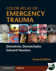
Color Atlas of Emergency Trauma
-
- Published online:
- 07 October 2011
- Print publication:
- 06 October 2011
Index
-
- Book:
- Color Atlas of Emergency Trauma
- Published online:
- 07 October 2011
- Print publication:
- 06 October 2011, pp 308-313
-
- Chapter
- Export citation
4 - Thoracic Injury
-
-
- Book:
- Color Atlas of Emergency Trauma
- Published online:
- 07 October 2011
- Print publication:
- 06 October 2011, pp 75-118
-
- Chapter
- Export citation
8 - Burn Injuries
-
-
- Book:
- Color Atlas of Emergency Trauma
- Published online:
- 07 October 2011
- Print publication:
- 06 October 2011, pp 253-270
-
- Chapter
- Export citation
Contributors
-
-
- Book:
- Color Atlas of Emergency Trauma
- Published online:
- 07 October 2011
- Print publication:
- 06 October 2011, pp x-x
-
- Chapter
- Export citation
Preface
-
-
- Book:
- Color Atlas of Emergency Trauma
- Published online:
- 07 October 2011
- Print publication:
- 06 October 2011, pp xiii-xiii
-
- Chapter
- Export citation
Contents
-
- Book:
- Color Atlas of Emergency Trauma
- Published online:
- 07 October 2011
- Print publication:
- 06 October 2011, pp vii-ix
-
- Chapter
- Export citation
Acknowledgments
-
- Book:
- Color Atlas of Emergency Trauma
- Published online:
- 07 October 2011
- Print publication:
- 06 October 2011, pp xiv-xiv
-
- Chapter
- Export citation
11 - Disaster Medicine
-
-
- Book:
- Color Atlas of Emergency Trauma
- Published online:
- 07 October 2011
- Print publication:
- 06 October 2011, pp 301-307
-
- Chapter
- Export citation
Copyright page
-
- Book:
- Color Atlas of Emergency Trauma
- Published online:
- 07 October 2011
- Print publication:
- 06 October 2011, pp iv-iv
-
- Chapter
- Export citation
