





Introduction
Leishmaniasis is an infectious disease caused by parasites of the genus Leishmania, a member of the Trypanosomatidae family, and is transmitted through the bite of sandflies (Reithinger et al., Reference Reithinger, Dujardin, Louzir, Pirmez, Alexander and Brooker2007). This disease presents a wide spectrum of clinical symptoms ranging from self-healing skin ulcers to severe visceral forms, sometimes leading to death. Generally, visceral leishmaniasis and mucocutaneous leishmaniasis require prompt treatment, but in the case of cutaneous leishmaniasis, treatment may not always be necessary depending on the species of the disease agent and the patient’s condition (Bailey and Lockwood, Reference Bailey and Lockwood2007). Currently, the treatment cost, drug toxicity and drug resistance in the treatment of leishmaniasis are among the major concerns of physicians. Recently, new drug delivery methods have been developed that can deliver the drug directly to target cells. The use of drug delivery systems that deliver anti-leishmanial agents directly into reticuloendothelial system cells such as macrophages has been highly regarded as an effective strategy for prevention and treatment (Hepburn, Reference Hepburn2000). Nanoparticles are recognized as foreign bodies by the body, engulfed by macrophages, and act as a specific drug delivery system to Leishmania residing in macrophages (Minodier and Parola, Reference Minodier and Parola2007). Liposomes offer several advantages, including low toxicity, the ability to co-deliver hydrophilic and hydrophobic drugs, and prolonged retention at the site of administration compared to conventional formulations. The introduction of nanoliposomes in antileishmanial therapy has shown potential for an increase in the therapeutic efficacy of several drugs (Want et al., Reference Want, Islammudin, Chouhan, Ozbak, Hemeg, Chattopadhyay and Afrin2017; Fattahi Bafghi et al., Reference Fattahi Bafghi, Haghirosadat, Yazdian, Mirzaei, Pourmadadi, Pournasir, Hemati and Pournasir2021; Khan et al., Reference Khan, Dar, Aib, Khalid, Iqbal and Razaque2024).
The aim of this study is to achieve an effective treatment for cutaneus leishmaniasis using nanoliposome technology.
Materials and methods
Animals and parasites
Number 82 female BALB/c inbred mice at 8–6 weeks of age in seven groups are infected with promastigotes of L. major in the stationary growth phase through subcutaneously into the upper area of the tail. At 4 weeks postinfection, the lesions were measured with calipers in two dimensions, the mean diameters were determined. The group of mice including infected mice with Leishmania disease receiving of nanoliposomal quercetin (QN), nanoliposomal quercetin-paromomycin (QPN), quercetin-paromomycin and clindamycin (QPC), nanoliposomal quercetin- clindamycin and paromomycin (QCPN), paromomycin (P) as treatments, compared to healthy group mice with no interference as a negative control. Then, in the fourth week after infecting the mice with L. major, they are locally treated twice a day with ointment for up to 8 weeks. The measurement of the ulcers will be based on the study by Jaafari et al. (Reference Jaafari, Bavarsad, Fazly Bazzaz, Samiei, Soroush, Ghorbani, Lotfi Heravi and Khamesipour2009). Lesions were measured weekly during treatment, and the measurement process be stopped in the 8 week after treatment.
Preparation quercetin-nanoliposomal
The preparation method for QN involved using the ultrasonic thin-layer dispersion method. A specific amount of quercetin in 10 mL of methanol solution was dissolved, which was then magnetically stirred to make the solution particle-free and transparent (at room temperature, 220 revolutions per minute). A certain amount of lecithin and cholesterol in 20 mL of chloroform solution was dissolved, which was also magnetically stirred to make the solution particle-free and transparent (at room temperature, 220 revolutions per minute). The two above solutions were uniformly mixed until they turned into an oil-in-water state and were then transferred to a 250-mL rotary evaporation flask. The organic solvents were removed by rotary evaporation at a temperature of 30 °C and 30 revolutions per minute until a bright yellow layer appeared in the rotary evaporation flask. Deionized water (40 mL) was added to the evaporation flask for hydration, and the layer was removed with ultrasound for 20 min. The quercetin nanoliposomes were labelled QN-1. Simultaneously, charge-free nanoliposomes were prepared and labelled QN-0. QN-1 and QN-0 were stored for future use at 4 °C.
Determination of the encapsulation efficiency of quercetin nanoliposomes
QN-1 (0.5 mL) was diluted with PBS solution to a volume of 10 mL. It was centrifuged at 8000 rpm for 30 min. The absorbance of the upper liquid at a wavelength of 370 nm was measured to obtain the concentration of free quercetin C1. QN-1 (0.5 mL) was dissolved in 4.5 mL absolute ethanol and diluted with PBS solution to a volume of 10 mL. The total concentration of quercetin C2 was obtained by centrifugation and measuring the absorption. Among these, samples of neutral nanoliposomes were used as the control group. The encapsulation rate was calculated according to the following formula: Encapsulation rate (%) = (C1 − C2) ÷ C2 × 100%, where C1 is the content of free quercetin in QUE-NL-1 and C2 is the content of quercetin after breaking the QUENL-1 membrane. Nanoliposome curcumin dried by freeze-drying (1.5 mg) was milled and mixed uniformly with 1% potassium bromide powder. This nanoliposome was pressed to prepare a thin sample sheet with an approximate thickness of 2 mL, which was then measured with an infrared spectrometer. Infrared spectral information was obtained in the frequency range of 4000–400 cm−1.
Preparation of an oil/water cream containing quercetin nanoliposomes
Vaseline (10%) and cetyl alcohol (6.8%) were heated to 77–82 °C as an oil phase. quercetin nanoliposomes (0.5%) was added to the lipid phase at the end of the melting process. Tween 80 (2.8%) and water up to 100% were heated as the aqueous phase with constant stirring until complete solubilization. The aqueous phase was added to the oil phase and stirred until cooled to room temperature.
Characterization of quercetin nanoliposomes
Negative staining of nanoliposomes was used to observe the fine structure of QN-1. The quercetin nanoliposome solution was diluted. Particle size distribution (scattering index) was measured by Mastersizer 3000 in the range of 0.1–1000 micrometers. Then, a specific zeta potential dish was replaced and potentiometric measurement was performed.
Analysis of the promastigote form and determination of EC50
The effects of the formulations on the viability of Leishmania promastigotes were assessed by monitoring MTT metabolism after a 48-h culture period in the presence of the formulations. Parasites were harvested at stationary phase of culture, and 400 000 promastigotes were added to each well of 96-well flat-bottom plates containing different concentrations of the formulations; triplicate wells were used for each concentration. The plates were incubated at 25 °C for 48 h prior to the addition of MTT (40 L/well of 5 mg/mL in PBS), and then the plates were incubated in the dark at 37 °C for a further 4 h. The formation of formazan was evaluated by adding 50 L/well 20% sodium dodecyl sulphate and incubating the plates overnight at 37 °C, and the relative absorbance was photometrically measured with an enzyme-linked immunosorbent assay reader (Statfax-2100; Awareness Technology) at 545 nm. The relative absorbance was correlated to the number of promastigotes per well by using a standard curve that consisted of the results for different numbers of promastigotes treated with the MTT dye, as explained above. The 50% effective concentration (EC50) for each formulation was calculated by the Litchfield-Wilcoxon method with PCS (version 4) software (Jaafari et al., Reference Jaafari, Bavarsad, Fazly Bazzaz, Samiei, Soroush, Ghorbani, Lotfi Heravi and Khamesipour2009)
Evaluation of synthesized ointment in vivo (animal phase study)
In vivo experiments L. major promastigotes (MHROM/IR/75/ER) at stationary phase (2 × 10⁶) were inoculated sub-cutaneously into the upper area of the tail in BALB/c mice (seven groups, n = 12 per group). No significant differences (p > 0.05) were observed in lesion size among the different groups. Following lesion development, mice were treated topically by ointment at a dose of 50 mg twice daily. As positive control (group 5), the mice were treated with pentostam. As negative control (group 6), the mice were treated with PBS. As additional negative control (group 7), the mice remain untreated. After 4 weeks, the size of cutaneous lesions was measured by colis in 2 diameters. The smears from skin lesion were prepared for microscopic examinations and parasitological assessment.
The toxicity of quercetin nanoliposomes in living organisms
The toxicity of QN synthesized in healthy BALB/c mice at the highest therapeutic dose of this drug was evaluated after 24 h of injection. This evaluation was done by measuring serum levels of liver markers such as AST, ALT and ALP.
Statistical analysis
Statistical analysis One-way ANOVA statistical test was used to assess the significance of the differences among various groups, p < 0.05 was considered as statistically significant.
Results
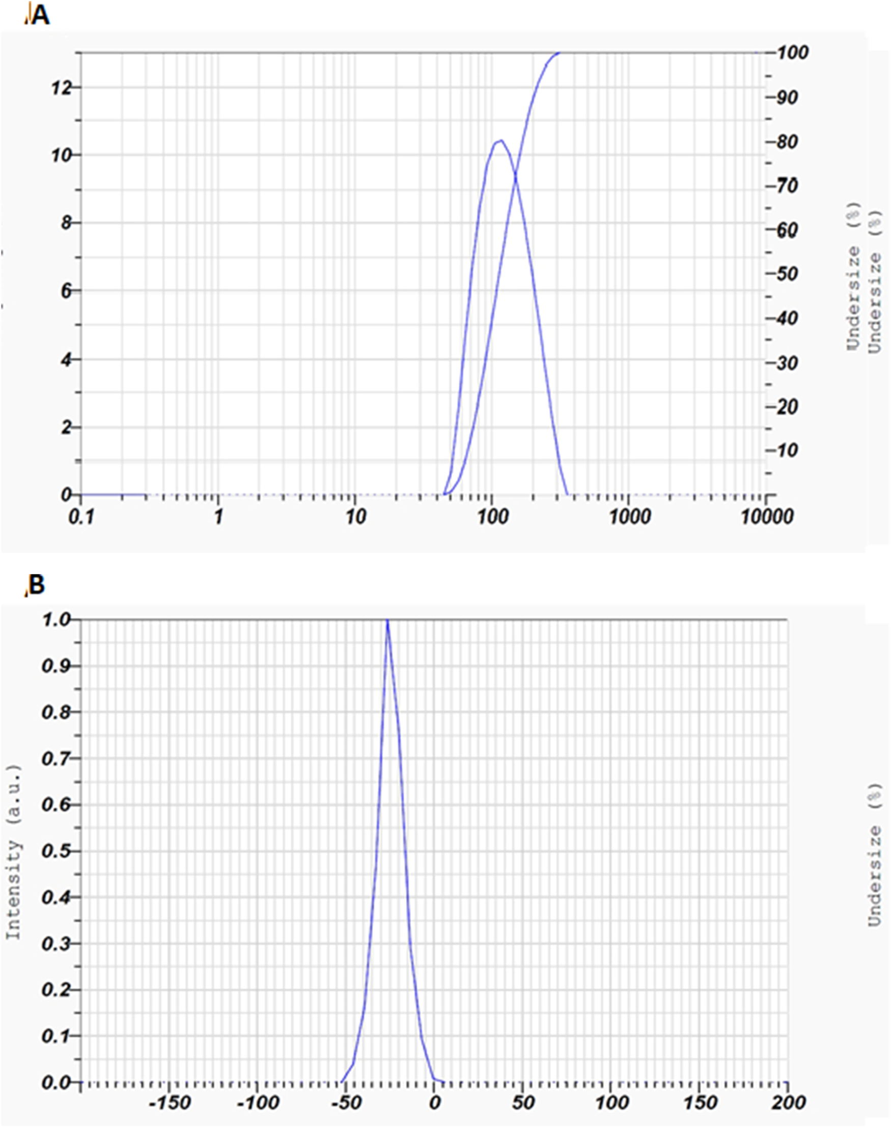
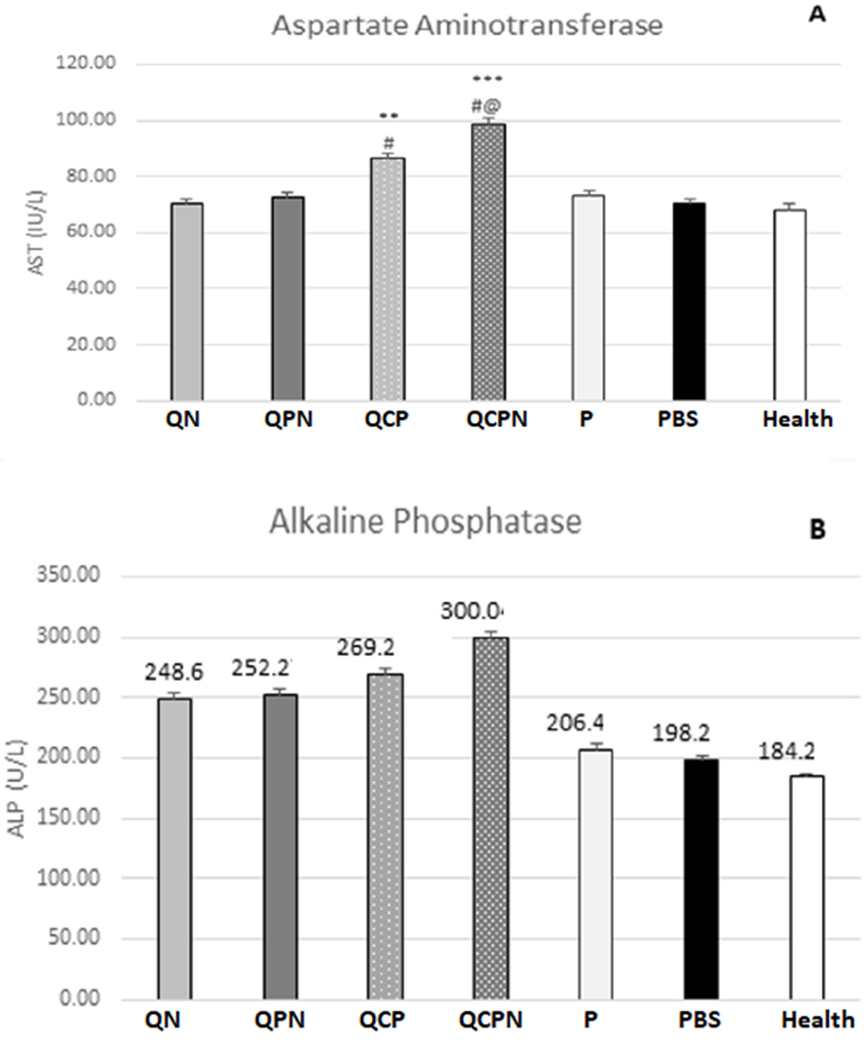
The average particle size in the nanoliposome containing Quercetin NanoCAV was 74.8 nm. The dispersibility indices were equal to 0.61 (Figure 1A). The zeta potential of the nanoliposome was 24.2 mV (Figure 1B). These results indicate that the formulated liposome was in the nanometer scale and the particles were heterogeneously dispersed. ED50 levels of QN, QPN, QCP, QCPN and P against L. major promastigotes were 70 ± 2, 57 ± 2, 51 ± 9, 43 ± 8 and 41 ± 8 micromolar, respectively. There was no significant difference in the activity of liposomal formulations against L. major promastigotes. Hepatic enzyme activity in serum after treatment of mice with QN, QPN, QCP, Q-C-N and P compared to control mice is shown in Figure 2.
Particle size and distribution index (A); Zeta potential for quercetin nanoliposomes (B), Anti-leishmanial activity of synthesized compounds.

The activity of liver enzymes aspartate aminotransferase (A), alkaline phosphatase (B), in mice infected with Leishmania disease receiving Q-N, Q-P-N, P-C-Q, Q-C-N P, and P, PBS, as treatments, compared to healthy group mice with no interference.

Infection with L. major did not increase AST activity in mice in the negative control group (PBS) compared to mice receiving standard drug. Additionally, treatment with QN, QPN, and QCP had no increasing effect on serum AST levels. On the other hand, AST activity increased in mice treated with QCPN (Figure 2A). It is worth mentioning that no significant difference in ALT activity was observed between the different groups of mice (Figure 2B).
Evaluation of synthesized compounds on a mouse model
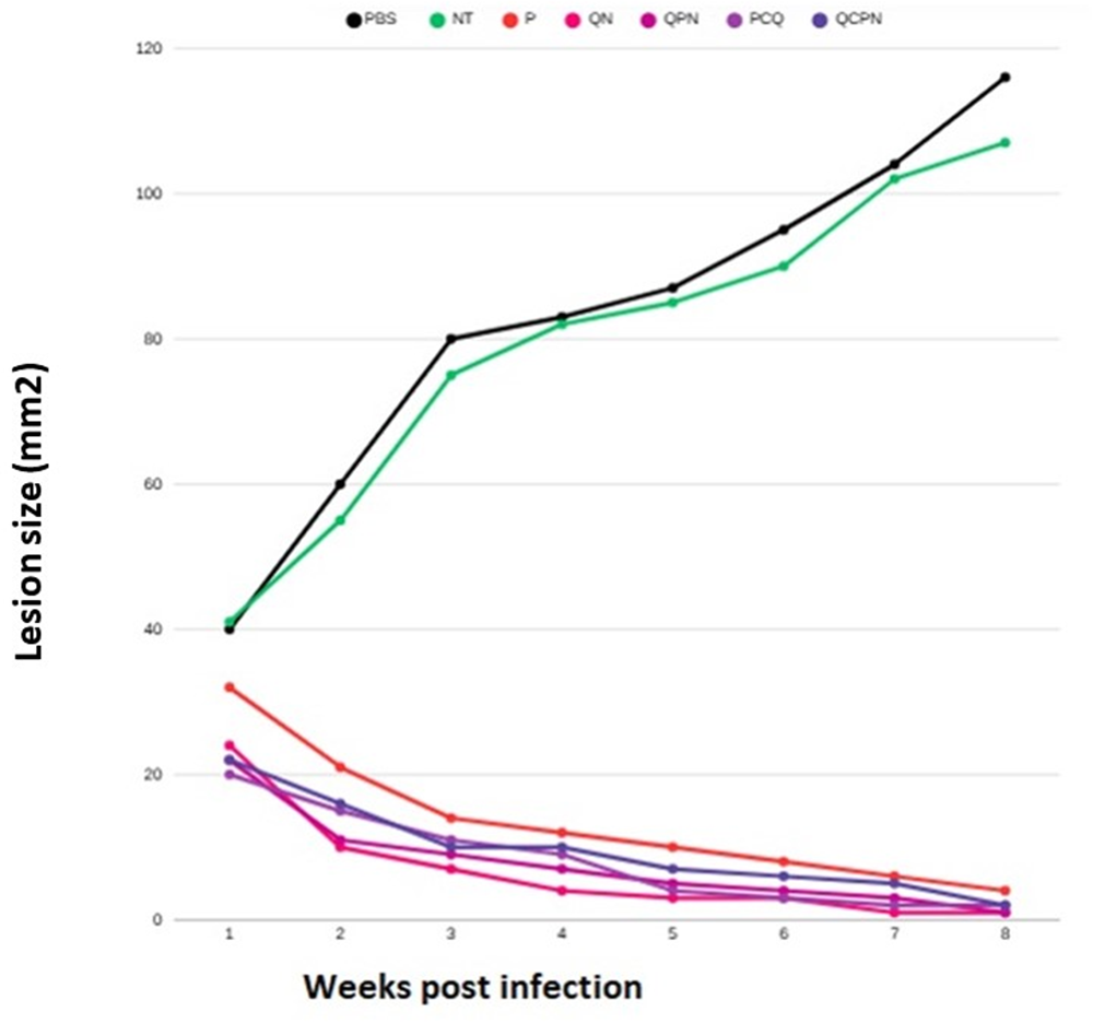
There were no significant differences (p ≤ 0.05) in the lesion sizes among the different groups before the initiation of the treatment (Figure 2; week 4 postinfection). The topical application of all treatments groups compare to PBS and untreated group caused significant reductions in the lesion sizes. At week 12 postinfection, the effects of QN, QPN, QCP, QCPN and P in reducing the lesion size were significant (p < 0.001) when compared to PBS and untreated groups. No significant differences (Figure 3) were observed in the two control groups that received PBS or untreated (p > 0.05).
Effect of topical liposomal substance and PBS on the progress of the lesion size in the BALB/c mouse model of CL caused by L. Major. At week 4 post-infection, lesion size was measured and mean diameters were determined. Mice were topically treated with formulations twice daily for 8 weeks. At the beginning of treatment there were no significant (p > 0.05) differences in the size of lesions among various groups; *, *** represents (p < 0.05) and (p < 0.003), respectively.

Discussion
The current study was designed to prepare and characterize QN and then determine the effects of the QN in vitro against L. major promastigotes and in vivo against ulcers induced by L. major infection in susceptible BALB/c mice. The primary medication utilized for treating leishmaniasis is pentavalent antimony (SbV), known as Glucantime in Iran (Sharifi et al., Reference Sharifi, Khosravi, Aflatoonian, Salarkia, Bamorovat, Karamoozian, Moghadam, Sharifi, Afshar and Afshari2023). Because of the adverse effects caused by the aforementioned medications, it is crucial to discover a treatment for leishmaniasis that has minimal side effects (Parkash et al., Reference Parkash, Ashwin, Dey, Sadlova, Vojtkova, Van Bocxlaer, Wiggins, Thompson, Dey and Jaffe2024). One of the best extract from plant is Oryan et al. (Reference Oryan, Bemani and Bahrami2023). Quercetin is a plant flavonol from the flavonoid group of polyphenols and is presented in many plants and food sources (Clemente et al., Reference Clemente, Murillo, Garro, Arbeláez, Pineda, Robledo and Ravetti2024). Studies of quercetin showed that quercetin is an exceptional natural compound and can be a source of hope for the treatment of important infections such as malaria and leishmaniasis (Bashir et al., Reference Bashir, Shabbir, Ud Din, Khan, Ali, Khan, Kim and Khan2023). The effectiveness of in biological settings is typically influenced by their physicochemical characteristics, particularly their reactivity and membrane permeability, which are crucial in determining their performance on the skin. Comprehensive physicochemical analyses are essential for ensuring the stability and effectiveness of liposomes. Hence, parameters such as particle size and ζ-potential serve as indicators of the physical stability of liposomes (Ramos et al., Reference Ramos, Vallejos, Borges, Almeida, Alves, Aguiar, Fernandes, Guimarães, Fujiwara and Loiseau2022).
The reported dispersibility index (PDI = 0.61) of this study indicates a highly polydisperse system, reflecting a heterogeneous particle size distribution. Additionally, the measured zeta potential (24.2 mV) suggests moderate electrostatic stability, which may be insufficient to ensure long-term colloidal stability. Nanotechnology has been shown to be a successful method for accelerating wound healing, while nanoliposomes serve as effective drug delivery systems that enhance the solubility of hydrophobic drugs in both plasma and skin tissue (Frézard et al., Reference Frézard, Aguiar, Ferreira, Ramos, Santos, Borges, Vallejos and De Morais2022). They also help in reducing the rate of drug release. In this particular study, nanoliposomal quercetin was utilized in the treatment of cutaneous leishmaniasis. The findings revealed that nanoliposomal quercetin effectively slowed down the progression of lesions caused by Leishmania infection in BALB/c mice. Furthermore, it exhibited promising anti-leishmanial activity. In conjunction with other drugs, nanoliposomal formulations have shown positive outcomes (Want et al., Reference Want, Islammudin, Chouhan, Ozbak, Hemeg, Chattopadhyay and Afrin2017). For instance, the nanoliposomal miltefosine displayed stronger anti-leishmanial effects compared to regular miltefosine, likely due to its improved ability to penetrate dermal-infected macrophages. Interestingly, Akbari et al. conducted a study using a topical nanoliposomal formulation of curcumin against leishmaniasis in a BALB/C mouse model (Akbari et al., Reference Akbari, Saeedi, Enayatifard, Morteza-Semnani, Hashemi, Babaei, Rahimnia, Rostamkalaei and Nokhodchi2020). Their results indicated no significant difference in lesion size between treated and control BALB/c mice, contrary to the outcomes of our research. Additionally, they reported that there was no significant alteration in the splenic parasite burden among the tested animal groups. Intriguingly, when compared to the outcomes seen in size of infected skins of mice treated with quercetin with other standard drugs such as panotostam and paromomycin, there was not any significant difference. The size of liposomes plays a crucial role in their performance and stability within the body, with liposomes measuring less than 0.6 µm able to penetrate the skin effectively (Abtahi-Naeini et al., Reference Abtahi-Naeini, Hadian, Sokhanvari, Hariri, Varshosaz, Shahmoradi, Feizi, Khorvash and Hakamifard2021). ED50 levels of QN, QPN, QCP, QCPN and P against L. major promastigotes were 70 ± 2, 57 ± 2, 51 ± 9, 43 ± 8 and 41 ± 8 micromolar, respectively. There was no significant difference in the activity of liposomal formulations against L. major promastigotes. When comparing the impact of various concentrations of nanoliposomal on L. major promastigotes, all concentrations exhibited a reduction in the number of promastigotes compared to control samples. However, the antiparasitic effect was more potent with paromomycin. These results demonstrate the no enhanced anti-parasitic efficacy of nanoliposomal compared to traditional Drugs.
Assessment of hepatic enzyme activity is a critical indicator of potential hepatotoxicity associated with experimental treatments. In this study, serum levels of AST and ALT were evaluated following treatment with QN, QPN, QCP, QCPN and P in Leishmania-infected mice, and compared with control groups. This suggests a favourable hepatic safety profile for these treatments. Importantly, treatment with QN, QPN, and QCP and P did not result in elevated AST and ALT levels, indicating that these formulations did not exert hepatotoxic effects. Unlike our study, the previous study indicated that paromomycin can cause biochemical alterations of liver enzymes (Davachi et al., Reference Davachi, Nahrevanian, Omidinia, Hajihosseini, Amini, Farahmand, Mirkhani and Javadian2009). The elevated AST observed with QCPN highlights the need for further investigation, including histopathological analysis and additional biochemical markers. Lesion size is a key clinical indicator of disease progression and therapeutic efficacy in cutaneous leishmaniasis. Following treatment, all topical formulations produced a significant reduction in lesion size compared with both the PBS-treated and untreated control groups. Further studies incorporating parasite burden assessment, histopathological analysis, and relapse monitoring would be valuable to better characterize the therapeutic potential of these treatments. One limitation of the present study is that it employed L. major parasites; therefore, validation of these results in other Leishmania species is necessary. BALB/c mice were selected based on the assumption that successful protection or cure in this susceptible strain would imply potential efficacy in more resistant mouse strains. Previous work has outlined key stages of anti-leishmanial drug discovery using both the BALB/c – L. major and golden hamster – L. panamensis models (Grogl et al., Reference Grogl, Hickman, Ellis, Hudson, Lazo, Sharlow, Johnson, Berman and Sciotti2013). However, due to pathological differences between murine models and human disease, extrapolation from the BALB/c model to humans has inherent limitations. In this context, the golden hamster may represent a more appropriate in vivo model, and the reliance on BALB/c mice should be regarded as a limitation of the present findings.
Data availability statement
All data are available in the publication. Raw data may be obtained from the University of AJA.
Acknowledgements
The authors would like to express the deepest thanks from AJA University of Medical Sciences, AJA.
Author contributions
MD: Study design, data analysis, writing and revision; MM, ZH, RS, HSh, MM and MCh: data analysis, writing and revision, writing and revision, sample collection.
Financial support
Not applicable.
Competing interests
The authors declare there are no conflicts of interest.
Ethical standards
This study was reviewed and approved by ethical commited of AJA University of medical university, Tehran, Iran (IR.AJAUMS.REC.1403.031).

 Open access

