

Fe deficiency anaemia (IDA) is a nutritional disorder afflicting large population groups in the world( 1 ). It is prevalent amongst vulnerable infants, adolescent girls, pregnant women and the aged in both developed and developing countries. The challenge of increased physiological requirements of Fe for growth and reproduction, within these population groups is compounded by inadequate intake and poor Fe bioavailability from foods( Reference Aspuru, Villa and Bermejo 2 ). This is particularly evident in populations subsisting predominantly on vegetables or plants for their sources of Fe. Substantive evidence has shown that IDA has debilitating effects on cognition, mental function, work performance and pregnancy outcomes( Reference Munoz and Humeres 3 , Reference Rufer, Tran and Attridge 4 ). Consequently, Fe supplementation and fortification of staple foods in different countries( Reference Gera, Sachdev and Boy 5 ) have been practical approaches to alleviate this important nutritional disorder. Fe compounds used for such purposes include ferrous sulphate, ferric pyrophosphate, fumarate, gluconate and ferric ammonium citrate among a host of others. Some of these Fe formulations, because they are redox active, cause irritations or disturbances to the gastrointestinal tract by inducing stomach upset, abdominal pain, constipation or nausea( Reference Tiwari, Mahdi and Chandyan 6 – Reference Tolkien, Stecher and Mander 8 ). Fe supplements, as a locus of high concentration of Fe in the gastrointestinal lumen, could cause DNA damage by generating reactive oxygen species in Fenton reaction. Strategies and initiatives aimed at ameliorating some of the adverse effects of Fe supplements led to the synthesis of nano Fe compounds such as a nano particulate ligand modified Fe(III) polyoxo-hydroxide( Reference Pereira, Bruggraber and Faria 9 ) and nanoparticulate Fe pyrophosphates( Reference Hilty, Arnold and Hilbe 7 ), which were shown to have high bioavailability. This promising outcome underlines the need to exploit and explore other formulations or compounds that exhibit rigid chemical structures, are redox inert, and are highly soluble to maintain colloidal Fe for absorption in the milieu of the gastrointestinal tract. Consequently, Fe multi-amino acid chelate (IMAAC) was formulated and found to be better tolerated than FeSO4 in a randomised, double-blind placebo-controlled trial (RCT) in healthy premenstrual women( Reference Fouad, Evans and Sharma 10 ). There is currently no information on the metabolism and the mechanism of Fe absorption from IMAAC. The present study, therefore, investigates the bioavailability, characteristics and metabolism of IMAAC using both in vitro cell culture and in vivo mice models. It is hypothesised that Fe absorption from IMAAC will be comparable with that of standard FeSO4. To test this hypothesis, anaemic mice were administered IMAAC or FeSO4 to evaluate Hb regeneration, serum, liver and spleen Fe levels and Fe-related genes in the tissues.
Methods
Reagents and chemicals
Unless otherwise stated chemicals and reagents were obtained from Sigma-Aldrich. Standard American Institute of Nutrition diet (AIN-76A) without Fe (low-Fe diet) and the Fe-replete diet contained 48 mg/kg Fe were provided to the mice. IMAAC was prepared and supplied by Biotron Laboratories, Centerville UT, USA. IMAAC used contained 10 % elemental Fe chelated in an aqueous solution to low molecular weight peptides, polypeptides and amino acids derived from enzymatically hydrolysed soya protein isolates using non-GM organism Food chemical codex/food grade enzymes. The preparation is then dried and milled to a fine powder. IMAAC preparation with excipients was used in a RCT to test safety and tolerability( Reference Fouad, Evans and Sharma 10 ).
Animal studies
Three-week-old CD1 strain male mice (Charles Rivers) were used for the studies. Mice were housed in a light- and a temperature-controlled room with ad libitum access to diet and water. Fe-deficient diet (TD 80396, 3/kg Fe, and Fe-replete diet TD 80396, 48 mg/kg ferric citrate (Harlan Teklad) were used for the experiment and the diets composition, a modification of AIN-76A is given by Chaudhury et al. ( Reference Chaudhury, Kim and Mehnaz 11 ).
Hb repletion study
In all, fifteen CD1 mice were made Fe deficient on a low Fe diet of 3 mg/kg, for 3 weeks. Five mice were also placed on a normal Fe-replete diet (48 mg/kg) to serve as a control. Following this, blood was withdrawn from the tails to determine the initial Hb levels of the mice. The Fe-depleted mice were then divided into three treatment groups based on similar Hb levels. These fifteen mice were maintained on the low-Fe diet in groups of five, of which one group did not receive any Fe supplementation (low-Fe diet), the two other groups were gavaged daily liquid solutions of 150 µg of Fe as IMAAC or FeSO4. This was done for 10 d, and the total food consumption of the mice was measured. After 10 d of Fe supplementation, the mice were weighed, anaesthetised and blood samples were taken for Hb and serum Fe determinations. The mice were then killed, and the spleen, duodenum, kidney and liver samples were excised, snap frozen in liquid N2 and stored in −80°C for further analysis. The experiment was approved by the Institutional (King’s College London) Animal Welfare and Ethics Board. All procedures were approved and conducted in accordance with the UK Animals (Scientific Procedures) Act, 1986.
Measurement of Hb, serum iron and tissue non-haem Fe contents
Hb concentrations were calculated from the change in optical density at 540 nm, following the addition of 5 µl of whole blood to Drabkin’s reagent (Sigma-Aldrich) and centrifugation (Heraeus Biofuge Pico) at 13 000 rpm for 5 min. Serum Fe was measured with a liquid ferrozine-based Fe reagent (Thermo Electron).
Tissue non-haem iron
Tissue samples were weighed and homogenised (1:5, w/v) in 0·15 m-NaCl in 10 mm-NaOH-Hepes buffer (pH 7·0) using a 1 ml glass Dounce homogenizer (Wheaton Scientific). An aliquot of the homogenate was then analysed for non-haem Fe content as described by Simpson & Peters( Reference Simpson and Peters 12 ). The Fe values were expressed as either content (µmol Fe/organ) or concentration (nmol Fe/mg wet weight).
Quantitative real-time reverse transcription PCR
Total RNA was extracted from tissue samples using TRIZOL reagent (Invitrogen) according to manufacturer’s instructions. Quantitative RT-PCR was carried out using an ABI Prism 7000 (Applied Biosystems) detection system in a two-step protocol with Roche Universal primers and probes. Quantitative measurement of each gene was normalised to the cycle threshold value for ribosomal protein L (ribosomal protein L19 (RPL 19)).
Sequences of mouse primers used, forward and reverse, respectively, are as follows:

Cell studies
Duodenal HuTu 80 cells were obtained from the American Type Culture Collection. Cells were cultured in Dulbecco’s Modified Eagle Medium (DMEM; Life TechnologiesTM) supplemented with 10 % fetal calf serum (Sigma-Aldrich) and with 100 kU/l of penicillin and 100 mg/l streptomycin. Cells were maintained at 37°C in an atmosphere of 5 % CO2 and 95 % air at a relative humidity of approximately 95 %. Cells were passaged at 70 % confluence using Gibco® Versene Solution (Life TechnologiesTM).
Iron uptake
A balanced salt solution (BSS) containing 130 mm-NaCl, 10 mm-KCl, 1 mm-MgSO4, 5 mm-glucose and 1·8 mm-CaCl2 in 10 mm-piperazine-N,N′-bis(2-ethanesulfonic acid) buffer (pH 6·5) that was reported to prevent precipitation or aggregation of nano-Fe(III), Fe compound was used for cell uptake studies( Reference Pereira, Mergler and Faria 13 ). Unless otherwise stated the Fe concentration was 10 µm. Confluent HuTu 80 cells were exposed to serum-free DMEM for 4 h before Fe uptake studies. The cells were washed with pre-warmed PBS before incubation with the different Fe compounds or the non-supplemented BSS control for 1 h at 37°C. After the Fe incubation period, the uptake medium was decanted, and cells were washed three times with PBS–EDTA (2 mM) to remove Fe that bound non-specifically to the cell membrane. Cells were suspended in fresh serum-free DMEM and incubated for a further 23 h for ferritin synthesis. Following this incubation period, cells were washed with PBS and lysed with Mammalian Protein Extraction Reagent (Thermo Fisher Scientific). The cell lysate was centrifuged (5 min, 16 000 g ) to remove cell debris and the supernatant used for ferritin and protein analysis.
Ferritin ELISA assay in cell lysates
The Spectro Ferritin MT ELISA Kit (ATI Atlas) was used to determine cellular ferritin content as described in the manufacturer’s protocol. Cellular protein concentration was determined according to Bio-Rad assay protocol (Bio-Rad Laboratories). Experiments were carried out in triplicates, and data are expressed as ng ferritin per mg protein.
Fe uptake and the effect of ferrous Fe inhibition were determined in cells by co-incubating ferrozine (1 mM) with 20 µM Fe from IMAAC and FeSO4 for 1 h. Following this, cells were washed with versene (PBS–EDTA solution) and incubated with serum-free DMEM for 23 h to allow ferritin synthesis.
Ferric reductase assay
Ferric reductase assay was performed on HuTu 80 cells plated into twelve well plates. The cells were washed three times with PBS, before incubation in a BSS buffer supplemented with either IMAAC or ferric citrate (different concentrations) and 200 µM 3-(2-pyridyl)-5,6-diphenyl 1,2,4-triazine − 40 400-disulfonic acid sodium salt (ferrozine). Ferric reduction of Fe was measured by the formation of the coloured Fe(II)-ferrozine complex and monitoring the change in absorbance at 562 nm.
Statistical analysis
All values are expressed as means with their standard errors from at least three different experiments. Statistical differences between means were calculated using Student’s t test in correcting for differences in sample variance. When multiple comparisons were necessary, one-way or two-way ANOVA was performed, using GraphPad with Tukey’s post hoc test. Differences were considered significant at P<0·05.
Results
In vivo bioavailability of Fe multi-amino acid chelate in a mouse feeding study
There were no statistical differences (P>0·05) in initial and final body weights between the experimental groups over the intervention period (Table 1). Average feed intake was 71·76 g and there were no differences between the groups (Table 1). Fe intakes of mice expressed as mg/kg body weight are, respectively, 0·62 (sd 0·02), 4·27 (sd 0·11), 4·54 (sd 0·17) and 9·88 (sd 0·33) for Fe-deficient, FeSO4, IMAAC and Fe-replete diets.
Initial and final body weights and feed intake of the experimental groupsFootnote * (Mean values and standard deviations, n 5)
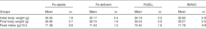
IMAAC, Fe multi-amino acid chelate.
* Data were analysed by two-way ANOVA followed by the Tukey’s post hoc test. There were no significant differences in the parameters between the groups.
Efficacy of Hb repletion was employed to compare IMAAC and FeSO4 following diet-induced Fe deficiency. Mice fed a diet low in Fe (Fe-deficient group) for 4 weeks showed significantly (P<0·001) lower blood Hb levels than control mice kept on the Fe-replete diet (Fe-replete group) throughout (Fig. 1(a)). Supplementation of the Fe-deficient diet with either IMAAC or FeSO4 for 10-d significantly increased Hb levels in comparison with mice fed the Fe-deficient diet throughout (P<0·001 and P<0·005), respectively (Fig. 1(a)). Furthermore, Fig. 1(b) shows that only supplemented mice had a significant gain in Hb levels from baseline. Moreover, serum Fe levels were significantly higher in the mice fed the Fe-replete diet (P<0·005) or supplemented Fe (P<0·001) than the mice on the Fe deficient diet (Fig. 1(c)). There was no significant difference in serum Fe between mice, which were given FeSO4 or IMAAC supplementation (P>0·05).
Intragastric administration of iron multi-amino acid chelate (IMAAC) or FeSO4 and haematic responses of anaemic mice. Hb levels of male CD1 mice following 10-d of administering the test diets (a), Hb gains (b) and serum iron levels in the mice (c). Iron-replete (![]() ) control diet contained 48 mg Fe/kg diet; the iron-deficient (
) control diet contained 48 mg Fe/kg diet; the iron-deficient (![]() ) diet contained approximately 3 mg Fe/kg diet. Mice were maintained on the iron-deficient diet and were supplemented daily by oral gavage with 150 µg Fe as IMAAC (
) diet contained approximately 3 mg Fe/kg diet. Mice were maintained on the iron-deficient diet and were supplemented daily by oral gavage with 150 µg Fe as IMAAC (![]() ) or FeSO4 (
) or FeSO4 (![]() ). Values are means (n 5), with their standard errors. * P<0·05, ** P<0·01, *** P<0·001.
). Values are means (n 5), with their standard errors. * P<0·05, ** P<0·01, *** P<0·001.

Tissue non-haem iron of mice
Mice receiving Fe supplementation had significantly higher liver non-haem Fe levels (Fig. 2(a)) than the mice receiving the Fe-replete diet (P<0·005) and the Fe-deficient diet (P<0·001). Mice on the control Fe-replete diet had significantly higher liver Fe levels that the Fe-deficient group (P<0·05) as expected. There was no difference in liver levels of Fe between FeSO4 and IMAAC supplementation (P>0·05). Similarly, mice on Fe replete or FeSO4 or IMAAC supplementation exhibited significantly higher (P<0·001) splenic Fe levels than mice fed Fe-deficient diet (Fig. 2(b)). As shown in Fig. 2(c), although the duodenal Fe levels increased in mice fed FeSO4 or IMAAC supplements, the difference only reached significance level (P<0·05) in the mice on the replete Fe diet compared with mice on the Fe-deficient diet. This trend is similar to the observation in the kidney as shown in Fig. 2(d).
Intragastric administration of iron multi-amino acid chelate (IMAAC) or FeSO4 and tissue iron distribution of anaemic mice. Non-haem iron levels in the liver (a), spleen (b) duodenum (c) and kidney (d) of male CD1 mice following 10-d feeding with different test diets. Iron-replete (![]() ) control diet contained 48 mg Fe/kg diet; the iron-deficient (
) control diet contained 48 mg Fe/kg diet; the iron-deficient (![]() ) diet contained approximately 3 mg Fe/kg diet. Mice were maintained on the Fe-deficient diet and were supplemented daily by oral gavage with 150 µg Fe as IMAAC (
) diet contained approximately 3 mg Fe/kg diet. Mice were maintained on the Fe-deficient diet and were supplemented daily by oral gavage with 150 µg Fe as IMAAC (![]() ) or FeSO4 (
) or FeSO4 (![]() ). Values are means (n 5), with their standard errors. * P<0·05, ** P<0·01, *** P<0·001
). Values are means (n 5), with their standard errors. * P<0·05, ** P<0·01, *** P<0·001

.
Gene expression in the duodenum and liver tissues of mice
As expected Fig. 3(a) indicates that hepcidin mRNA expression in the liver was up-regulated significantly in animals fed Fe replete and supplemented Fe and significantly down-regulated in animals on the Fe-deficient diet (P<0·001). Consequently, duodenal cytochrome b (Dcytb) mRNA levels (Fig. 3(b)) increased in the duodenum of the mice were given the Fe-deficient diet. Moreover, a trend of increase was observed in the duodenal levels of divalent metal transporter 1 (DMT1) and Fpn mRNA of Fe-deficient mice (Fig. 3(c) and (d)). Fe supplementation, however, significantly (P<0·01) down-regulated Dcytb mRNA levels in the duodenum of mice. Down-regulation of Fe transport genes was more responsive to FeSO4 than IMAAC administration (Fig. 3(c) and (d)).
Intragastric administration of iron multi-amino acid chelate (IMAAC) or FeSO4 and mRNA levels of iron metabolism genes of anaemic mice. Hepcidin mRNA levels in the liver (a) of male CD1 mice following 10-d feeding with different test diets as described above. Dcytb mRNA expression (b), DMT1 (c) and ferroportin (Fpn) (d) levels in the duodenum of mice on experimental diets. ![]() , iron replete;
, iron replete; ![]() , iron deficient;
, iron deficient; ![]() , FeSO4;
, FeSO4; ![]() , IMAAC. * P<0·05, ** P<0·01, *** P<0·001.
, IMAAC. * P<0·05, ** P<0·01, *** P<0·001.

In vivo iron uptake and ferric iron reduction studies in HuTu 80 cells
Consistent with the observation in the in vivo mouse studies, IMAAC and FeSO4 exhibited comparable Fe uptake values in HuTu 80 cells (Fig. 4(a)). However, Fe(II) chelator ferrozine, significantly inhibited Fe uptake and utilisation from FeSO4 (P<0·05), the effect on IMAAC was not significant (Fig. 4(b)). Ferric citrate reduction in HuTu 80 cells exhibited pH-dependent activity that is higher than that of IMAAC (Fig. 4(c) and (d)). Reductase activity was significantly different (P<0·001) between pH 3·5 and 7·4 for both ferric citrate and IMAAC. However, reduction of Fe and dissolution in the presence of ascorbic acid was higher with IMAAC (Fig. 4(c) and (d)). Ascorbic acid significantly enhanced (P<0·01) ferric reduction in both Fe compounds.
Iron uptake and in vitro solubilisation of iron multi-amino acid chelate (IMAAC) in HuTu 80 cells. Cellular ferritin levels in HuTu 80 cells (a) after a 1-h exposure to 50 µm (Fe) as IMAAC, ferrous sulphate (FeSO4) or co-incubation with ferrozine (1 mm) (b). Effect of varying pH and on reductive iron dissolution from IMAAC (c) or ferric citrate (d). HuTu 80 cells were exposed to 50 µm (Fe) as IMAAC, FeSO4 in balanced salt solution following which (Fe(II) was measured using ferrozine. Total reducible iron was subsequently determined by the addition of ascorbic acid (1 mm). Values are means, and standard deviations of three independent experiments with three replicate wells per experiment. ![]() , Untreated cells;
, Untreated cells; ![]() , IMAAC;
, IMAAC; ![]() , IMAAC+ferrozine;
, IMAAC+ferrozine; ![]() , FeSO4;
, FeSO4; ![]() , FeSO4+ferrozine;
, FeSO4+ferrozine; ![]() , IMAAC;
, IMAAC; ![]() , IMAAC+ascorbic acid;
, IMAAC+ascorbic acid; ![]() , ferric citrate;
, ferric citrate; ![]() , ferric citrate+ascorbic acid.
, ferric citrate+ascorbic acid.

Discussion
Fe supplementation and food fortification are panaceas aimed at improving Fe nutrition( Reference Prentice, Mendoza and Pereira 14 , Reference Latunde-Dada, Pereira and Tempest 15 ) particularly to the vulnerable groups of the population. These strategies, however, are associated with diverse issues limiting effectiveness and as such are subjects of continuing research and investigations. FeSO4, the standard Fe supplement is highly reactive, potentially toxic and prone to causing gastrointestinal disturbances. Alternatives such as IMAAC was reported to be better tolerated than FeSO4 in a RCT in healthy premenstrual women( Reference Fouad, Evans and Sharma 10 ). The bioavailability, metabolism and the mechanism of Fe absorption from IMAAC were consequently investigated in Fe-deficient mice in the present study. In general, studies in humans revealed that differences in diets that vary in bioavailability are apparent in Fe-deficient rather than in Fe-replete subjects( Reference Hulten, Gramatkovski and Gleerup 16 ).
The data demonstrate the comparable efficiency of both IMAAC and ferrous sulphate in replenishing Hb in Fe-deficient mice and similar Fe uptake in vitro in intestinal HuTu 80 cells. This agrees with the observation of which Fe glycine chelate compared with FeSO4 in Hb repletion of chicks( Reference Ma, Sun and Zhou 17 ) and in a study that examines ferrous carbamoyl glycine with FeSO4 in rats( Reference Zhang, Sun and Xie 18 ). Furthermore, Fe glycine chelate was equally efficacious in treating cancer patients with mild IDA( Reference Ferrari, Nicolini and Manca 19 ). Remarkably, however, IMAAC is a repository of Fe and several amino acids. Consequently, tolerability of IMAAC possibly accrues from combined antioxidant properties of the constituent multiple amino acids( Reference Elias, Bridgewater and Vachet 20 ).
The study next compared the metabolism of Fe in IMAAC with FeSO4 in mice. During Fe deficiency, liver and spleen Fe stores are rapidly depleted to maintain Fe homoeostasis and functional Fe requirements. Consequently, repletion of these tissues during Fe supplementation is a relative marker of bioavailability. IMAAC and FeSO4 supplementation had equivalent levels of liver Fe repletion (Fig. 2(a)) compared with the Fe-deficient diet (P<0·001). Furthermore, in the spleen (Fig. 2(b)) the level of non-haem Fe was higher in the mice given IMAAC (P<0·005) than FeSO4 compared with the Fe-deficient diet. In fact, FeSO4 caused a similar level of splenic non-haem Fe to the Fe-replete diet. The spleen is the last tissue in the body to replenish its Fe stores after Fe deficiency( Reference Chua-anusorn, Webb and Macey 21 ). This observation was also evident in pregnant pigs that were given hydrolysed soya protein Fe AAC supplement for 4 weeks before parturition. On analysis, there was 34·5 % greater non-haem Fe levels in the liver and 8·5 % higher Fe in the spleen of piglets whose mothers were given the Fe amino acid chelate supplement compared with the control group( Reference Brady, Ku and Ullrey 22 ). In addition to tissue Fe, IMAAC modulation of Fe metabolism genes in mice was also evident in the present study.
Remarkably, though, administration of IMAAC supplement for 10 d replenished Fe stores of anaemic mice to the same level as the normal healthy mice kept on the Fe replete diet despite significant difference (P<0·001) in Fe intake (4·54 (sd 0·17) v. 9·88 (sd 0·33) mg/kg body weight). Fe intake (4·27 (sd 0·11) mg/kg body weight) of mice that were given FeSO4 was comparable with those on IMAAC. This attests to the superior Fe bioavailability of IMAAC and FeSO4 than ferric citrate Fe component of the Fe-replete diet (TD 80396, 48 mg/kg ferric citrate (Harlan Teklad). Contrary to some concerns( Reference Allen 23 ) that Fe chelates if absorbed intact could bypass the normal homoeostatic regulatory mechanisms, Fe amino acid chelates were reported to be regulated by the body’s Fe status in humans( Reference Olivares, Pizarro and Pineda 24 ). Expression of hepcidin and other Fe metabolism genes were mostly appropriately modulated by IMAAC and FeSO4 supplementation in the present study. As expected from gene expression studies, hepcidin mRNA levels (Fig. 3(a)) was down-regulated in mice given the Fe-deficient diet and up-regulated by both Fe supplements (P<0·005). Consequently, DMT1, ferroportin and, in particular, Dcytb mRNA levels (Fig. 3(b)–(d)) were up-regulated in mice on the Fe-deficient diet and down-regulated in both groups on supplements (P<0·001). These results reflect the effectiveness of both Fe supplements in resolving IDA. Furthermore, increased hepcidin expression will exert a negative feedback regulation of the genes involved in Fe transport machinery until Fe homoeostasis is restored. Hb repletion and hepatic hepcidin mRNA levels exhibited a trend that reflected responsiveness to bioavailability of IMAAC and FeSO4 in mice (Figs. 1(a) and 3(a)). This might be similar to the luminal endocytic uptake of nanoparticulate Fe(3+) poly oxo-hydroxide (nanoFe(3+)), which differs from the uptake mechanism of inorganic ferrous ion( Reference Pereira, Bruggraber and Faria 9 ), but both share a ferroportin-dependent abluminal efflux mechanism into the circulation( Reference Aslam, Frazer and Faria 25 ). This is consistent with the concept of the labile Fe pool( Reference Jacobs 26 ) in the cytosol, where Fe is released from inorganic or organic complexes into the common matrix for efflux into circulation as regulated by ferroportin/hepcidin interaction.
The chemical nature of IMAAC and the mechanism of the Fe uptake process were studied in duodenal HuTu 80 cells (Fig. 4(a)). The data suggest that the content of ferrous chelatable Fe is more negligible in IMAAC than FeSO4 (Fig. 4(b)). Food Fe sources and Fe salts are liable to autoxidation to the ferric form as they traverse the neutral pH section of the gastrointestinal tract. Consequently, the rate of dissolution of Fe in different formulations( Reference Harju 27 ) is critical in the absorption efficiency. We next investigated the reductive dissolution of IMAAC in the presence of ascorbic acid and compared this to ferric citrate. While ferric citrate exhibited a pH-dependent reductive dissolution of Fe (Fig. 4(c) and (d)), the dissolution of IMAAC seems independent of pH. Ferrozine chelable Fe was higher for FeSO4 than that of IMAAC, thus implying the availability of less redox active species in the later. This perhaps explains the reported higher tolerability of IMAAC over FeSO4 in a RCT in human subjects( Reference Fouad, Evans and Sharma 10 ). However, negligible free ferric Fe component of IMAAC could be easily reduced by ascorbic acid (Fig. 4(c)). Chelation, sequestration and encapsulation of Fe by amino acids and peptides possibly prevent redox reactivity in the lumen of the gastrointestinal tract. For inorganic Fe salts, however, the rate of autoxidation and dissolution of the ferric Fe in different formulations( Reference Harju 27 ) could be critical in the absorption efficiency in the proximal duodenal tract. However, the resultant consequences on microbial population and the generation of reactive Fe species in the distal duodenum are subject of continuing investigation( Reference Pereira, Aslam and Frazer 28 ).
IMAAC has been shown to compare favourably well with FeSO4 in Hb regeneration of anaemic mice and in vitro HuTu 80 intestinal Fe uptake studies. It has potential, therefore, to be used widely as an alternative to FeSO4 in the management of IDA as it has been shown to be better tolerated than FeSO4 in humans. In vitro cell culture studies offer opportunities for screening Fe compounds such as pyrophosphate, gluconate, ammonium citrate and bis glycinate, etc. as the relative stability of various Fe compounds in vivo has been a matter of uncertainty to the nutrition industry. On the other hand, animal studies may be useful for evaluating relative bioavailability and understanding the mechanisms of Fe absorption; the results may not wholly translate to what occurs in humans and interpretations as such should be treated with caution( Reference Reddy and Cook 29 ). Future studies on IMAAC could be for a longer duration to investigate the balance of the highly bioavailable Fe species and the potentials of pathogenic microbes to pilfer this Fe source for proliferation in the lower segment of the gastrointestinal tract. Fe absorption in mice and cells was comparable for both IMAAC and FeSO4 and both compounds induced and regulated Fe metabolism genes in a similar way.
Acknowledgements
G. O. L.-D. acknowledges funding support of the Diabetes and Nutritional Sciences Division, King’s College London. The IMAAC supplement called FerriActiv was prepared by Biotron Laboratories, Centerville, USA. The authors are grateful to Dr Gameil Fouad for providing IMAAC supplement.
G. O. L -D. designed and conducted the research, analysed the data and wrote the manuscript; N. K., C. B., A. C. and G. T. performed the experiments, data collection and statistical analysis.
All authors declare that they have no conflicts of interest.


