





Introduction
Cryptogonimidae Osborn, 1903 (Digenea: Opisthorchioidea) comprises >70 genera and 200 nominal species that infect poikilothermic vertebrates worldwide (Miller and Cribb, Reference Miller and Cribb2007, Reference Miller and Cribb2008a, Reference Miller, Cribb, Bray and Gibson2008b, Reference Miller and Cribb2009, Reference Miller and Cribb2013; Miller et al., Reference Miller, Bray, Goiran, Justine and Cribb2009; Kmentová et al., Reference Kmentová, Bray, Koblmüller, Artois, De Keyzer, Gelnar, Vanhove and Georgieva2020; Montes et al., Reference Montes, Barneche, Gonzalez, Cavallo, Shimabukuro, Moncada and Martorelli2025). A vast majority of cryptogonimids mature in fishes (Miller and Cribb, Reference Miller, Cribb, Bray and Gibson2008b), and only 7 accepted genera (∼10%) include species that infect amphibians and reptiles: Andrincola Zhang and Yang, 1993 and Andritrema Zhang and Yang, 1993 from amphibians in China (Zhan and Yang, Reference Zhan and Yang1993); Proctocaecum Baugh, Reference Baugh1957 and Caimanicola Teixeira de Freitas and Lent, 1938 from reptiles in the Americas, Africa, Australia and Asia (Brooks, Reference Brooks1980; Miller and Cribb, Reference Miller, Cribb, Bray and Gibson2008b); Timoniella Rebecq, 1960, Acanthostomum Looss, 1899 and Neochasmus Van Cleave and Mueller, 1932, from fishes and reptiles in the Americas, Africa and Asia (Brooks, 1970; Miller and Cribb, Reference Miller, Cribb, Bray and Gibson2008b). Relevant for the present study are the Proctocaecum species (Table 1). Specifically, cryptogonimids infecting the American alligator (Alligator mississippiensis [Daudin, 1802] [Alligatoridae]): Proctocaecum diploporum (Stunkard, Reference Stunkard1931) Baugh, Reference Baugh1957 (type species), Proctocaecum coronarium (Cobbold, Reference Cobbold1861) Brooks, Reference Brooks1980 and Caimanicola pavida (Brooks and Overstreet, Reference Brooks and Overstreet1977) Brooks, Reference Brooks1980.
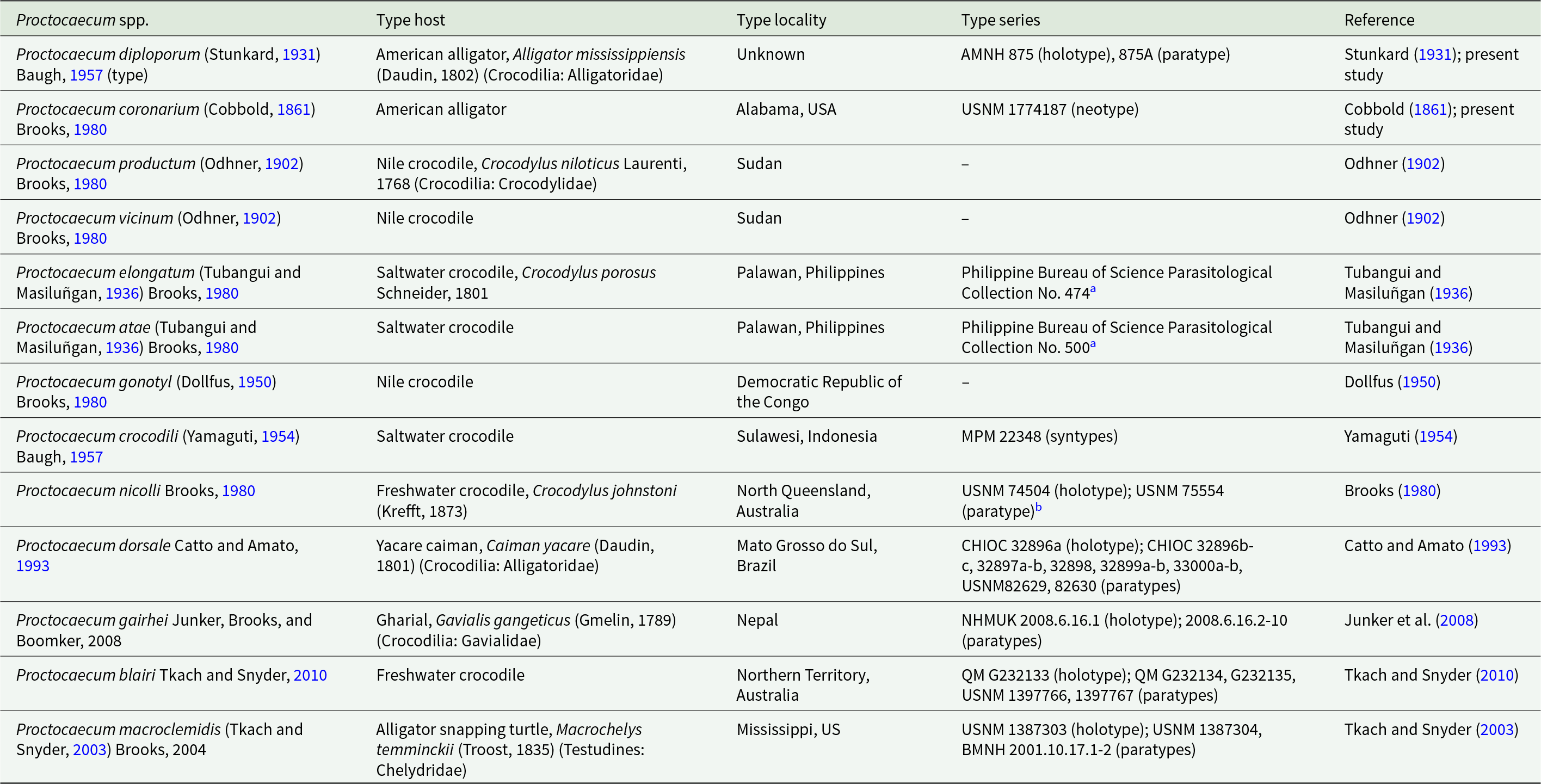
Proctocaecum Baugh, Reference Baugh1957 species with associated type host, type locality and museum type series

Table 1 Long description
The table catalogs 13 Proctocaecum species and links each to its type host, type locality, museum type series identifiers, and the original reference. Most species are reported from crocodilians, especially American alligator, Nile crocodile, and saltwater crocodile, with additional records from yacare caiman, gharial, freshwater crocodile, and one turtle host (alligator snapping turtle). Localities span the United States, Sudan, Democratic Republic of the Congo, the Philippines, Indonesia, Brazil, Nepal, and Australia; one entry lists an unknown locality. Several species have no surviving or listed type series, while others provide specific holdings such as AMNH, USNM, MPM, CHIOC, NHMUK, and QM catalog numbers. One species uses a neotype designation (USNM 1774187) for Proctocaecum coronarium. Some entries note that original type material was destroyed and that additional paratypes exist in other museums, so completeness of type series information varies by species.
a Type series destroyed during World War II (Brooks, Reference Brooks1980; p. 356).
b Additional paratypes in the collections of North Queensland Museum, and South Australian Museum.
Abbreviations: American Museum of Natural History, Division of Invertebrate Zoology, New York City, NY (AMNH); Natural History Museum, Parasitic Worms Collection, London, UK (NHMUK); Coleção Helmintológica do Instituto Oswaldo Cruz, Rio de Janeiro, Brazil (CHIOC); Meguro Parasitological Museum, Tokyo, Japan (MPM); Queensland Museum, Brisbane, Australia (QM); National Museum of Natural History’s Invertebrate Zoology Collection (USNM; Smithsonian Institution, Washington, DC).
Although >25 life cycles for fish-infecting cryptogonimids have been described, most of those are partial and/or experimental life cycles, did not emphasize morphological identification, or lack nucleotide information (Lundahl, Reference Lundahl1941; Cable and Hunnien, Reference Cable and Hunnien1942; Maillard, Reference Maillard1973, Reference Maillard1974; Cribb, Reference Cribb1986; Ostrowski de Núñez and Gil de Pertierra, Reference Ostrowski de Núñez and Gil de Pertierra1991; Scholz et al., Reference Scholz, Lavadores, Vargas, Mendoza, Rodriguez and Vivas1994; El-Darsh and Whitfield, Reference El-Darsh and Whitfield1999; Ostrowski de Núñez et al., Reference Ostrowski de Núñez, Semenas, Brugni, Viozzi and Flores1999; Simões et al., Reference Simões, Das Neves and Santos2008; Quintana and Ostrowski de Núñez, Reference Quintana and Ostrowski de Núñez2014; Quintana and Ostrowski de Núñez, Reference Quintana and Ostrowski de Núñez2016; Kvach et al., Reference Kvach, Bryjová, Sasal and Winkler2018; Jithila and Prasadan, Reference Jithila and Prasadan2022). To our knowledge, the study of Vélez-Sampedro et al. (Reference Vélez-Sampedro, Uruburu and Lenis2022) is the only complete life cycle of naturally infected (non-experimental) host using morphology and associated nucleotide information for a fish-infecting cryptogonimid.
In contrast to the fish-infecting cryptogonimids, current knowledge on the life histories of reptile-infecting cryptogonimids is scarce and incomplete. It comprises the experimental infection of 3 crocodilians (spectacled caiman, Caiman crocodilus Linnaeus, 1758 [Alligatoridae], Orinoco crocodile, Crocodylus intermedius Graves, 1819, and Morelet’s crocodile, Crocodylus moreletii [Duméril and Bibron, 1851] [Crocodylidae]) with metacercariae from freshwater fishes from Venezuela and Mexico (Ostrowski de Núñez, Reference Ostrowski de Núñez1984; Salgado-Maldonado and Aguirre-Macedo, Reference Salgado-Maldonado and Aguirre-Macedo1991), 1 snake (asiatic water snake, Fowlea piscator [Schneider, 1799] [Colubridae]) with metacercariae from a freshwater fish from India (Roopa and Janardanan, Reference Roopa and Janardanan1998), 1 hypothetical snake-infecting cercaria shed from the thorny tower snail, Mieniplotia scabra (Müller, 1774) (Gastropoda: Thiaridae) used to experimentally expose tadpoles of the Asian common toad, Duttaphrynus melanostictus (Schneider, 1799) (Anura: Bufonidae) from Sri Lanka (Jayawardena et al., Reference Jayawardena, Tkach, Navaratne, Amerasinghe and Rajakaruna2013, Reference Jayawardena, Rohr, Amerasinghe, Navaratne and Rajakaruna2017), and 2 hypothetically crocodilian-infecting metacercariae infecting fish from México (Martínez-Aquino et al., Reference Martínez-Aquino, Vidal-martínez and Aguirre-Macedo2017). Ostrowski de Núñez (Reference Ostrowski de Núñez1987) successfully characterized for the first time a complete experimental life cycle of a reptile-infecting cryptogonimid, Caimanicola brauni (Mañé-Garzón and Gil, 1961) Brooks, Reference Brooks1980, by feeding a Hilaire’s side-necked turtle Phrynops hilarii (Dumeril and Birbon, 1835) (Chelidae) metacercariae recovered from a 10 spotted live-bearer, Cnesterodon decemmaculatus (Jenyns, 1842) (Actinopterygii: Poeciliidae), infected with cercariae from a cochliopid snail, Heleobia castellanosae (Gaillard, 1974) (Gastropoda: Cochliopidae) from a Buenos Aires Zoo (Argentina).
Our work herein is the first to characterize a complete naturally occurring life cycle for a reptile-infecting cryptogonimid, the first to be completed in North America, and the first to provide life history and nucleotide information for any species of Proctocaecum. In doing so, we provide a redescription of P. diploporum, supplemental description of P. coronarium, and discuss the taxonomy of C. pavida infecting the American alligator, based on morphology, 28S nucleotide information and museum specimens.
Materials and methods
Specimen collection, preparation and deposition
Snails and fish were collected (7 Oct 2024, 12 Nov 2024 and 6 Mar 2025) from a saltmarsh pond that holds American alligators and that is intermittently connected to Bon Secour Bay (Mobile Bay, Alabama, north-central Gulf of America) during spring tides or storm events. Snails were collected from pond vegetation using kick nets, kept alive in pond water from the same pond they were collected, and transported to the Southeastern Cooperative Fish Parasite and Disease Laboratory (SCFPDL) at Auburn University (Auburn, AL, USA), where they were individually isolated in 6-well plates containing pond water from the same pond they were collected. The water was monitored daily under a stereo-dissecting microscope (Meiji RZ 3288, Meiji Techno Co., Saitama, Japan) to obtain naturally shed cercariae and subsequently the snails were cracked open for morphological identification and recovery of rediae. Fish were captured using standard minnow traps placed in shallow areas of the pond for 1–2 h, kept alive in pond water with aeration, and transported to the SCFPDL for identification and necropsy.
Fish were identified using morphological keys for the area (Boschung and Mayden, Reference Boschung and Mayden2004); snails were identified based on shell and penis morphology following Heard et al. (Reference Heard, Overstreet and Foster2002).
Ten fish were whole fixed in 10% neutral-buffered formalin (n.b.f.) for histopathology. Fixed fish intended for histopathological examination were grossed to fit standard tissue processing cassettes, yielding 60 portions of tissue that were rinsed with distilled water for 2 h, immersed in Ethylenediaminetetraacetic acid (EDTA) for 3 weeks, processed routinely for histology by dehydration in an ethanol series, embedded in paraffin, sectioned at 4–5 μm, mounted on glass slides, dried and stained with hematoxylin and eosin (H&E) for tissue morphology (Luna, Reference Luna1992; Gordillo-González et al., Reference Gordillo-González, Ksepka, Dutton, Cajiao-Mora, Brule, Kuhajda, George and Bullard2026).
Adult trematodes were collected opportunistically from American alligators on 2 occasions. The first collection occurred during an alligator hunt organized by the Alabama Department of Conservation and Natural Resources during 12–13 August 2022. Eight alligators were necropsied in the field at the weigh-in station in Spanish Fort (Mobile Bay area, Alabama, USA), where hunters delivered the alligators. The second collection occurred on 31 May 2023 where 3 fresh-dead adult alligators were collected and killed by personnel of the South Carolina Department of Conservation and Natural Resources at the Dennis Wildlife Center Fish Hatchery (Santee Cooper Reservoir, Cooper River drainage, South Carolina, USA). These specimens were transported to the SCFPDL for necropsy following SCFPDL standard necropsy procedures for intestinal parasite collection (Cajiao-Mora et al., Reference Cajiao-Mora, Brule, Warren, Ksepka, Dutton and Bullard2024b).
Regarding larval stages collection, rediae and cercariae were observed alive in temporary mounts by pipetting them into a glass slide with pond water and a cover slip using an Olympus BX51 compound microscope (Olympus, Tokyo, Japan) equipped with differential interface contrast (DIC) optical components and a Jenoptik Gryphax camera (Jenoptik AG, Jena, Germany). Metacercariae were classified by size during necropsy as ‘large cyst’ (cyst diameter >240 μm) and ‘small cyst’ (<200 μm).
Specimens intended for morphological identification were heat-killed and fixed in 10% n.b.f. All other specimens were preserved in 95% non-denatured ethanol (EtOH) for DNA extraction. Fixed specimens were rinsed in water, stained overnight in Van Cleave’s and Ehrlich’s hematoxylins following Cajiao-Mora et al. (Reference Cajiao-Mora, Brule, Dutton and Bullard2024a), drawn using an Olympus BX51 compound microscope equipped with DIC and a drawing tube (Olympus U-DA Drawing Tube, Olympus, Tokyo, Japan), and selectively measured using a Jenoptik Gryphax camera. Measurements are reported in micrometres (μm), unless otherwise stated as the range followed by the mean in parentheses. Standard deviation is included when more than 10 characters are measured. Fixed specimens for scanning electron microscopy (SEM) were washed with de-ionized water, dehydrated through a graded EtOH series, pipetted onto a 45 μm mesh cut to fit within a 30 μm microporous specimen capsule, critical point dried in liquid CO2, mounted on SEM aluminium stubs with double-sided carbon tape, sputter-coated with gold palladium (19.32 g/cm3; 25 mA), and viewed with a Zeiss EVO 50VP SEM (Warren and Bullard, Reference Warren and Bullard2019). Nomenclature for Cryptogonimidae follows Miller and Cribb (Reference Miller, Cribb, Bray and Gibson2008b). Morphology follows Cajiao-Mora et al. (Reference Cajiao-Mora, Brule, Dutton, Caicedo-Portilla and Bullard2025) and Truong et al. (Reference Truong, Warren, Ksepka, Curran and Bullard2021; oesophagus length comprises the oesophagus from the posterior end of the oral sucker to the oesophageal bifurcation).
We examined the following museum specimens: vouchers of P. coronarium and Timoniella loossi (Viguera, 1957) Salgado-Maldonado and Aguirre-Macedo, Reference Salgado-Maldonado and Aguirre-Macedo1991, and type series of Caimanicola pavidus (Brooks and Overstreet, Reference Brooks and Overstreet1977) Brooks, Reference Brooks1980 held in the National Museum of Natural History’s Invertebrate Zoology Collection (USNM, Smithsonian Institution, Washington, DC) and in the Harold W. Manter Laboratory of Parasitology (HWML, University of Nebraska, Lincoln, NE); vouchers of Caimanicola marajoara Teixeira de Freitas and Lent, 1938, type series of Proctocaecum nicolli Brooks, Reference Brooks1980, and Proctocaecum macroclemidis (Tkach and Snyder, Reference Tkach and Snyder2003) Brooks, 2004, and paratypes of Proctocaecum dorsale Catto and Amato, Reference Catto and Amato1993, and Proctocaecum blairi Tkach and Snyder, 2010 held in the USNM; type series of P. diploporum held in the American Museum of Natural History, Division of Invertebrate Zoology (AMNH, New York City, NY); microphotographs of the holotype of C. marajoara from the Helminthological Collection of the Oswaldo Cruz Institute (CHIOC, Rio de Janeiro, Brazil). Vouchers of each life stage obtained and described herein were deposited in the USNM; accession numbers are in Table 2.
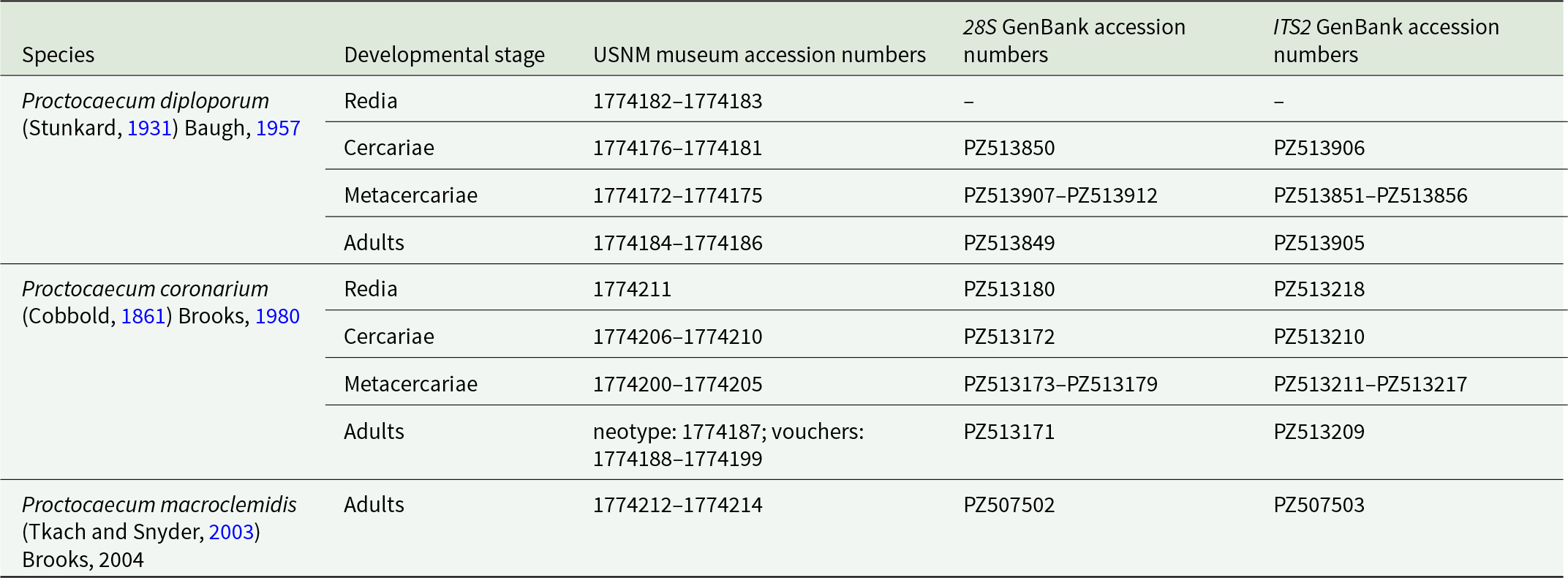
Museum and GenBank accession numbers of Cryptogonimidae species generated in this study

Table 2 Long description
The table lists three Cryptogonimidae species and, for each developmental stage sampled, provides USNM museum accession ranges plus GenBank accession numbers for 28S and ITS2 sequences. Proctocaecum diploporum includes redia, cercariae, metacercariae, and adults; redia have only USNM numbers, while cercariae, metacercariae, and adults have both 28S and ITS2 accessions. For diploporum, cercariae have one 28S accession and one ITS2 accession, metacercariae have multiple accessions for both markers, and adults have one accession for each marker. Proctocaecum coronarium also includes all four stages, and each stage has both 28S and ITS2 accessions. Coronarium adults are represented by a neotype plus voucher specimens, with one 28S accession and one ITS2 accession. Proctocaecum macroclemidis is represented only by adults, with one 28S accession and one ITS2 accession. Dashes indicate that no GenBank accession number is provided for that marker and stage.
Abbreviation: National Museum of Natural History’s Invertebrate Zoology Collection (USNM; Smithsonian Institution, Washington, DC).
DNA extraction
A total of 3 rediae, 4 cercariae, 12 small cyst metacercariae, 12 large cyst metacercariae, and 5 adults preserved in EtOH were used for DNA extraction and sequencing. Extraction was performed using the DNeasyTM Blood and Tissue kit (Qiagen) following the manufacturer’s protocol, except that the proteinase-K incubation period was extended overnight. Once extracted, DNA concentration was measured using a NanoDrop-One Microvolume Spectrophotometer (Thermo Fisher Scientific), diluted to 50 ng/μL, and stored at −20°C. The partial 28S gene was amplified using the forward primer U178 (5’-GCACCCGCTGAAYTTAAG-3’), and reverse primer L1642 (5’-CCAGCGCCATCCATTTTCA-3’), sequencing primers 300 F (5’-CAAGTACCGTGAGGGAAAGTTG-3’) and 1200 R (5’-GCATAGTTCACCATCTTTCGG-3’) (Lockyer et al., Reference Lockyer, Olson and Littlewood2003). The ITS2 region was amplified using forward primer GA1 (5’-AGAACATCGACATCTTGAAC-3’; Anderson and Barker, Reference Anderson and Barker1998) and reverse primer ITS2.2 (5’-CCTGGTTAGTTTCTTTTCCTCCGC-3’; Cribb et al., Reference Cribb, Adlard and Bray1998). Polymerase chain reaction (PCR) reactions were performed following Truong et al. (Reference Truong, Warren, Ksepka, Curran and Bullard2021). DNA amplification was verified with a 1% agarose gel stained with ethidium bromide. PCR products were purified using the QIAquick PCR Purification Kit (Qiagen) according to the manufacturer’s protocol, except that the last elution step was performed with autoclaved nanopure water. DNA sequencing was performed by Genewiz (South Plainfield, New Jersey). Sequence assembly and analysis of chromatograms were performed with Geneious prime version 2023.2.1. Forward and reverse sequences were aligned using MAFFT tool (Katoh and Standley, Reference Katoh and Standley2013) and low-quality read-ends were trimmed. All sequences were deposited in GenBank; accession numbers are in Table 2.
Phylogenetic analysis
We performed maximum likelihood tree inference using 28S sequences. The ITS2 phylogeny did not show useful resolution and therefore is not shown herein. The ingroup taxa of the 28S analysis comprised our newly generated sequences, including a sequence of a specimen of Proctocaecum macroclemidis (Tkach and Snyder, Reference Tkach and Snyder2003) Brooks, 2004 that the authors collected from an alligator snapping turtle (Macrochelys temminckii Troost,1835 [Chelydridae]) from Alabama (the identification was based on comparisons with the holotype USNM 1387303; see description in Tkach and Snyder, Reference Tkach and Snyder2003), and those ascribed to Cryptogonimidae following Kmentová et al. (Reference Kmentová, Bray, Koblmüller, Artois, De Keyzer, Gelnar, Vanhove and Georgieva2020), Yong et al. (Reference Yong, Martin and Smit2023) and Cajiao-Mora et al. (Reference Cajiao-Mora, Brule, Dutton, Caicedo-Portilla and Bullard2025). We excluded sequences shorter than 1000 base pairs (bp). The outgroup follows Yong et al. (Reference Yong, Martin and Smit2023). Sequences were aligned using the multiple alignment using fast Fourier transform (MAFFT) tool (Katoh and Standley, Reference Katoh and Standley2013), and the alignment was trimmed to our shortest sequence (1145 bp). The phylogeny was inferred using IQTREE v.1.16.12 (Nguyen et al., Reference Nguyen, Schmidt, von Haeseler and Minh2015). Substitution model testing was done with ModelFinder (Kalyaanamoorthy et al., Reference Kalyaanamoorthy, Minh, Wong, von Haeseler and Jermiin2017) as implemented in IQTREE. After model testing, tree inference was done using best-fitting substitution models (Chernomor et al., Reference Chernomor, von Haeseler and Minh2016). Default tree search parameters were used, except perturbation strength was set to 0.2 and 500 iterations had to be unsuccessful to stop the tree search. Tree inference was done 20 times with only the tree with the best log-likelihood score reported. Support for relationships was measured with 1000 ultrafast bootstrap replicates (UFBoot) (Hoang et al., Reference Hoang, Chernomor, von Haeseler, Minh and Vinh2018). The inferred phylogenetic tree was visualized using FigTree v1.4.4 (Rambaut et al., Reference Rambaut, Suchard, Xie and Drummond2018) and further edited for visualization purposes with Adobe Illustrator (Adobe Systems).
Results
Proctocaecum coronarium (Cobbold, Reference Cobbold1861) Brooks, Reference Brooks1980 (Cryptogonimidae) (Figures 1, 2, 3A, 4A and 4C)
Redia (Figure 1A)
Host: Littoridinops monroensis (Frauenfeld, 1863) (Gastropoda: Cochliopidae).
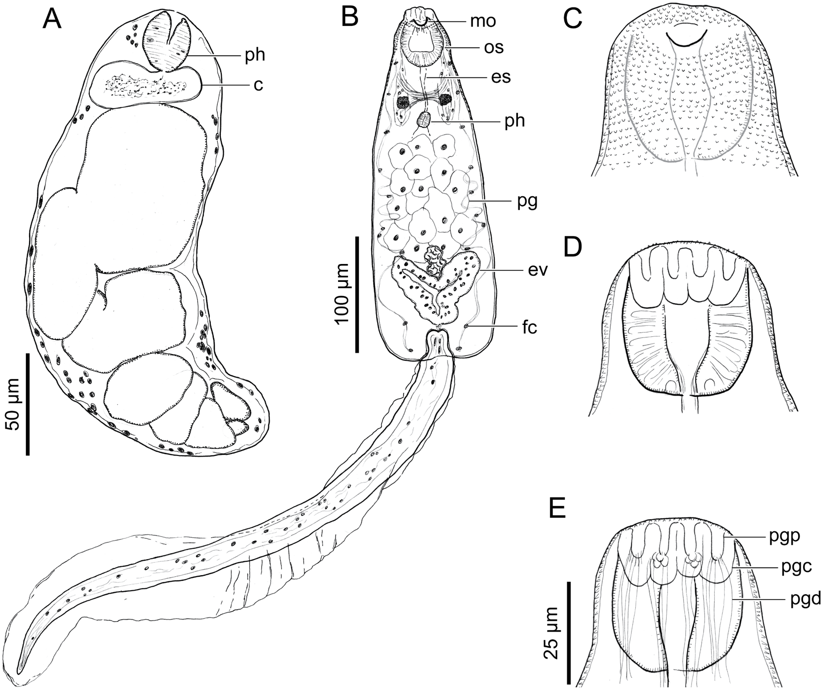
Proctocaecum coronarium (Cobbold, Reference Cobbold1861) Brooks, Reference Brooks1980 (Digenea: Cryptogonimidae) from Littoridinops monroensis (Frauenfeld, 1863) (Gastropoda: Cochliopidae) from a saltmarsh pond in the Bon Secour Bay area, Alabama, USA. Scale value aside each bar. (A) Redia, ventral view (voucher USNM 1774211). (B) Cercaria, ventral view (voucher USNM 1774210). (C) Cercaria, anterior end, ventral view (voucher USNM 1774207). (D) Cercaria, anterior end, medial view (voucher USNM 1774207). (E) Cercaria, anterior end, dorsal view (voucher USNM 1774207). Abbreviations: cecum (c); oesophagus (es); excretory vesicle (ev); flame cells (fc); mouth (mo); oral sucker (os); penetration glands (pg); penetration gland channel (pgc); penetration gland duct (pgd); penetration gland pore (pgp); pharynx (ph).

Locality: Bon Secour Bay, Alabama (USA).
Site in host/tissue infected: Digestive gland.
Prevalence and intensity: 3 of 300 (1%) snails were infected with 2–5 rediae each.
Specimens deposited: USNM 1774211.
Representative DNA sequences: 28S GenBank accession No. PZ513180, ITS2 GenBank accession No. PZ513218.
Description (based on 1 heat-killed, stained and permanently whole-mounted redia plus 3 live, wet-mounted rediae): Body ovoid in outline, 253–456 (3) in total length, 83–164 (3) in maximum width. Oral opening terminal. Pharynx ovoid, 27 (1) long, 22 (1) wide. Cecum sac-like, immediately post-pharyngeal, transversally elongated. Birth pore posterior to pharynx, lateral to cecum. Body containing several developing cercariae; larger cercariae in anterior portion (Figure 1A).
Cercaria (Figures 1B–E)
Host: Littoridinops monroensis (Frauenfeld, 1863) (Gastropoda: Cochliopidae).
Locality: Bon Secour Bay, Alabama (USA).
Site in host/tissue infected: Digestive gland.
Prevalence and intensity: 3 of 300 (1%) snails shed 5–10 cercariae each in 7 days; each snail was infected with 10–20 cercariae upon being necropsied.
Specimens deposited: USNM 1774206–1774210.
Representative DNA sequences: 28S GenBank accession No. PZ513172, ITS2 GenBank accession No. PZ513210.
Description (based on 5 heat-killed, stained and permanently whole-mounted cercariae plus 10 live, wet-mounted cercariae): Pleurolophocercous. Body ovoid in outline, 775–819 (794; 4) in total length, 96–112 (106; 5) in maximum width, 7 × longer than wide. Tegument spinose, containing few hair-like sensory structures. Tegumental spines visible in anterior end of body, becoming smaller posteriad, extending close to eyespots level (Figure 1C). Hair-like sensory structures 25–30 (1) in total length, distributing along ventral lateral body margin, herein indistinct except in live cercariae observed with DIC. Tail 472–554 (502; 4) in total length, 32–40 (36; 4) in maximum width, having dorsoventral fin-folds (Figure 1B); fin-folds extending from anterior quarter of tail to end of tail, becoming confluent at end of tail (Figure 1B). Primordial ventral sucker located immediately anterior to excretory vesicle (Figure 1B). Oral sucker subterminal, ovoid in outline (Figures 1B–E), 37–47 (44; 5) in total length, 34–37 (35; 5) in maximum width. Oesophagus narrow (Figure 1B). Pharynx ovoid in outline (Figure 1B). Ceca not observed. Eye spots 2, in anterior portion of body, flanking oesophagus, between pharynx and oral sucker (Figure 1B). Penetration glands occupying middle third of body (Figure 1B), at least 7 pairs observed, ovoid to irregular in outline, having spheroid nucleus (Figure 1B), with ducts extending anteriad; penetration gland ducts dorsal to oral sucker, becoming confluent at level of oral sucker, forming 4 channels (Figures 1D and 1E); channels having a terminal pore (Figures 1D and 1E). At least 10 flame cells per side of body observed in live cercariae, distributing along lateral body margin (Figure 1B). Excretory vesicle Y-shaped, in posterior portion of body, posterior to penetration glands, (Figure 1B); excretory vesicle wall 11–14 (12; 3) thick. Cercarial behaviour comprising rapid, irregular tail movements in no particular vertical direction for 1–3 sec then a pause in activity and followed by cercarial sinking for 3–5 sec before restarting cycle.
Metacercaria (Figures 2A and 3A)
Hosts: Sheepshead minnow, Cyprinodon variegatus Lacepède, 1803 (Cyprinodontiformes: Cyprinodontidae); sailfin molly, Poecilia latipinna (Lesueur, 1821) (Cyprinodontiformes: Poeciliidae); rainwater killifish, Lucania parva (Baird and Girard, 1855) (Cyprinodontiformes: Fundulidae); gulf killifish, Fundulus grandis Baird and Girard, 1853 (Cyprinodontiformes: Fundulidae); bayou killifish, Fundulus pulvereus (Evermann, 1892); inland silverside, Menidia beryllina (Cope, 1867) (Atheriniformes: Atherinopsidae); striped mullet, Mugil cephalus Linnaeus, 1758 (Mugiliformes: Mugilidae).
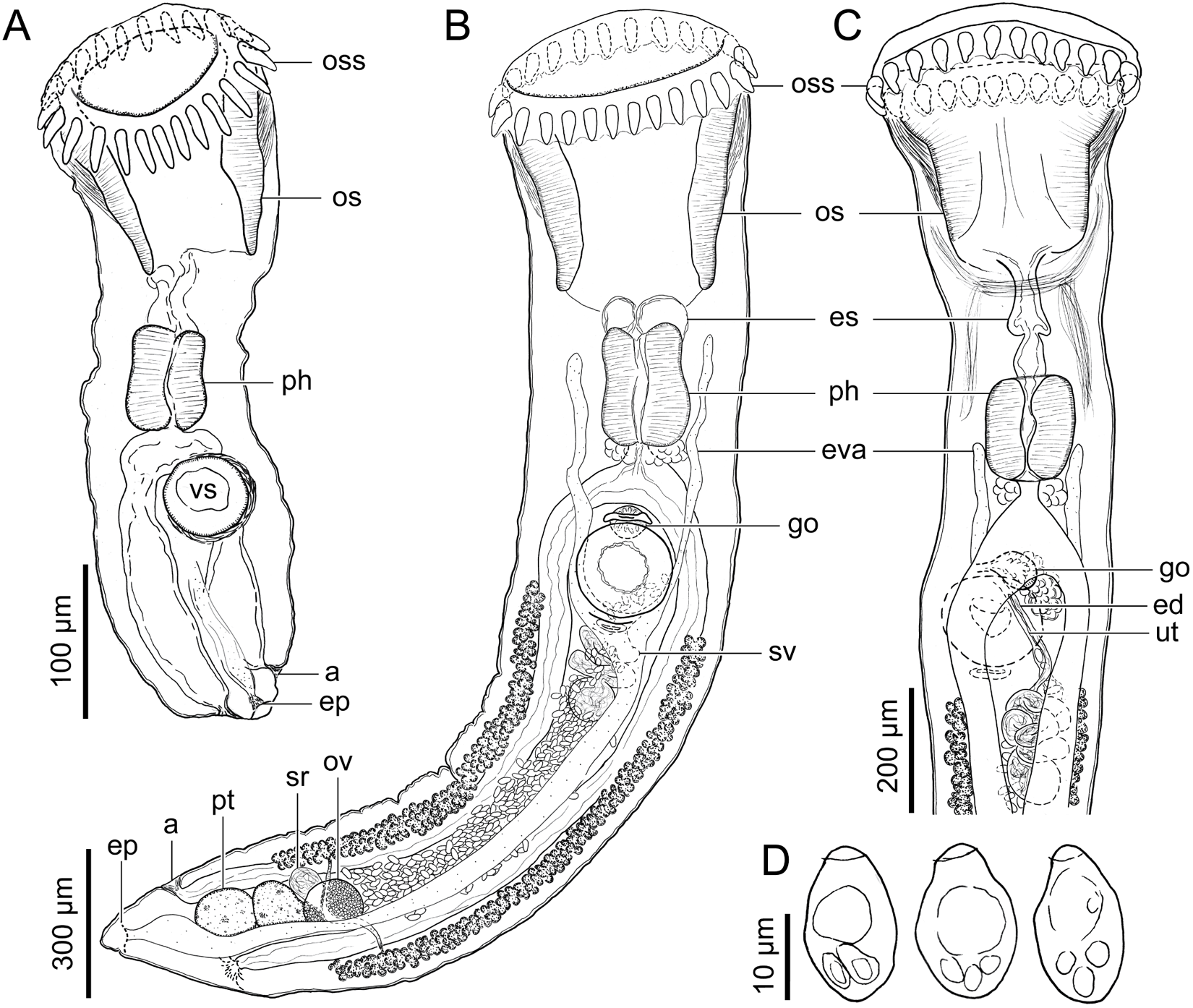
Proctocaecum coronarium (Cobbold, Reference Cobbold1861) Brooks, Reference Brooks1980 (Digenea: Cryptogonimidae). Scale value aside each bar. (A) Metacercaria (voucher USNM 1774202) excysted from within a scale of sheepshead minnow, Cyprinodon variegatus Lacepède, 1803 (Cyprinodontiformes: Cyprinodontidae) from a saltmarsh pond in the Bon Secour Bay area, Alabama, USA. Ventral view. (B) Adult (neotype USNM 1774187) infecting American alligator, Alligator mississippiensis (Daudin, 1802) (Crocodilia: Alligatoridae) from Alabama, USA. Ventral view. (C) Adult (voucher USNM 1774188) infecting American alligator, from Alabama, USA. Anterior end, dorsal view. (D) Eggs (voucher USNM 1774193). Abbreviations: anal pore (a); oesophagus (es); ejaculatory duct (ed); excretory pore (ep); excretory vesicle arms (eva); gonotyl (go); oral sucker (os); oral sucker spines (Oss); ovary (ov); pharynx (ph); posterior testis (pt); seminal receptacle (sr); seminal vesicle (sv); uterus (ut); ventral sucker (vs).

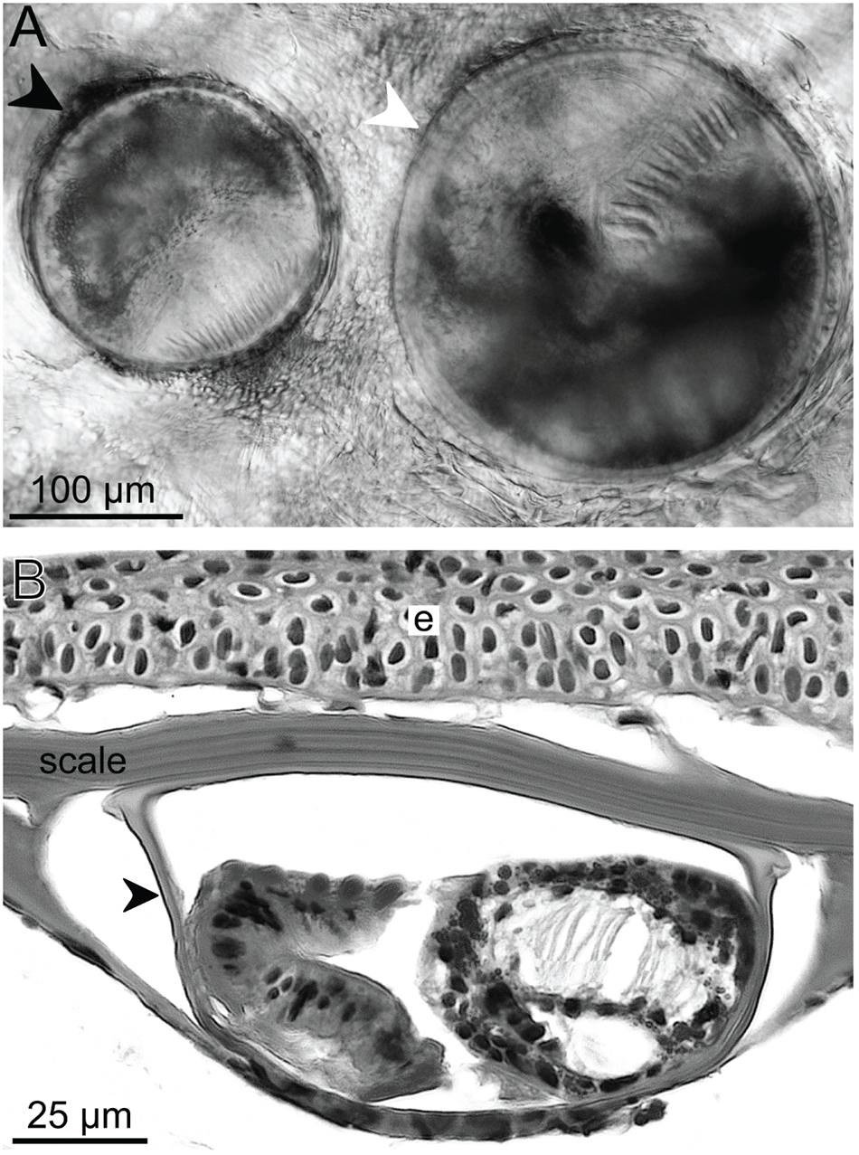
Encysted metacercariae. Scale value aside each bar. (A) Encysted metacercariae of Proctocaecum diploporum (Stunkard, Reference Stunkard1931) Baugh, Reference Baugh1957 (Digenea: Cryptogonimidae) (black arrowhead) and Proctocaecum coronarium (Cobbold, Reference Cobbold1861) Brooks, Reference Brooks1980 (white arrowhead) within the scale of a gulf killifish, Fundulus grandis Baird and Girard, 1853 (Cyprinodontiformes: Fundulidae) from a saltmarsh pond in the Bon Secour Bay area, Alabama, USA. (B) Histological section stained with hematoxylin and eosin of a scale of a sheepshead minnow, Cyprinodon variegatus Lacepède, 1803 (Cyprinodontiformes: Cyprinodontidae) from a saltmarsh pond in the Bon Secour Bay, with an encysted metacercaria of Proctocaecum sp. (black arrowhead). Abbreviations: epithelium (e).

Locality: Bon Secour Bay, Alabama (USA).
Site in host: Within scales, in the fins enveloped by lepidotrichia, and in the epidermis between fin rays.
Prevalence and intensity: We examined ∼5–20 specimens of each fish species; each individual of each fish species was infected (100% prevalence) with approximately 5–50 cysts.
Specimens deposited: USNM 1774200–1774205.
Representative DNA sequences: 28S GenBank accession No. PZ513173–PZ513179, ITS2 GenBank accession No. PZ513211–PZ513217.
Description (based on 22 encysted metacercariae, 4 excysted, heat-killed, stained and permanently whole-mounted metacercariae, and 10 excysted, live, wet-mounted metacercariae): Cyst spheroid, membranous-like, transparent (Figure 3A), 244–292 (265 ± 14; 22) by 232–292 (261 ± 15; 22), within fish scales and fin rays. Excysted metacercaria 420–471 (449; 4) in total length, 140–166 (156; 4) in maximum width. Tegument spinose; tegumental spines visible in anterior third of body. Ventral sucker spheroid, equatorial to slightly post-equatorial (Figure 2A), 49–65 (60; 4) in total length, 54–66 (60; 4) in maximum width. Oral sucker terminal, infundibular, 117–135 (127; 4) in total length, 140–166 (156; 4) in maximum width, having circumoral spines (Figure 2A); oral sucker spines 24 in number, widest at insertion, tapering posteriad (Figure 2A), 27–31 (29; 31) in total length, 6–8 (7; 31) width at insertion. Oesophagus medial (Figure 2A). Pharynx quadrate in outline, having blunt corners, 67–77 (74; 4) in total length, 35–50 (44; 4) in maximum width (Figure 2A). Intestine bifurcating after pharynx (Figure 2A). Ceca extending posteriad in parallel with lateral body margins; each cecum opening through a posterolateral pore (anal pore). Excretory vesicle Y-shaped, extending from posterior body end to posterior end of oral sucker, bifurcating posterior to ventral sucker; excretory pore sub-terminal, dorsal (Figure 2A).
Adult (Figures 2B–D, 4A and 4C; Table 3)
Type host and host reported herein: American alligator, Alligator mississippiensis (Daudin, 1802) (Crocodilia: Alligatoridae).
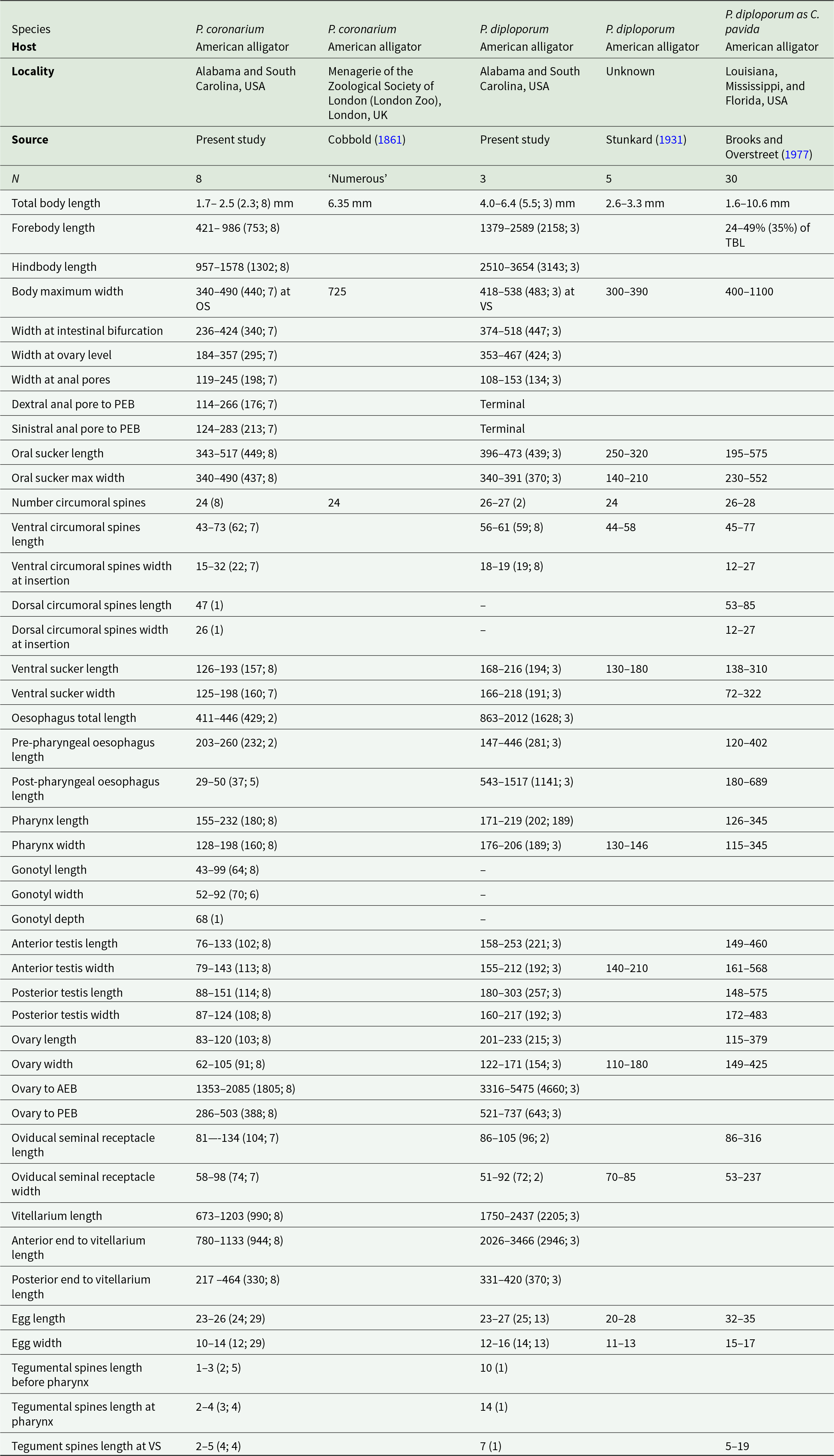
Comparative measurements of adult Cryptogonimidae: Proctocaecum coronarium (Cobbold, Reference Cobbold1861) Brooks, Reference Brooks1980, Proctocaecum diploporum (Stunkard, Reference Stunkard1931) Baugh, Reference Baugh1957, and P. diploporum (as Caimanicola pavida [Brooks and Overstreet, Reference Brooks and Overstreet1977] Brooks, Reference Brooks1980) infecting American alligator, Alligator mississippiensis (Daudin, 1802) (Crocodilia: Alligatoridae)

Table 3 Long description
The table compiles morphometric measurements for adult Proctocaecum coronarium and Proctocaecum diploporum from American alligators, comparing multiple sources and localities. Sample sizes range from 3 to 30 in modern studies, while one historical report lists an unspecified number of specimens. In the present study, P. coronarium has a total body length of about 1.7 to 2.5 mm, whereas P. diploporum is larger at about 4.0 to 6.4 mm; a separate report of P. coronarium lists a much longer single value, and the P. diploporum form reported as C. pavida spans a very wide length range. Forebody and hindbody lengths follow the same pattern, with P. diploporum showing notably longer segments than P. coronarium where reported. Body width differs by study and measurement point, but P. diploporum commonly reaches similar or greater maximum widths than P. coronarium, and the C. pavida report includes the broadest widths. Oral and ventral sucker dimensions overlap between species, but P. diploporum tends to have slightly larger ventral sucker measurements in the present study. Circumoral spine counts are close across taxa, with P. coronarium typically reported with 24 and P. diploporum commonly around 26 to 28, while dorsal spine measurements are missing for some sources. Egg sizes are broadly similar between P. coronarium and P. diploporum in the present study, but the C. pavida report lists larger eggs. Several rows are blank or marked as absent in some sources, so comparisons should be interpreted cautiously because not all traits were measured or reported consistently.
Abbreviations: anterior end of body (AEB); oral sucker (OS); posterior end of body (PEB); ventral sucker (VS).
Type locality: Alabama (USA)
Localities reported herein: Alabama (USA); Dennis Wildlife Center Fish Hatchery (Santee Cooper Reservoir, Cooper River drainage, South Carolina, USA)
Site in host: Intestine.
Prevalence and intensity: 3 of 11 (27%) alligators were infected with 5–10 P. coronarium
Specimens examined: vouchers of P. coronarium USNM 1381392, 1385103 1356162; HWML 20854
Specimens deposited: Neotype USNM 1774187; vouchers USNM 1774188–1774199.
Representative DNA sequences: 28S GenBank accession No. PZ513171, ITS2 GenBank accession No. PZ513209.
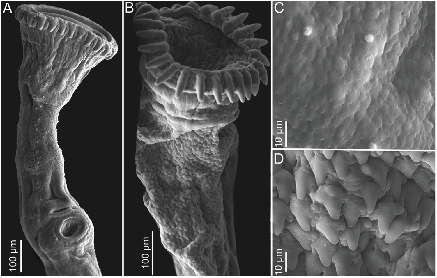
Supplemental description (based on 8 heat-killed, stained and permanently whole-mounted adults): Body elongate, having a pointed posterior end (Fig. 2B), 5–6× longer than wide. Forebody 20–38% (32%; 8) of total body length. Hindbody 52–63% (56%; 8) of total body length. Tegument spinose, having 2 tegumental infoldings (Figures 4A and 4C); body surface spines minute (Figure 4C), scale-like, becoming larger at level of ventral sucker, then becoming smaller and fewer posteriad, extending to near posterior end of body; tegumental infoldings anterior and posterior to ventral sucker (Figure 4A). Ventral sucker spherical, in first quarter of body (Figure 2B). Oral sucker terminal, infundibular, having circumoral spines (Figures 2B, 2C, 4A); oral sucker spines widest at insertion, tapering posteriad (Figures 2B, 2C and 4A). Oesophagus medial, representing 17–19% (18%; 2) of total body length, comprising pre-pharyngeal, pharyngeal and post-pharyngeal parts (Figures 2B and 2C). Pre-pharyngeal oesophagus retractile, 49–58% (54%; 2) of total oesophagus length. Pharynx quadrate in outline, having blunt corners (Figures 2B and 2C), surrounding oesophagus, representing 38–41% (39%; 2) of total oesophagus length. Post-pharyngeal oesophagus very short, surrounded by clusters of glandular-like cells (Figures 2B and 2C), 9–10% (9%; 2) of total oesophagus length. Intestine bifurcating shortly after pharynx (Figures 2B and 2C), 30–36% (33%; 8) of body length to anterior body end. Ceca slender, extending posteriad parallel to lateral body margins, asymmetrical in length, symmetrical in width, each cecum opening through posterolateral pore (anal pore); anal pores asymmetrical in position, 1 pore at level of posterior testis, other pore posterior to posterior testis (Figures 2B).
Scanning electron microscopy (SEM) of adult Proctocaecum coronarium (Cobbold, Reference Cobbold1861) Brooks, Reference Brooks1980 (Digenea: Cryptogonimidae) and Proctocaecum diploporum (Stunkard, Reference Stunkard1931) Baugh, Reference Baugh1957 from American alligators, Alligator mississippiensis (Daudin, 1802) (Crocodilia: Alligatoridae) from South Carolina and Alabama, USA. Scale value aside each bar. (A) Anterior end of P. coronarium. (B) Anterior end of P. diploporum. (C) Body surface spines from anterior end of body of P. coronarium. (D) Body surface spines from anterior end of body of P. diploporum.

Testes 2, spherical, medial, in last quarter of body, in tandem, contiguous with ovary (Figure 2B). Vasa efferentia obscured by dense mass of eggs through its length. Seminal vesicle long, sinuous, extending posterior to ventral sucker (Figures 2B and 2C); having convoluted proximal portion (Figure 2B and 2C), having distal portion enlarged, sac-like (Figures 2B and 2C); prostatic duct not observed; ejaculatory duct long, opening into genital pore through a hermaphroditic duct (Figure 2C). Genital pore medial, in anterior tegumental infolding, between ventral sucker and gonotyl (Figure 2C). Gonotyl spheroid, muscular, protractile, surrounded by clusters of glandular-like cells (Figures 2B and 2C)
Ovary spheroid (Figure 2B); pre-ovarian space 76–83% (78%; 8) of total body length; post-ovarian space 13–20% (17%; 8) of total body length. Oviduct emerging dorsally from ovary. Oviducal seminal receptacle sac-like, postero-dorsal to ovary. Laurer’s canal, oötype, and Mehlis’ gland not observed. Uterus coiling anteriorly between ceca, extending from ovary to common genital pore, having a convoluted proximal portion, having a sinuous to straight distal portion (Figures 2B and 2C), ventral to seminal vesicle, with numerous eggs (Figure 2B), confluent with hermaphroditic duct. Hermaphroditic duct opening into genital pore (Figure 2C). Eggs ovoid, operculate (Figure 2D). Vitellarium follicular, distributing in 2 bilaterally symmetrical fields, extending from anterior margin of anterior testis to posterior margin of ventral sucker (Figure 2B), occupying 36–50% (42%; 8) of total body length; pre-vitellarium space 34–50% (41%; 8) of total body length; post-vitellarium space 10–18% (14%; 8) of total body length. Transverse vitelline ducts emanating from posterior quarter of vitellarium, at level of ovary, forming vitelline reservoir; vitelline reservoir dorsal to ovary, connecting with oviduct (Figure 2B).
Excretory vesicle Y-shaped, ventral, extending from posterior body end to pharynx (Figures 2B and 2C), bifurcating posterior to ventral sucker (Figure 2B); excretory pore sub-terminal, dorsal (Figure 2B).
Proctocaecum diploporum (Stunkard, Reference Stunkard1931 ) Baugh, Reference Baugh1957 (Cryptogonimidae) (Figures 3A, 4B, 4D, 5 and 6A–B)
Redia (Figure 5A)
Host: Littoridinops monroensis (Frauenfeld, 1863) (Gastropoda: Cochliopidae).
Locality: Bon Secour Bay, Alabama (USA).
Site in host/tissue infected: Digestive gland.
Prevalence and intensity: 1 of 300 (0.3%) snails was infected.
Specimens deposited: USNM 1774182, 1774183.
Description (based on 1 heat-killed, stained and permanently whole-mounted redia): Body elongate, 619 (1) in total length, 106 (3) in maximum width. Oral opening terminal. Pharynx ovoid, 30 (1) long, 31 (1) wide. Cecum sac-like, immediately post-pharyngeal. Birth pore lateral, at level of cecum. Body containing several developing cercariae; larger cercariae in anterior portion (Figure 5A).
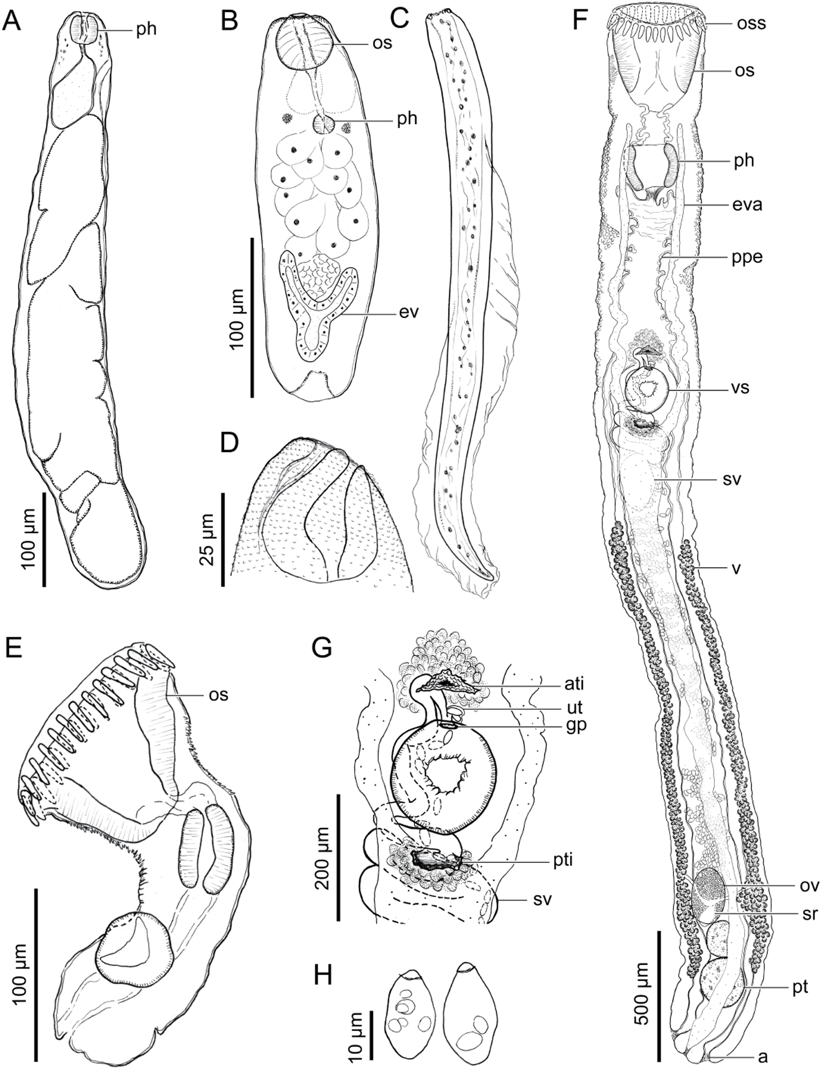
Proctocaecum diploporum (Stunkard, Reference Stunkard1931) Baugh, Reference Baugh1957 (Digenea: Cryptogonimidae). Scale value aside each bar. (A) Redia (voucher USNM 1774183) from Littoridinops monroensis (Frauenfeld, 1863) (Gastropoda: Cochliopidae) from a saltmarsh pond in the Bon Secour Bay area, Alabama, USA. Ventral view. (B) Cercarial body (voucher USNM 1774180) from L. Monroensis. Ventral view. (C) Cercarial tail (voucher USNM 1774179). (D) Anterior end of cercaria (voucher USNM 1774179). (E) Metacercaria (voucher USNM 1774175) excysted from within a scale of sheepshead minnow, Cyprinodon variegatus Lacepède, 1803 (Cyprinodontiformes: Cyprinodontidae) from a saltmarsh pond in the Bon Secour Bay area, Alabama, USA. Lateral view. (F) Adult (voucher USNM 1774186) infecting American alligator, Alligator mississippiensis (Daudin, 1802) (Crocodilia: Alligatoridae) from Alabama, USA. Ventral view. (G) Close view of male and female terminal genitalia (voucher USNM 1774186). Ventral view. (H) Eggs (voucher USNM 1774186). Abbreviations: anal pore (a); anterior tegumental infolding (ati); excretory vesicle (ev); excretory vesicle arms (eva); genital pore (gp); oral sucker (os); oral sucker spines (Oss); ovary (ov); pharynx (ph); post-pharyngeal oesophagus (ppe); posterior tegumental infolding (pti); posterior testis (pt); seminal receptacle (sr); seminal vesicle (sv); uterus (ut); ventral sucker (vs); vitellarium (v).

Cercaria (Figures 5B–D)
Host: Littoridinops monroensis (Frauenfeld, 1863) (Gastropoda: Cochliopidae).
Locality: Bon Secour Bay, Alabama (USA).
Site in host/tissue infected: Digestive gland.
Prevalence and intensity: 1 of 300 (0.3%) snails was infected with ∼11 cercariae.
Specimens deposited: USNM 1774176–1774181.
Representative DNA sequences: 28S GenBank accession No. PZ513850, ITS2 GenBank accession No. PZ513906.
Description (based on 3 heat-killed, stained and permanently whole-mounted cercariae): Pleurolophocercous. Body ovoid in outline (Figure 5B), 226–250 (239; 3) length without the tail, 63–70 (67; 4) in maximum width. Tegument spinose; tegumental spines scale like, visible in anterior end of body, becoming smaller and fewer posteriad, extending close to eyespots level (Figure 5D). Tail 360–364 (362; 2) in total length, 28–31 (30; 2) in maximum width, having dorsoventral fin-folds (Figure 5C); fin-folds extending from anterior quarter of tail to end of tail, becoming confluent at end of tail (Figure 5C). Primordial ventral sucker located immediately anterior to excretory vesicle (Figure 5B). Oral sucker subterminal, spheroid (Figures 5B and 5D), 34–36 (35; 3) in total length, 29–36 (33; 3) in maximum width. Oesophagus narrow (Figure 5B). Pharynx small (Figure 5B). Ceca not observed. Eye spots 2, in anterior portion of body, flanking oesophagus. Penetration glands occupying middle third of body (Figure 5B), ovoid to irregular in outline, having spheroid nucleus (Figure 5B), with ducts extending anteriad; penetration glands ducts dorsal to oral sucker, becoming confluent at level of oral sucker; ducts having a terminal pore (Figure 5D). Flame cells not observed. Excretory vesicle Y-shaped, in posterior portion of body, posterior to penetration glands, thick walled (Figure 5B). Cercarial behaviour not observed.
Metacercaria (Figures 3A and 5E)
Hosts: Sheepshead minnow, Cyprinodon variegatus Lacepède, 1803 (Cyprinodontiformes: Cyprinodontidae); sailfin molly, Poecilia latipinna (Lesueur, 1821) (Cyprinodontiformes: Poeciliidae); rainwater killifish, Lucania parva (Baird and Girard, 1855) (Cyprinodontiformes: Fundulidae); gulf killifish, Fundulus grandis Baird and Girard, 1853 (Cyprinodontiformes: Fundulidae); bayou killifish, Fundulus pulvereus (Evermann, 1892); inland silverside, Menidia beryllina (Cope, 1867) (Atheriniformes: Atherinopsidae); striped mullet, Mugil cephalus Linnaeus, 1758 (Mugiliformes: Mugilidae).
Locality: Bon Secour Bay, Alabama (USA).
Site in host: Within scales, in the fins enveloped by lepidotrichia, and in the epidermis between fin rays.
Prevalence and intensity: We examined ∼5–20 specimens of each fish species; each individual of each fish species was infected (100% prevalence) with approximately 5–50 cysts.
Specimens deposited: USNM 1774172–1774175.
Representative DNA sequences: 28S GenBank accession No. PZ513907–PZ513912, ITS2 GenBank accession No. PZ513851–PZ513856.
Description (based on 21 encysted specimens, 3 excysted, heat-killed, stained and permanently whole-mounted specimens, and 3 excysted specimens observed alive in temporary wet-mounts): Cyst spheroid, membranous-like, transparent (Figure 3A), 163–188 (178 ± 8; 21) by 158–196 (171 ± 10; 21), within fish scales and fin rays. Excysted metacercaria, 249–312 (289; 3) in total length, 97–119 (108; 3) in maximum width. Tegument spinose; tegumental spines visible in anterior third of body (Figure 5E). Ventral sucker spheroid, post-equatorial (Figure 5E), 31–44 (36; 3) in total length, 30–39 (36; 3) in maximum width. Oral sucker terminal, infundibular, 76–81 (79; 3) in total length, 97–119 (108; 3) in maximum width, having circumoral spines (Figure 5E); oral sucker spines 27 in number, widest at insertion, tapering posteriad (Figure 5E), 21 (3) in total length, 4 (3) width at insertion. Oesophagus medial (Figure 5E). Pharynx ovoid in outline, 42–47 (44; 3) in total length, 26–28 (27; 3) in maximum width (Figure 5E). Intestinal bifurcation not observed. Ceca extending posteriad parallel to lateral body margins; each cecum opening through a posterolateral pore (anal pore). Excretory vesicle not observed.
Adult (Figures 5F–H and 6A–B; Table 3)
Type host and host reported herein: American alligator, Alligator mississippiensis (Daudin, 1802) (Crocodilia: Alligatoridae).
Type locality: Unknown.
Localities reported herein: Alabama (USA); Dennis Wildlife Center Fish Hatchery (Santee Cooper Reservoir, Cooper River drainage, South Carolina, USA).
Site in host: Intestine.
Prevalence and intensity: 3 of 11 (27%) alligators were infected with 1–5 P. diploporum.
Specimens examined: Proctocaecum diploporum holotype AMNH 875, and paratype AMNH 875A.
Specimens deposited: USNM 1774184–1774186.
Representative DNA sequences: 28S GenBank accession No. PZ513849, ITS2 GenBank accession No. PZ513905.
Supplemental description (based on holotype AMNH 875, paratype AMNH 875A, and 3 heat-killed, stained and permanently whole-mounted adults):
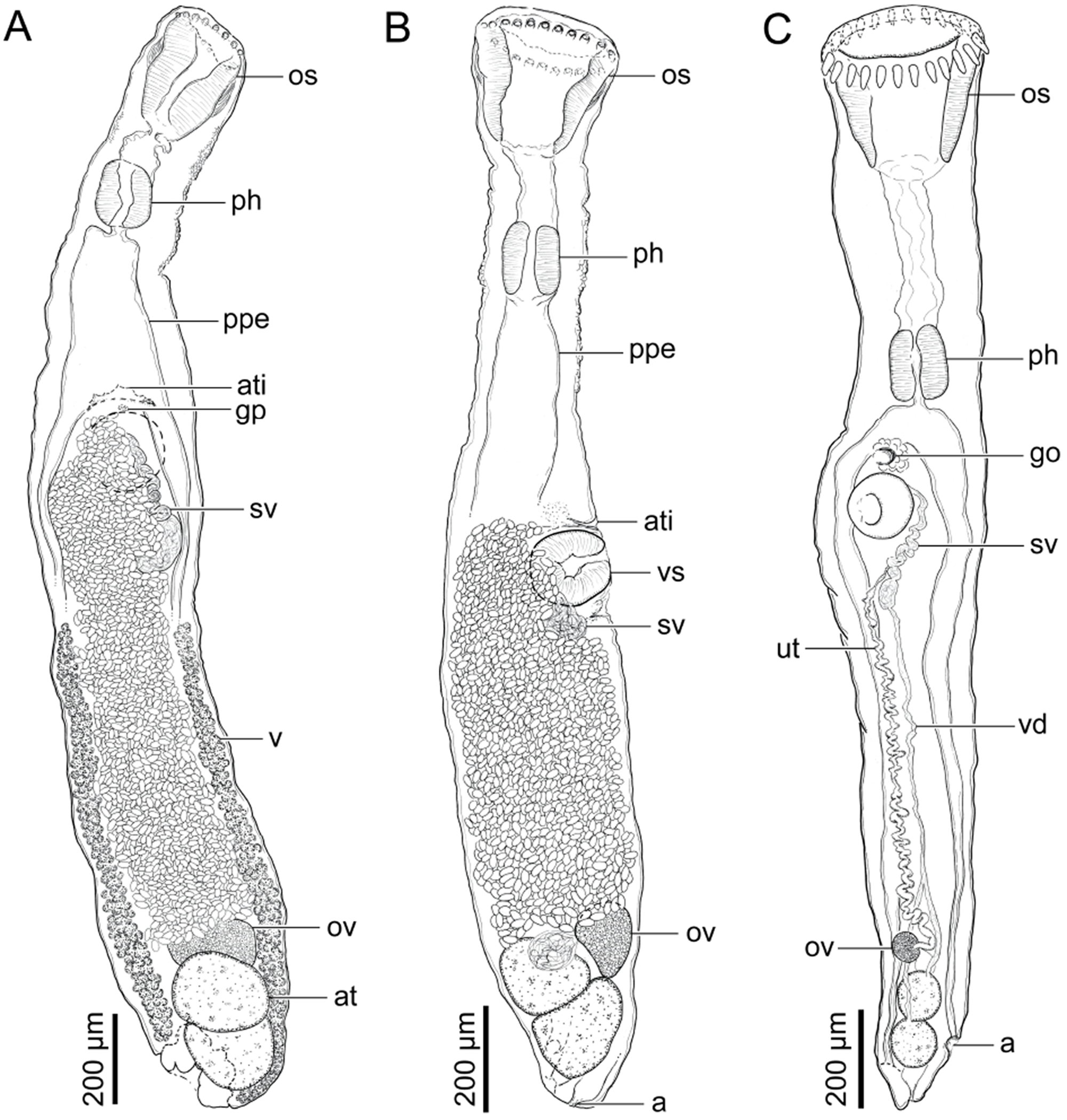
Body elongate, rounded posterior end (Figures 5F, 6A and 6B), 10–13 × longer than wide. Forebody 34–43% (39%; 3) of total body length. Hindbody 54–62% (57%; 3) of total body length. Tegument spinose, having 2 tegumental infoldings (Figures 5F, 5G, 6A and 6B); body surface spines big (Figures 5F, 4B and 4D), scale-like, becoming bigger at level of ventral sucker, then becoming smaller and fewer posteriad, extending to near posterior end of body; tegumental infoldings anterior and posterior to ventral sucker (Figures 5F, 5G, 6A and 6B). Ventral sucker spherical, in first quarter of body (Figures 5F, 6A and 6B). Oral sucker terminal, infundibular shaped, having circumoral spines; oral sucker spines forming a collar-like structure (Figures 5F, 6A and 6B), widest at insertion, tapering posteriad (Figure 5F). Oesophagus medial, representing 21–33% (28%; 3) of total body length, comprising pre-pharyngeal, pharyngeal and post-pharyngeal parts (Figures 5F, 6A and 6B). Pre-pharyngeal oesophagus retractile, 12–22% (17%; 3) of total oesophagus length. Pharynx quadrate in outline (Figures 5F, 6A and 6B), surrounding oesophagus, representing 11–20% (14%; 3) of total oesophagus length, followed by short muscular portion, sphincter-like, then followed by post-pharyngeal oesophagus. Post-pharyngeal oesophagus very long, thick-walled, 63–75% (69%; 3) of total oesophagus length (Figures 5F and 6A–B). Intestine bifurcating at level of ventral sucker (Figures 5F and 6A–B), 31–41% (36%; 3) of body length to anterior body end. Ceca slender, extending posteriad parallel to lateral body margins, symmetrical in length and width, each cecum opening through terminal pore (anal pore); anal pores terminal, flanking excretory pore (Figure 5F).
Types of Proctocaecum diploporum (Stunkard, Reference Stunkard1931) Baugh, Reference Baugh1957 (Digenea: Cryptogonimidae) from American alligator, Alligator mississippiensis (Daudin, 1802) (Crocodilia: Alligatoridae) from an indeterminate geographic locality. Scale value aside each bar. (A) Holotype (AMNH Platyhelminthes 875). Dorsal view. (B) Paratype (AMNH Platyhelminthes 875A). Lateral view. (C) Juvenile specimen of Proctocaecum coronarium (Cobbold, Reference Cobbold1861) Brooks, Reference Brooks1980 (AMNH Platyhelminthes 880). Ventral view. Abbreviations: anal pore (a); anterior tegumental infolding (ati); anterior testis (at); genital pore (gp); gonotyl (go); oral sucker (os); ovary (ov); pharynx (ph); post-pharyngeal oesophagus (ppe); seminal vesicle (sv); seminal vesicle (sv); uterus (ut); ventral sucker (vs); vitellarium (v).

Testes 2, spherical, medial, in last quarter of body, in tandem, contiguous with ovary (Figures 5F, 6A and 6B). Vasa efferentia obscured by dense mass of eggs through its length. Seminal vesicle long, sinuous, extending posterior to ventral sucker (Figures 5F, 5G, 6A and 6B); having convoluted proximal portion, having distal portion enlarged, sac-like (Figures 5F and 6A); prostatic duct short, weakly muscular, followed by ejaculatory duct. Ejaculatory duct short, opening into genital pore through a hermaphroditic duct (Figure 5F and 5G). Genital pore medial, between ventral sucker and anterior tegumental infolding (Figures 5F, 5G, 6A and 6B). Muscular gonotyl absent, but a dense mass of glandular-like cells is present in tegumental infoldings (Figures 5F, 5G, 6A and 6B).
Ovary spheroid (Figures 5F, 6A and 6B); pre-ovarian space 81–85% (84%; 3) of total body length; post-ovarian space 11–13% (12%; 3) of total body length. Oviduct emerging dorsally from ovary. Oviducal seminal receptacle sac-like, postero-dorsal to ovary. Laurer’s canal long, extending posterior to ovary, having a dorsal opening. Oötype, and Mehlis’ gland not observed. Uterus coiling anteriorly between ceca, extending from ovary to common genital pore (Figures 5F, 6A and 6B), dorsal to seminal vesicle, filled with numerous eggs (Figures 5F, 6A and 6B), confluent with hermaphroditic duct. Hermaphroditic duct opening into genital pore (Figures 5F and 5G). Eggs ovoid, having an operculum (Figure 5H). Vitellarium follicular, distributing in 2 bilaterally symmetrical fields, extending from anterior margin of anterior testis to equator of body (Figures 5F and 6A), occupying 38–43% (40%; 3) of total body length; pre-vitellarium space 50–55% (53%; 3) of total body length; post-vitellarium space 5–9% (7%; 3) of total body length. Transverse vitelline ducts emanating from posterior quarter of vitellarium, at level of ovary, forming vitelline reservoir; vitelline reservoir dorsal to ovary, confluent with oviduct (Figure 5F).
Excretory vesicle Y-shaped, ventral, extending from posterior body end to middle portion of pre-pharyngeal oesophagus (Figure 5F), bifurcating posterior to ventral sucker (Figure 5F); excretory pore terminal (Figure 5F).
Taxonomic remarks for P. coronarium and P. diploporum
Juveniles of P. coronarium and P. diploporum were morphologically similar and difficult to differentiate without having high quality, heat-killed and well-stained whole-mounted specimens and observations of live specimens with DIC. The redia were nearly identical. The cercaria and metacercaria of P. coronarium are 1.2–1.6 × larger than those of P. diploporum (see descriptions above). The adult of P. coronarium differ from P. diploporum by a combination of having smaller tegumental spines (2–5 long) in the forebody (vs 7–14 long), 24 oral sucker spines (vs 26–27), a short (29–50 long; 9–10% of oesophagus length) post-pharyngeal oesophagus (vs 543–1517 long; 63–75% of oesophagus length), asymmetrical ceca with anal pores at level of posterior testis (vs symmetrical ceca with anal pores in posterior end of body), a muscular gonotyl (vs absent), a vitellarium extending from the ovary to the posterior margin of the ventral sucker (vs extending from posterior testis to middle portion of body), excretory vesicle arms extending anteriad to level of the pharynx (vs extending anteriad to pre-pharyngeal oesophagus), and a dorsal excretory pore in the posterior body end (vs having a postero-terminal excretory pore).
The taxonomic history of Proctocaecum, its type species P. diploporum, and P. coronarium is convoluted and needs clarification. This confusion probably exists because these species are morphologically similar, they infect the same definitive host (the American alligator), their original descriptions contain errors, the type series of P. coronarium was lost or never deposited (Brooks and Overstreet, Reference Brooks and Overstreet1977; present study), and the type series for P. diploporum includes more than 1 species (see below). Brooks and Overstreet (Reference Brooks and Overstreet1977) asserted that Cobbold (Reference Cobbold1861) probably failed to note the presence of a pharynx and anal pores and that he misinterpreted ‘darkly-staining … parenchymal cells’ (Brooks and Overstreet, Reference Brooks and Overstreet1977; p. 1020) as vitelline follicles that extend anteriorly to the oesophageal bifurcation in P. coronarium. Based on our newly collected specimens and the examination of P. coronarium museum vouchers, we agree with Brooks and Overstreet (Reference Brooks and Overstreet1977) regarding the presence of a pharynx, anal pores and the extension of the vitellarium from anterior margin of anterior testis to posterior margin of ventral sucker in P. coronarium. Anal pores were erroneously omitted in several early descriptions of cryptogonimids. For example, when Acanthostomum was proposed by Looss (Reference Looss1899), he described the ceca as blind ending. Later, Khalil (Reference Khalil1963) and Moravec (Reference Moravec1976, Reference Moravec2001) asserted that Looss (Reference Looss1899) overlooked the anal pores and confirmed their presence in its type species Acanthostomum spiniceps (Looss, 1896). Baugh (Reference Baugh1957), evidently unaware of the presence of anal pores in Acanthostomum Looss, 1899, proposed Proctocaecum Baugh, Reference Baugh1957 and designated P. diploporum as its type species. Brooks and Overstreet (Reference Brooks and Overstreet1977) collected cryptogonimids from American alligators from Florida, Mississippi and Louisiana (USA). They considered P. diploporum (as Acanthostomum diploporus [Stunkard, Reference Stunkard1931] Stunkard, 1938) a junior subjective synonym of P. coronarium (as Acanthostomum coronarium [Cobbold, Reference Cobbold1861] Looss, Reference Looss1899) stating that specimens from Stunkard’s collection are identical to specimens they identified as P. coronarium. Later, in his revision of the Acanthostomidae, Brooks (Reference Brooks1980) established the new combination P. coronarium, erroneously listed Baugh (Reference Baugh1957) as the taxonomic authority, and considered it the type species of Proctocaecum. Instead of Baugh (Reference Baugh1957), Brooks (Reference Brooks1980) is the correct taxonomic revisor for P. coronarium.
Based on our newly collected specimens and our study of the type specimens, we accept P. diploporum as distinct, therefore P. diploporum is the type species of Proctocaecum because Baugh (Reference Baugh1957) originally designated it (International Commission on Zoological Nomenclature ICZN, 1999; Article 67.4). Brooks and Overstreet (Reference Brooks and Overstreet1977) also described C. pavida as Acanthostomum pavidum Brooks and Overstreet, 1977. Caimanicola pavida is morphologically identical to P. diploporum because it has large forebody spines (Table 3), 26–28 oral sucker spines, a long post-pharyngeal oesophagus (Table 3), symmetrical ceca with anal pores in the posterior body end, a vitellarium that extends from the posterior testis to the middle portion of the body, and a terminal excretory pore in the posterior body end as well as by lacking a muscular gonotyl. Therefore, we consider C. pavida a junior subjective synonym of P. diploporum. However, and importantly, future workers should be aware that the type series of P. diploporum and the type series of ‘C. pavida’ comprises some misidentified specimens. Specifics of these problems and the clarifications are below.
First, the type series of P. diploporum comprises 11 slides deposited in the AMNH collection. The holotype is mounted on the same slide as one of the paratypes (AMNH 875 and 875A), which are illustrated herein (Figures 6A and 6B, respectively). These specimens are indeed P. diploporum and are distinct from each of the remaining 10 paratypes (on 10 slides) that can only be morphologically diagnosed as P. coronarium (AMNH 876–881; AMNH 880 is Figure 6C). We herein identified them as immature specimens of P. coronarium because they have a short post-pharyngeal oesophagus, minute tegumental spines, 24 oral sucker spines, asymmetrical cecae, and anal pores at level of the posterior testis (vs the holotype [AMNH 875] and paratype [875A] of P. diploporum that have a long post-pharyngeal oesophagus, large tegumental spines, symmetrical ceca and terminal anal pores). We cannot determine the number of oral sucker spines in the holotype AMNH 875 and paratype 875A since they are highly contracted and have evidently lost the oral spines (Figures 6A and 6B). Stunkard (Reference Stunkard1931) described P. diploporum as having 24 oral sucker spines but his drawing shows 26. Additionally, Stunkard’s drawing of P. diploporum has a short post-pharyngeal oesophagus (see Figure 1 in Stunkard, Reference Stunkard1931) but we observed it to be long (Figure 6A). The misidentified paratypes (AMNH 876–881) should be regarded as vouchers of P. coronarium.
Second, the types of C. pavida were deposited in the USNM, the HWML and the Natural History Museum, Parasitic Worms Collection (NHMUK; London, UK). The holotype (USNM 74505) is identical to our specimens of P. diploporum. Regarding the paratypes, 3 of 26 slides under accession number HWML 20851 and 1 out of 6 slides under HWML 20852 are in fact P. coronarium.
Designation of a neotype for P. coronarium
Proctocaecum coronarium was described by Cobbold (Reference Cobbold1861), and later illustrated (Cobbold, Reference Cobbold1864) based on specimens he collected from an American alligator maintained at the Zoological Society of London Menagerie (the London Zoo). He did not designate a holotype, and no name-bearing type material exists to our knowledge: the USNM, AMNH and NHMUK lack a name bearing type for P. coronarium. Therefore, the type series of P. coronarium is considered lost or never deposited (Brooks and Overstreet, Reference Brooks and Overstreet1977; present study). The absence of a name-baring type has contributed to the long-standing taxonomic confusion involving P. coronarium, P. diploporum and C. pavida (see Taxonomic Remarks). Designation of a neotype therefore is warranted and necessary to clarify its taxonomic identity and to stabilize nomenclature within Proctocaecum (in accordance with International Commission on Zoological Nomenclature ICZN, 1999; Article 75.3). Although the original specimens of P. coronarium were collected from a host maintained in captivity in London, the American alligator native range is the southeastern USA. Since P. coronarium has been re-collected on several occasions from USA (Brooks and Overstreet, Reference Brooks and Overstreet1977; unpublished vouchers in USNM Invertebrate Zoology Collection; present study), we inferred that Cobbold’s (Reference Cobbold1861) specimens also came from within the natural range of the host. The neotype designated herein was collected from an American alligator within its native distribution in Alabama (southeastern USA), closely approximating the inferred type locality and host association of the original material.
Based on the careful consideration of the above information, we designate a neotype (an adult specimen) of P. coronarium (USNM 1774187). The specimen conforms to Cobbold’s (Reference Cobbold1861) original description and is consistent with the concept of P. coronarium as distinguished from its congeners (see Taxonomic Remarks). The neotype is designated as the name-bearing type to ensure long-term nomenclature stability and accessibility for future study as per the ICZN.
Histology of metacercarial infections in fish scales
Our histological sections confirmed that the metacercarial cyst resides within the scale, not within the epidermis covering the scale nor within the dermis beneath the relatively transparent scale (Figure 3B). A thin basophilic structure corresponding to the metacercaria cyst wall is clear (Figure 3B). No inflammatory response was observed. The metacercariae was identified as belonging to a species of Proctocaecum because it has conspicuous spines in the oral sucker.
Phylogenetic results
Our 28S and ITS2 sequences representing all developmental stages of P. coronarium and the adults and metacercariae of P. diploporum comprised 1620 bp and 402 bp, respectively. Our 28S sequences of the cercariae of P. diploporum comprised 1303 bp. We herein provide the first nucleotide information for P. macroclemidis (voucher USNM 1774212–1774214; see Materials and Methods), its 28S sequence comprised 1620 bp. GenBank accession numbers are in Table 2. All life history stages were identical to their corresponding morphologically identified adult, except for the cercaria of P. diploporum that was 99.9% similar (1 bp different) to its corresponding adult. We regard this as intra-specific variation. Six metacercarial cysts of each size class (see Materials and Methods) were sequenced. We found that the size of the cyst was a reliable objective character to differentiate the 2 species. The large cysts (>240 μm) had identical 28S sequences and also matched those of the morphologically identified adult of P. coronarium. Likewise, the small cysts (<200 μm) had identical 28S sequences and also matched those of the morphologically identified adult of P. diploporum. Remarkably, in some instances the cysts of both species were encysted mm apart and infecting the same individual scale on the same host individual (Figure 3A).
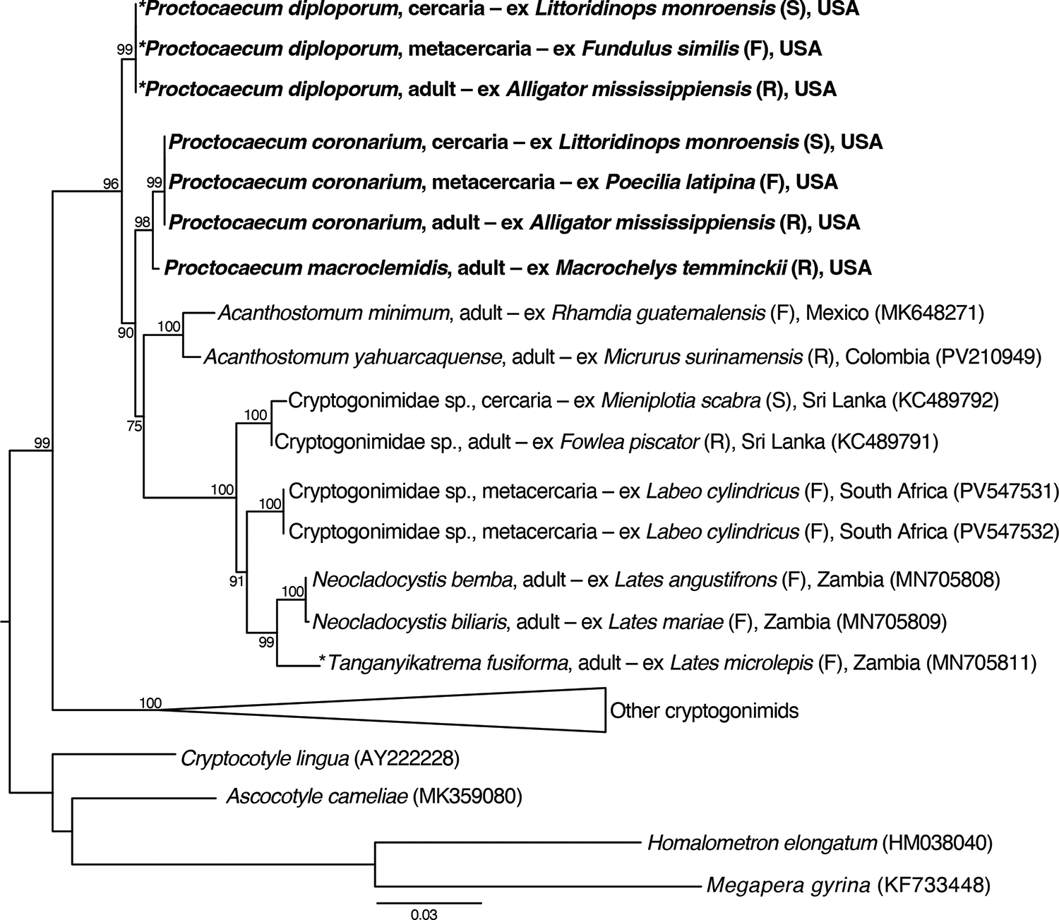
Our sequences of P. coronarium and P. diploporum were 98.6% similar (19–22 bp differences) to each other. Both species were more similar to our sequence of P. macroclemidis (P. coronarium 99.5%, 8 bp difference; P. diploporum 98.9%, 18 bp differences). Our 28S tree topology (Figure 7) resembled that of Cajiao-Mora et al. (Reference Cajiao-Mora, Brule, Dutton, Caicedo-Portilla and Bullard2025) and Martínez-Aquino et al. (Reference Martínez-Aquino, Vidal-martínez and Aguirre-Macedo2017). However, we included for the first time sequences ascribed to species of Proctocaecum and its type species P. diploporum. We recovered a paraphyletic Proctocaecum with our sequence of P. diploporum sister to the clade containing P. coronarium + P. macroclemidis, that clade being sister to the paraphyletic Acanthostomum and several other fish-infecting cryptogonimids. The low support values in our phylogenetic analysis could be the result of low taxon sampling.
28S maximum likelihood phylogenetic analysis inferred tree for Cryptogonimidae. Values aside nodes are ultrafast bootstrap percentage. Scale bar is in substitution per site. GenBank accession numbers are in parenthesis following each taxon. Type species are indicated by asterisk (*). Newly generated sequences are bolded.

Figure 7 Long description
The diagram is a phylogenetic tree representing the maximum likelihood analysis of Cryptogonimidae. The tree is oriented vertically, with branches extending from a common ancestor at the bottom. Each branch represents a taxon, with labels indicating species names, life stages and host information. GenBank accession numbers are in parentheses following each taxon. Type species are marked with an asterisk. Newly generated sequences are bolded. Bootstrap values are shown beside nodes, indicating the support for each branch. The scale bar at the bottom represents substitutions per site. The tree includes various species such as ′Proctoecum diplobulbum′ and ′Proctoecum coronarium′ from different hosts and locations, as well as other genera like ′Acanthostomum′ and ′Neocladocystis′. The tree also includes outgroups such as ′Cryptopygop linga′ and ′Ascocotyle carmelae′.
Discussion
We herein describe, for the first time, the life cycle of 2 closely related, sympatric, and morphologically similar reptile-infecting cryptogonimids. Interestingly, both species share the same hosts. We found that metacercariae of both species exhibit low host-specificity, infecting the scales of 7 fish species across Cyprinodontidae, Poeciliidae, Fundulidae, Menidiinae and Mugilidae. The adults, to date, are only known to mature in the American alligator.
The current literature on the life history of reptile-infecting cryptogonimids is scarce. Four studies attempted to characterize, by doing experimental infections, the life cycle of cryptogonimids infecting 3 crocodilians (Ostrowski de Núñez, Reference Ostrowski de Núñez1984; Salgado-Maldonado and Aguirre-Macedo, Reference Salgado-Maldonado and Aguirre-Macedo1991), 1 turtle (Ostrowski de Núñez, Reference Ostrowski de Núñez1987), and 1 snake (Roopa and Janardanan, Reference Roopa and Janardanan1998). Ostrowski de Núñez (Reference Ostrowski de Núñez1984) collected mojarra amarilla, Caquetaia kraussii (Steindachner, 1878) (Actinopterygii: Cichlidae), from Lake Valencia (Aragua River), Venezuela, infected with metacercariae of T. loossi (as Acanthostomum loossi) and C. marajoara (as Acanthostomum marajoara), in the ‘skin of the head and the finrays’. The cysts were fed to 1 spectacled caiman, and 1 Orinoco crocodile, to later collect adults. Similarly to our study herein, Ostrowski de Núñez (Reference Ostrowski de Núñez1984) documented concurrent infections by 2 cryptogonimids (metacercariae) infecting fish that were initially distinguished by ‘size, color, and location’ (site of infection in host) (Ostrowski de Núñez, Reference Ostrowski de Núñez1984; p 179). Salgado-Maldonado and Aguirre-Macedo (Reference Salgado-Maldonado and Aguirre-Macedo1991) collected Mayan cichlids, Mayaheros urophthalmus (Günther, 1862) (Actinopterygii: Cichlidae) from the Yucatan Peninsula, Mexico, infected with metacercariae of T. loossi and fed them to a Morelet’s crocodile in the hopes of recovering adults. Only one type of metacercaria was described in Salgado-Maldonado and Aguirre-Macedo (Reference Salgado-Maldonado and Aguirre-Macedo1991). The cercaria and first intermediate host were not described in either of those studies.
Jayawardena et al. (Reference Jayawardena, Tkach, Navaratne, Amerasinghe and Rajakaruna2013, Reference Jayawardena, Rohr, Amerasinghe, Navaratne and Rajakaruna2017) used nucleotide information to identify cercariae shed from a thorny tower snail from Siri Lanka as Acanthostomum burminis (Bhalerao, 1926) Bhalerao, 1936, a cryptogonimid that infects snakes. They then experimentally exposed Asian common toad tadpoles to assess the effects associated with metacercarial development. However, they provided no morphological identification, deposited no voucher specimen, and did not attempt to obtain adult worms. Consequently, the identity of those specimens is not reliable and should be regarded as Cryptogonimidae sp.
Martínez-Aquino et al. (Reference Martínez-Aquino, Vidal-martínez and Aguirre-Macedo2017) used nucleotide information and morphology to identify metacercariae of a Mayan cichlid from the Yucatan Peninsula. They identified the metacercariae as Timoniella cf. loossi and Acanthostomum cf. americanum because they had spines in the oral sucker but lacked additional morphology. They acknowledge that identifications are not reliable without the adults but that the definitive host could be a species of Crocodylus (see Martínez-Aquino et al., Reference Martínez-Aquino, Vidal-martínez and Aguirre-Macedo2017). An additional study (see Scholz et al., Reference Scholz, Lavadores, Vargas, Mendoza, Rodriguez and Vivas1994), on the life cycle of Oligogonotylus manteri Watson, 1976, a fish-infecting cryptogonimid in Yucatan, Mexico, reported that the Mayan cichlid is either a second intermediate host (metacercaria infecting gill, fins, body surface, intestine wall) or a definitive host (adults infecting intestinal lumen).
In the Buenos Aires Zoo (Argentina), Ostrowski de Núñez (Reference Ostrowski de Núñez1987) collected H. castellanosae infected with cercariae as well as several freshwater fishes infected with metacercariae of C. brauni from a pond holding several species of crocodilians and turtles. Ostrowski de Núñez (Reference Ostrowski de Núñez1987) experimentally infected a 10 spotted live-bearer with cercaria shed from H. castellanosae and fed it to a Hilaire’s side-necked turtle to recover adults. The complete life cycle was successfully elucidated therein. Roopa and Janardanan (Reference Roopa and Janardanan1998) collected red-rimmed Melania, Melanoides tuberculata (Müller, 1774) (Gastropoda: Thiaridae), from Kerala, India, infected with cercariae of Acanthostomum burminis (Bhalerao, 1926) Bhalerao, 1936 and experimentally infected several freshwater fishes, characterized the metacercariae, and then fed the Asiatic water snake with the infected fish to characterize the adults.
We herein provide the first histopathology of a cryptogonimid metacercarial infection. Interestingly, we found that the metacercaria of both P. diploporum and P. coronarium encysts within the scale itself, i.e., not within the epidermis or dermis. The scale envelopes the metacercarial cyst (Figure 3B). Previous studies report cryptogonimid metacercarial cysts as being beneath the scale or beneath the skin (Ostrowski de Núñez, Reference Ostrowski de Núñez1987; Simões et al., Reference Simões, Das Neves and Santos2008; Quintana and Ostrowski de Núñez, Reference Quintana and Ostrowski de Núñez2016). Metacercarial cysts were enveloped by lepidotrichia and observed in the epidermis between fin rays (Lundahl, Reference Lundahl1941; Ostrowski de Núñez, Reference Ostrowski de Núñez1984, Reference Ostrowski de Núñez1987; Simões et al., Reference Simões, Das Neves and Santos2008; Quintana and Ostrowski de Núñez, Reference Quintana and Ostrowski de Núñez2014; Quintana and Ostrowski de Núñez, Reference Quintana and Ostrowski de Núñez2016). Other cryptogonimid metacercariae reportedly infect the buccal cavity, gill, muscle, intestinal wall and liver of fishes (Vélez-Sampedro et al., Reference Vélez-Sampedro, Uruburu and Lenis2022; Simões et al., Reference Simões, Das Neves and Santos2008; Ostrowski de Núñez et al., Reference Ostrowski de Núñez, Semenas, Brugni, Viozzi and Flores1999; Scholz et al., Reference Scholz, Lavadores, Vargas, Mendoza, Rodriguez and Vivas1994; Salgado-Maldonado and Aguirre-Macedo, Reference Salgado-Maldonado and Aguirre-Macedo1991; Maillard, Reference Maillard1973; Cribb, Reference Cribb1986).
Phylogenetically related and sympatric trematodes that use the same invertebrate and vertebrate hosts, like the ones studied here, could be a good model to further explore the mechanisms of digenean speciation and host specificity.
Acknowledgements
We thank Dr Anna Phillips, Chad Walter, Kathryn Ahlfeld and Amanda Robinson (all Smithsonian Institution, Washington, DC) for accepting and curating our voucher specimens and assisting with specimen loans. We also thank Drs Gabor Racz and Scott L. Gardner (Harold W. Manter Laboratory of Parasitology, University of Nebraska, Lincoln, NE), and Estefanía Rodríguez and Lily Berniker (American Museum of Natural History, Division of Invertebrate Zoology, New York City, NY) for assisting with specimen loans. We thank Gail Barton and the Department of Resource Sharing (Auburn University, Auburn, AL) for assisting with inter-library loan.
Author contributions
K.C.-M.: field work, specimen preparation and identification, deposition of holotype and paratypes, taxonomic and phylogenetic analyses, conception of manuscript, revisions, corresponding author duties. S.S.C.: field work, specimen preparation and identification, review of draft. J.H.B.: field work, review of draft. D.G.-G.: field work, histopathological analysis, review of draft. H.D.: field work, specimen preparation, review of draft. M.B.W.: field work, review of draft. S.A.B.: field work, taxonomic and phylogenetic analyses, funding, editing and review of draft.
Financial support
This study was supported by The Southeastern Cooperative Fish Parasite and Disease Project, U.S. Fish and Wildlife Service (Department of Interior), U.S. Department of Agriculture (National Institute of Food and Agriculture), Federal Aid in Sport Fish Restoration (Alabama Department of Conservation and Natural Resources), and the Alabama Agricultural Experiment Station (Auburn University, College of Agriculture).
Competing interests
The authors declare no conflict of interest.
Ethical standards
All applicable institutional, national and international guidelines for the care and use of animals were followed.

 Open access

