





Introduction
Schistosomiasis is a neglected tropical disease (NTD) caused by infection with parasitic worms of the genus Schistosoma that are transmitted by freshwater snail vectors (intermediate hosts). An estimated 253.7 million people, ∼90% of whom live in Africa, are infected with schistosomes and require preventative treatment to curb morbidity associated with chronic intestinal and urogenital schistosomiasis (WHO, 2026). Several human- and animal-infecting schistosome species are endemic across tropical and sub-tropical climates in Africa, Asia, South America and even focal regions in Europe (Boissier et al., Reference Boissier, Moné, Mitta, Bargues, Molyneux and Mas-coma2015), where the appropriate snail hosts are present. Public health measures aimed at reducing human schistosomiasis consist primarily of mass drug administration of praziquantel (Kokaliaris et al., Reference Kokaliaris, Garba, Matuska, Bronzan, Colley, Dorkenoo, Ekpo, Fleming, French, Kabore, Mbonigaba, Midzi, Mwinzi, N’goran, Polo, Sacko, Tchuem Tchuenté, Tukahebwa, Uvon, Yang, Wiesner, Zhang, Utzinger and Vounatsou2022), whilst approaches aimed at reducing transmission through snail control are seldom utilized in contemporary control programmes due to the significant costs and difficulties in effective implementation (King and Bertsch, Reference King and Bertsch2015; Garba Djirmay et al., Reference Garba Djirmay, Yadav, Guo, Rollinson and Madsen2024).
Unguja and Pemba, collectively known as Zanzibar, the Tanzanian semi-autonomous archipelago of islands off the east coast of Africa in the Indian Ocean, are endemic for urogenital schistosomiasis, caused by infection with Schistosoma haematobium, originally believed to be allopatric across the islands transmitted through a single snail host Bulinus globosus (Stothard et al., Reference Stothard, Loxton, Rollinson, Mgeni, Khamis, Ameri, Ramsan and Savioli2000), until the recent identification of Schistosoma bovis shed from B. globosus and S. haematobium infecting Bulinus nasutus (Pennance et al., Reference Pennance, Ame, Amour, Suleiman, Allan, Rollinson and Webster2018, Reference Pennance, Ame, Amour, Suleiman, Muhsin, Kabole, Ali, Archer, Allan, Emery, Rabone, Knopp, Rollinson, Cable and Webster2022). Unguja and Pemba have a long history of urogenital schistosomiasis research and control (Trippler et al., Reference Trippler, Knopp, Welsche, Webster, Stothard, Blair, Allan, Ame, Juma, Kabole, Ali, Rollinson and Pennance2023) and are targeted for elimination through ongoing efforts of the Zanzibar NTD programme and operational research projects trialling integrated interventions on mass drug administration and surveillance-response approaches (Knopp et al., Reference Knopp, Mohammed, Ali, Khamis, Ame, Albonico, Gouvras, Fenwick, Savioli and Colley2012, Reference Knopp, Person, Ame, Mohammed, Ali, Khamis, Rabone, Allan, Gouvras and Blair2013, Reference Knopp, Ame, Person, Hattendorf, Rabone, Juma, Muhsin, Khamis, Hollenberg, Mohammed, Kabole, Ali and Rollinson2019a, Reference Knopp, Person, Ame, Ali, Hattendorf, Juma, Muhsin, Khamis, Mohammed, Utzinger, Hollenberg, Kabole and Rollinson2019b; Trippler et al., Reference Trippler, Hattendorf, Ali, Ame, Juma, Kabole and Knopp2021a, Reference Trippler, Ali, Masoud, Mohammed, Amour, Suleiman, Ame, Kabole, Hattendorf and Knopp2024). Although prevalence has significantly reduced across the islands in the selected shehias (the smallest administrative region in Zanzibar), transmission has yet to be interrupted, leaving focal endemicity in hot spot areas that require new methods of surveillance and tailored interventions (Pennance et al., Reference Pennance, Person, Muhsin, Khamis, Muhsin, Khamis, Mohammed, Kabole, Rollinson and Knopp2016; Knopp et al., Reference Knopp, Person, Ame, Ali, Hattendorf, Juma, Muhsin, Khamis, Mohammed, Utzinger, Hollenberg, Kabole and Rollinson2019b, Reference Knopp, Ali, van Dijk, Emery, Pennance, Webster and Coffeng2026; Trippler et al., Reference Trippler, Ame, Hattendorf, Juma, Abubakar, Ali, Kabole, Rollinson and Knopp2021b, Reference Trippler, Knopp, Welsche, Webster, Stothard, Blair, Allan, Ame, Juma, Kabole, Ali, Rollinson and Pennance2023).
Infection with urogenital schistosomiasis in Unguja and Pemba occurs when humans contact freshwater bodies containing infected snail vector(s) of the genus Bulinus. Four Bulinus spp. are present in Unguja and Pemba: B. globosus, B. nasutus, B. forskalii and an undescribed B. forskalii group species (Stothard and Rollinson, Reference Stothard and Rollinson1997a; Stothard et al., Reference Stothard, Loxton and Rollinson2002a; Kane et al., Reference Kane, Stothard, Emery and Rollinson2008; Pennance et al., Reference Pennance, Ame, Amour, Suleiman, Muhsin, Kabole, Ali, Archer, Allan, Emery, Rabone, Knopp, Rollinson, Cable and Webster2022). Although it has long been established that B. forskalii spp. were likely to play no role in S. haematobium transmission in Unguja and Pemba (Cawston, Reference Cawston1927; Mansfield-Aders, Reference Mansfield-Aders1928; McCarthy, Reference McCarthy1930; Mozley, Reference Mozley1939; Webbe, Reference Webbe1962), with potentially some role in S. bovis transmission (Ame et al., Reference Ame, Juma, Juhász, Ali, Suleiman, Gobert, Cunningham, Cawley, Atkins, Jones, LaCourse, Kabole and Stothard2025), decades of contradictory reports regarding the role of B. globosus and B. nasutus in transmission have ‘muddied the waters’. Complications arose due to the significant degree of morphological overlap between B. globosus and B. nasutus in Zanzibar (Wright, Reference Wright1961; Goatly and Jordan, Reference Goatly and Jordan1965; Mandahl-Barth, Reference Mandahl-Barth1965), where even detailed shell morphometric analysis provided little aid in distinguishing them (Stothard et al., Reference Stothard, Mgeni, Alawi, Savioli and Rollinson1997). Molecular differentiation of the 2 species has established B. globosus as the predominant vector of S. haematobium in Zanzibar, with B. nasutus likely playing a minor role (Stothard et al., Reference Stothard, Loxton, Rollinson, Mgeni, Khamis, Ameri, Ramsan and Savioli2000; Pennance et al., Reference Pennance, Ame, Amour, Suleiman, Muhsin, Kabole, Ali, Archer, Allan, Emery, Rabone, Knopp, Rollinson, Cable and Webster2022). It is noteworthy that B. nasutus is a vector of S. haematobium on nearby coastal regions of Kenya (Kariuki et al., Reference Kariuki, Clennon, Brady, Kitron, Sturrock, Ouma, Ndzovu, Mungai, Hoffman, Hamburger, Pellegrini, Muchiri and King2004) and Tanzania (Webbe, Reference Webbe1962; Loker et al., Reference Loker, Moyo and Gardner1981; Sarda et al., Reference Sarda, Simonsen and Mahikwano1985; Angelo et al., Reference Angelo, Buza, Kinung’hi, Kariuki, Mwanga, Munisi and Wilson2018), in addition to the closely related snail species Bulinus productus (a currently disputed species classified as a form of B. nasutus (Brown, Reference Brown1994) but with molecular evidence supporting its species status (Pennance, Reference Pennance2020) in mainland Tanzania (McCullough et al., Reference McCullough, Eyakuze, Msinde and Nditi1968; Babbitt et al., Reference Babbitt, Laidemitt, Mutuku, Oraro, Brant, Mkoji and Loker2023). Bulinus globosus has also been incriminated for its role in endemic S. bovis transmission on Pemba (Pennance et al., Reference Pennance, Ame, Amour, Suleiman, Allan, Rollinson and Webster2018).
With the exception of now dated and not routinely used molecular assays (Stothard and Rollinson, Reference Stothard and Rollinson1996, Reference Stothard and Rollinson1997b; Stothard et al., Reference Stothard, Mgeni, Alawi, Savioli and Rollinson1997, Reference Stothard, Llewellyn-hughes, Griffin, Hubbard, Kristensen and Rollinson2002b), no rapid diagnostic test exists to easily differentiate B. globosus and B. nasutus. With the current targets moving towards the elimination of urogenital schistosomiasis in Zanzibar, and the newly established potential role of B. nasutus in schistosome transmission (Ame, Reference Ame2018; Pennance et al., Reference Pennance, Ame, Amour, Suleiman, Muhsin, Kabole, Ali, Archer, Allan, Emery, Rabone, Knopp, Rollinson, Cable and Webster2022), accurately establishing the risk of schistosomiasis infection at freshwater contact sites based on compatible snail vectors is pertinent. Here, we aimed to develop a simple and robust assay to differentiate these 2 snail species using basic molecular laboratory techniques (PCR and gel electrophoresis) that can be used in tandem with rapid diagnostic assays for Schistosoma xenomonitoring (Pennance et al., Reference Pennance, Archer, Lugli, Rostron, Llanwarne, Ali, Amour, Suleiman, Li, Rollinson, Cable, Knopp, Allan, Ame and Webster2020a) and Schistosoma spp. differentiation (Webster et al., Reference Webster, Rollinson, Stothard and Huyse2010) to inform and bolster malacological surveys and interventions in Zanzibar.
Materials and methods
Molecular diagnostic assay to rapidly distinguish B. globosus and B. nasutus
Ribosomal DNA (rDNA) sequence data were available from a single B. globosus and B. nasutus specimen collected from Unguja available through the Schistosomiasis Collection at the Natural History Museum (Emery et al., Reference Emery, Allan, Rabone and Rollinson2012). The sequence data were obtained from a MiSeq (600 cycles) run of multiple snail samples as part of a collaborative Natural History Museum research project (Pennance, Reference Pennance2020). The rDNA fragments were assembled by mapping them to available ribosomal sequence data on GenBank for Bulinus and annotated (Briscoe et al., Reference Briscoe, Bray, Brabec and Littlewood2016). The 5.8S rDNA and the internal transcribed spacer 2 (ITS2) were extracted (635 bp for B. globosus and 628 bp for B. nasutus) from the dataset and, following annotation, sequence alignment was performed in Geneious v.11.1.4 (Kearse et al., Reference Kearse, Moir, Wilson, Stones-havas, Cheung, Sturrock, Buxton, Cooper, Markowitz, Duran, Thierer, Ashton, Meintjes and AJ2012) using MAFFT v7.388 (Katoh and Standley, Reference Katoh and Standley2013).
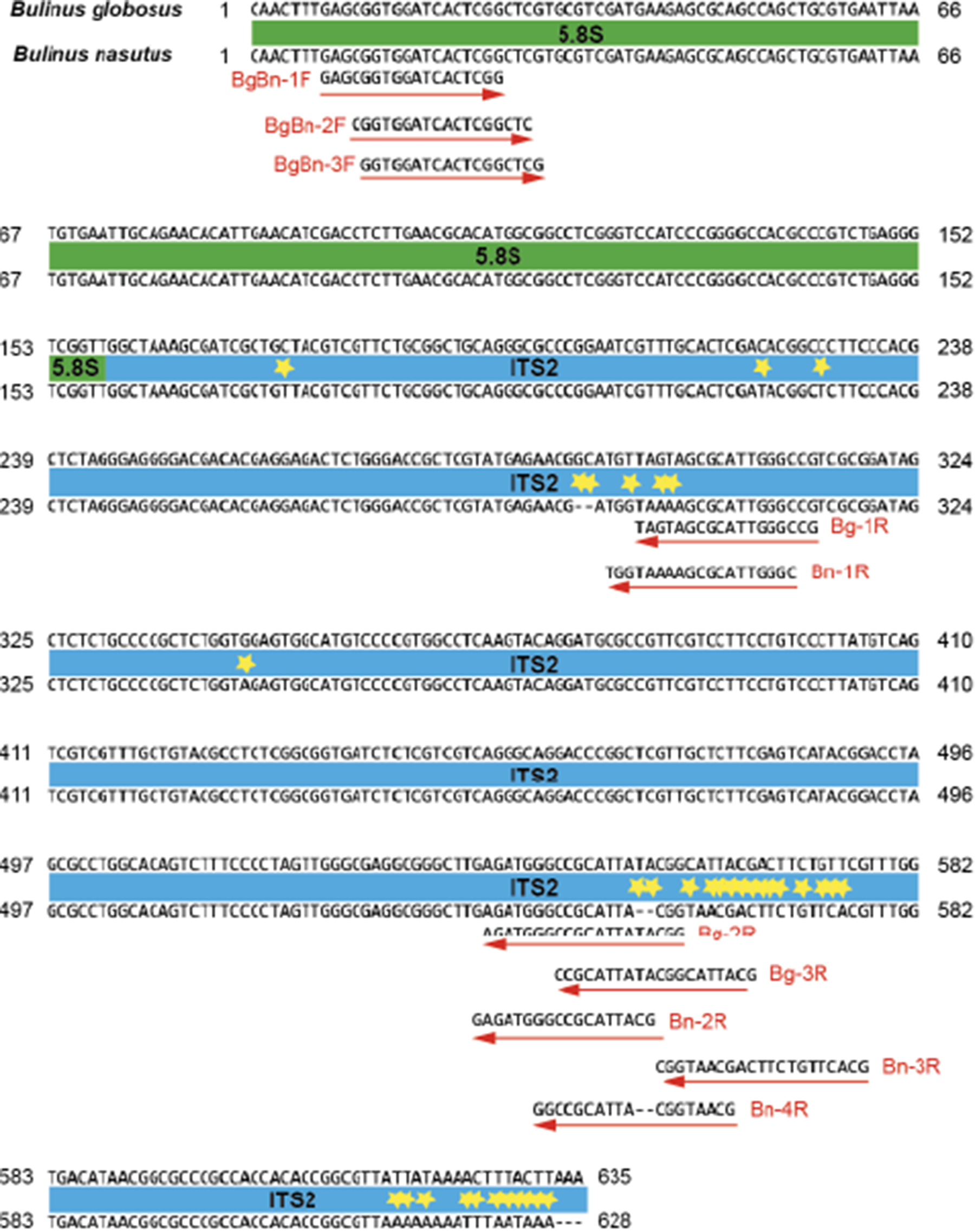
The alignment was inspected for regions of interspecies nucleotide variability and conservation to enable the design of species-specific and universal primers for the development of an amplicon-based diagnostic assay (Figure 1). The 5.8S rDNA region was conserved across species, and therefore universal forward primers were selected within this region. ITS2 regions containing single nucleotide polymorphisms (SNPs) (Figure 1) were targeted for species-specific reverse primer design. Primer design was performed using Primer3 v2.3.7 (Untergasser et al., Reference Untergasser, Cutcutache, Koressaar, Ye, Faircloth, Remm and Rozen2012). Three suitable universal forward primers were identified in the 5.8S gene region, and 7 ‘species-specific’ reverse primers from 2 regions of high SNP density in the ITS2 (Figure 1 and Table 1) were also deemed suitable (high %GC, containing a ‘GC clamp’ at the 3′ end and melting temperature within 3 °C of each other). The custom oligos were produced by Sigma-Aldrich (UK).
Sequence alignment of 5.8S rDNA and ITS2 of B. globosus and B. nasutus (Zanzibar isolates) showing the positions of universal forward and species-specific reverse primers. Reverse primers labelled Bn- and Bg- represent those designed specifically for B. nasutus and B. globosus, respectively. Yellow stars indicate single nucleotide polymorphisms (SNPs) or indels differentiating the 2 sequences.

Figure 1 Long description
Two aligned nucleotide sequence rows are shown, labeled Bulinus globosus and Bulinus nasutus. Position numbers appear at the left and right ends of multiple line blocks, including 66, 152, 238, 324, 410, 496, 582, 635 and 628. Regions are labeled internal transcribed spacer 1, 5.8S and internal transcribed spacer 2. Multiple red arrows label primer positions and names, including BgBn 1F, BgBn 2F, BgBn 3F, Bg 1R, Bg 3R, Bn 1R, Bn 2R, Bn 3R, Bg 4R and Bn 4R. Yellow star symbols mark several positions along the sequences that are single nucleotide polymorphisms.
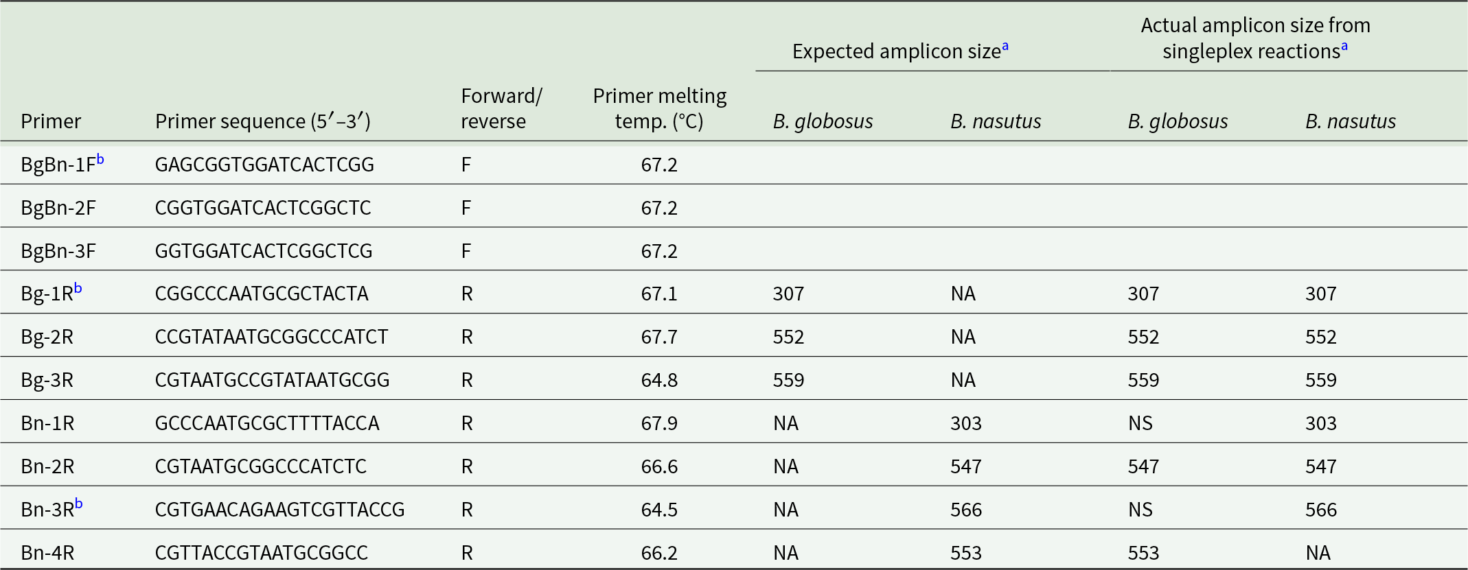
Primer sequences and expected amplicon lengths for B. nasutus and B. globosus. Forward primers are universal for both B. nasutus and B. globosus
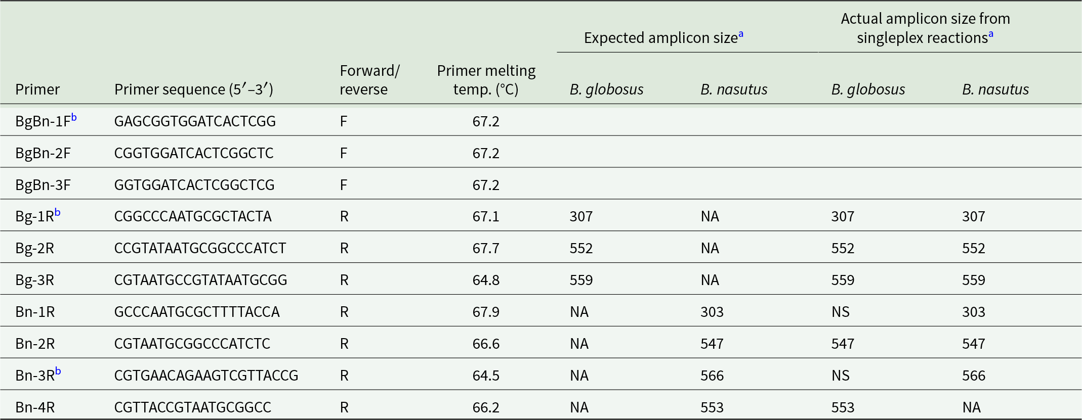
Table 1 Long description
Primer names, sequences, direction, and melting temperatures are listed alongside expected and observed amplicon sizes for Bulinus globosus and Bulinus nasutus in singleplex PCR. Three forward primers (BgBn-1 F, BgBn-2 F, BgBn-3 F) are universal and have melting temperatures of 67.2 degrees C, with no amplicon sizes reported for forward primers alone. Reverse primers labeled Bg are intended for B. globosus and show expected and observed products of 307 bp (Bg-1 R), 552 bp (Bg-2 R), and 559 bp (Bg-3 R) for B. globosus, while B. nasutus shows no amplification in the expected-size column for these Bg primers. In the observed singleplex results, those same Bg reverse primers produce the listed band sizes for both species, indicating cross-amplification for B. nasutus at 307, 552, and 559 bp. Reverse primers labeled Bn are intended for B. nasutus and show expected products of 303 bp (Bn-1 R), 547 bp (Bn-2 R), 566 bp (Bn-3 R), and 553 bp (Bn-4 R) for B. nasutus, while B. globosus shows no amplification in the expected-size column for these Bn primers. Observed singleplex results for Bn primers include non-specific amplification for B. globosus with Bn-1 R and Bn-3 R, a specific 547 bp product for both species with Bn-2 R, and a 553 bp product for B. globosus but no amplification for B. nasutus with Bn-4 R. Overall, melting temperatures cluster in the mid to high 60s degrees C, and several reverse primers show either cross-amplification or non-specific products, so species specificity should be interpreted cautiously.
a Amplicon size is shown for when the reverse primers are used in conjunction with the forward primer BgBn-1F and includes the primer sequence. NA = no amplification. NS = non-specific amplification.
b These primers were used in the final iteration of the duplex diagnostic assay.
Reverse primers labelled with ‘Bn-’ and ‘Bg-’ were designed to be specific for B. nasutus and B. globosus, respectively
Singleplex and duplex assay testing
All 21 forward and reverse primer combinations were trialled with single samples of B. globosus from Kizimbani (Pemba) and B. nasutus from Uwandani (Pemba), previously extracted and identified through cox1 barcoding (Pennance et al., Reference Pennance, Ame, Amour, Suleiman, Muhsin, Kabole, Ali, Archer, Allan, Emery, Rabone, Knopp, Rollinson, Cable and Webster2022), to provide an overview of primer performance with both species (Supplementary Table 1). PCR amplifications were performed in a total reaction volume of 25 µL using GE Healthcare (Amersham, Buckinghamshire, UK) ‘Ready-To-Go’ PCR beads, 1 µL of each 10 µM primer, 1 µL of template DNA and 22 µL of RNA-free PCR water. A negative control was also performed for each primer set reaction using 1 µL of RNA-free PCR water in replacement of template DNA. Thermocycling was performed in an Applied Biosystems GeneAmp PCR system 9700 thermal cycler (ThermoFisher, Waltham, MA, USA) with PCR conditions for all primer combinations involving denaturing at 95 °C for 5 min followed by 40 cycles of 95 °C for 30 sec, 60 °C for 30 sec, 72 °C for 45 sec, followed by a final 7 min extension at 72 °C. The melting temperature of all primers ranged from 64.5 to 67.7 °C, so a 60 °C annealing temperature was chosen to maximize specificity over lower annealing temperatures. A 4 µL aliquot of each PCR amplicon was loaded with 1 µL of a Bioline (London, UK) 5× DNA Loading Buffer Blue/gel red mix on a 2% agarose gel for 1 h at 90 V and imaged using a GBOX-Chemi-XRQ gel documentation system (Syngene, Cambridge, UK). All gels were run with BioLine (London, UK) HyperLadder IV to assess band sizes in 100 bp increments.
Selected PCR amplicons giving expected amplicon band size were purified using AxyPrep Bead Magnetic Bead Purification Kits (Axygen, part of Corning group, New York, USA). Bi-directional Sanger sequencing was performed on successful amplicons on an Applied Biosystems 3730xl DNA analyser, using a dilution of the original PCR primers. Sequences were manually edited, aligned and trimmed before being compared to the reference sequence data to confirm that amplicons were indeed the target amplicon and snail species.
After screening all singleplex primer combinations and confirming band identity through sequencing, those that gave robust, reproducible and desirable species-specific band sizes for B. globosus and B. nasutus from Pemba (Table 1) were selected for testing with further field isolates from Pemba and other Bulinus species from East Africa (mainland Tanzania) and West Africa (Niger) (Table 2 and Supplementary Table 1). To check for primer cross-reactivity with Schistosoma, these tests included snails with patent S. haematobium or S. bovis infections, the schistosome species of which were identified through the molecular identification of the shed cercariae (see Pennance et al., Reference Pennance, Ame, Amour, Suleiman, Muhsin, Kabole, Ali, Archer, Allan, Emery, Rabone, Knopp, Rollinson, Cable and Webster2022) (Supplementary Table 1). Duplex reactions were trialled utilizing a single universal forward primer (BgBn-1F) and each reverse primer. Duplex reactions were performed using the same singleplex thermocycling conditions (described above) but with the addition of 3 primers instead of 2 (1 universal forward primer and 2 species-specific reverse primers), therefore reducing the RNA-free PCR water in the reaction to 21 µL to account for the extra 1 µL of primer.
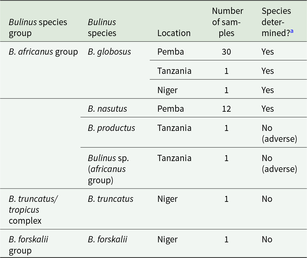
Summary of the Bulinus species used and those that could be determined during the species diagnostic primer trials
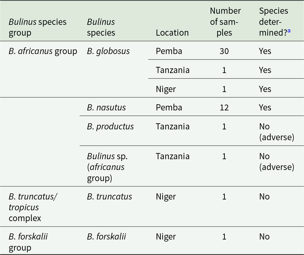
Table 2 Long description
The table summarizes snail samples by Bulinus species group, species, collection location, sample count, and whether the diagnostic primers produced a usable species identification. The largest set was Bulinus globosus from Pemba with 30 samples, all identified, plus one identified sample each from Tanzania and Niger. Bulinus nasutus from Pemba had 12 samples, all identified. Three single-sample entries were not identified: Bulinus productus from Tanzania and an unspecified Bulinus in the africanus group from Tanzania were marked as adverse outcomes, and Bulinus truncatus from Niger was not determined. Bulinus forskalii from Niger also had one sample that was not determined. Overall, identifications were common for the Pemba collections, while several single samples from Tanzania and Niger could not be resolved, including cases flagged as adverse based on gel band patterns.
a Adverse species determination was samples that produced PCR amplicons of the same or similar molecular weight as those observed from B. globosus or B. nasutus, judged by gel electrophoresis.
Specificity assessment with Bulinus spp. from Sub-Saharan Africa
Primer combinations were tested to validate their performance on B. globosus and B. nasutus samples from endemic regions other than Pemba. This included samples of B. globosus, B. nasutus, B. productus, B. africanus sp., B. truncatus and B. forskalii from Niger (Pennance et al., Reference Pennance, Allan, Emery, Rabone, Cable, Garba, Hamidou, Webster, Rollinson and Webster2020b) and mainland Tanzania (see Supplementary Table 1), previously identified by cox1 molecular analysis. The specimens included additional B. globosus and B. nasutus, B. productus and unidentified B. africanus group species closely related to B. nasutus and B. globosus, respectively (Kane et al., Reference Kane, Stothard, Emery and Rollinson2008), and more distantly related Bulinus spp., B. truncatus and B. forskalii (Table 2). Adverse or false positive reactions were noted as those which produced PCR amplicons of the same molecular weight as those observed from B. globosus or B. nasutus.
Results
Molecular diagnostic assay primer design
The 5.8S gene (158 bp) of B. globosus and B. nasutus was conserved between species, allowing the design of 3 universal forward primers at the 5′ end of this region (Figure 1 and Table 1). Two regions within the ITS2 (470 bp B. nasutus and 477 bp B. globosus) showed inter-species mutations that enabled the design of 7 (4 for B. nasutus and 3 for B. globosus) species-specific primers (Figure 1 and Table 1). Two primers were designed to generate a ‘short’ amplicon for B. globosus (Bg-1R) and B. nasutus (Bn-1R), and 5 primers were designed to generate a ‘long’ amplicon for B. globosus (Bg-2R and Bg-3R) and B. nasutus (Bn-2R, Bn-3R, Bn-4R) when used in conjunction with one of the universal forward primers identified in 5.8S (Figure 1). Primer sequences and desired amplicon lengths for the selected primer pairs for B. globosus or B. nasutus are displayed in Table 1.
Singleplex and duplex assay testing
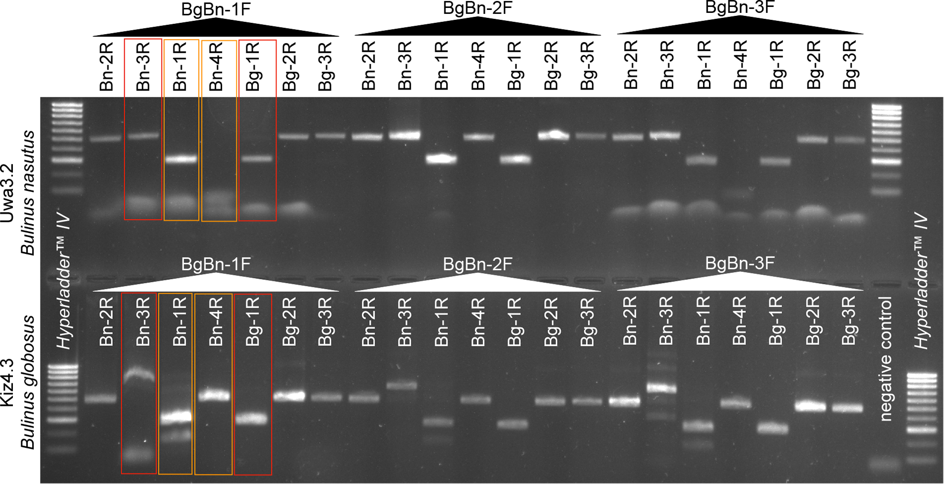
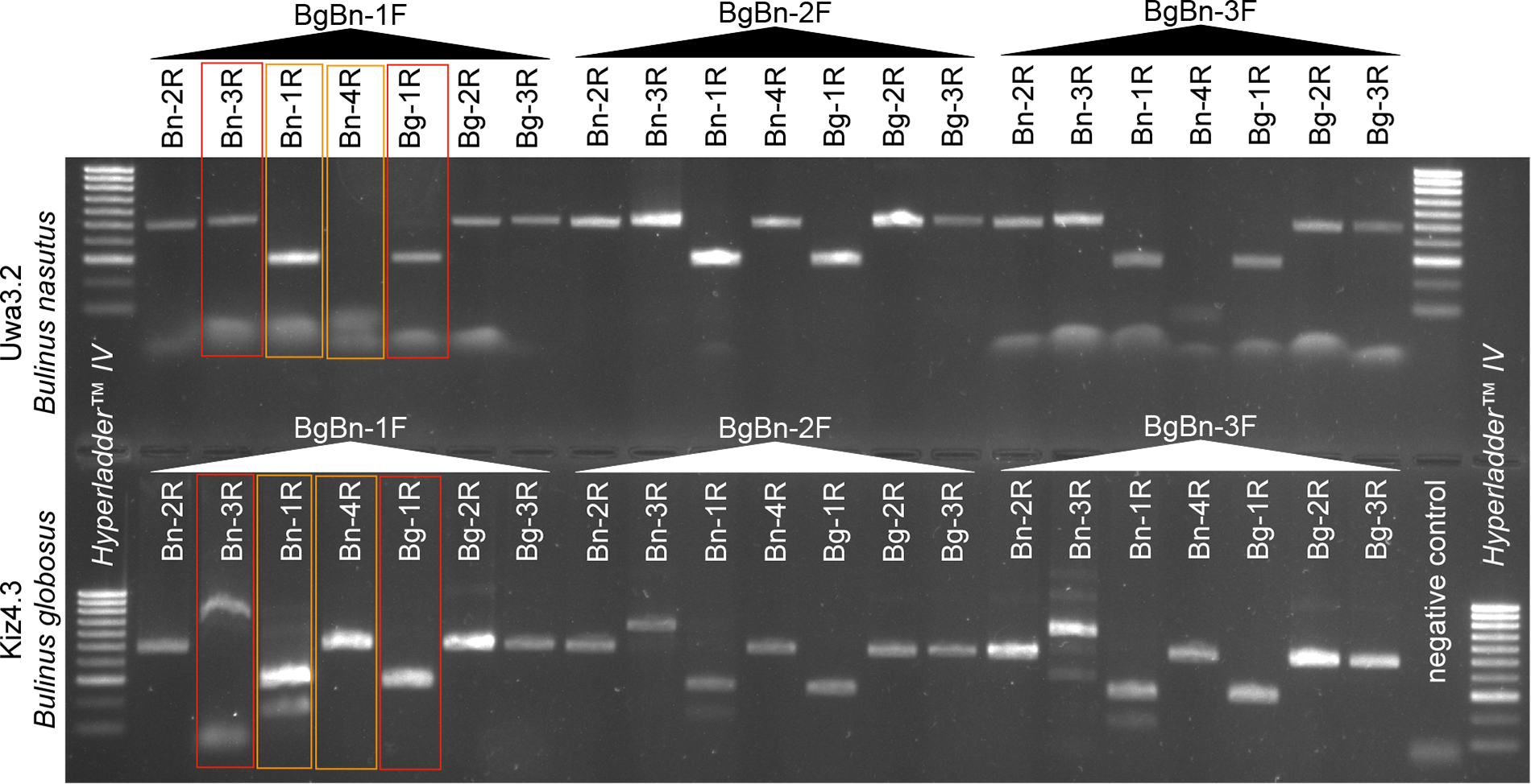
The majority of the 21 primer combinations tested generated PCR products for both B. globosus and B. nasutus samples from Pemba (Table 1 and Figure 2). As well as amplifying the targeted B. globosus rDNA, the B. globosus-specific primers (Bg-1R − Bg-3R) also generated amplicons of the same size for B. nasutus (Table 1). Two of the B. nasutus-specific primers (Bn-1R and Bn-3R) resulted in non-specific banding for B. globosus samples but expected amplicons for B. nasutus, another (Bn-2R) giving the same amplicon size for both B. globosus and B. nasutus, and the final primer (Bn-4R) unexpectedly amplified B. globosus instead of B. nasutus with the desired amplicon size (orange box, Figure 2). From these results, Bn-1R and Bn-4R were considered to give B. globosus and B. nasutus specific bands, importantly of different amplicon sizes. However, further trialling of these primers on other B. globosus and B. nasutus samples showed that amplification of B. nasutus and B. globosus also occurred with these primers in duplex reactions and therefore species could not be differentiated (results not shown). Bn-4R was replaced by Bn-3R to increase specificity for B. nasutus; however, this then failed to amplify B. globosus samples from mainland Tanzania and gave multiple bands for B. nasutus (Figure 3).
Gel electrophoresis showing PCR amplicon products of the partial 5.8S and ITS2 genes of B. nasutus (top row) and B. globosus (bottom row) using 21 primer combinations aimed at rapidly distinguishing the 2 species. Forward primers are shown in horizontal text and reverse primers in vertical text for each reaction. Reverse primers in red boxes show those used in the final iteration of the diagnostic assay, whilst those in orange represent those with patterns of amplification different than expected (Table 1). HyperLadder™ IV = 100 bp bands. Uwa3.2 = B. nasutus from Pemba. Kiz4.3 = B. globosus from Pemba.
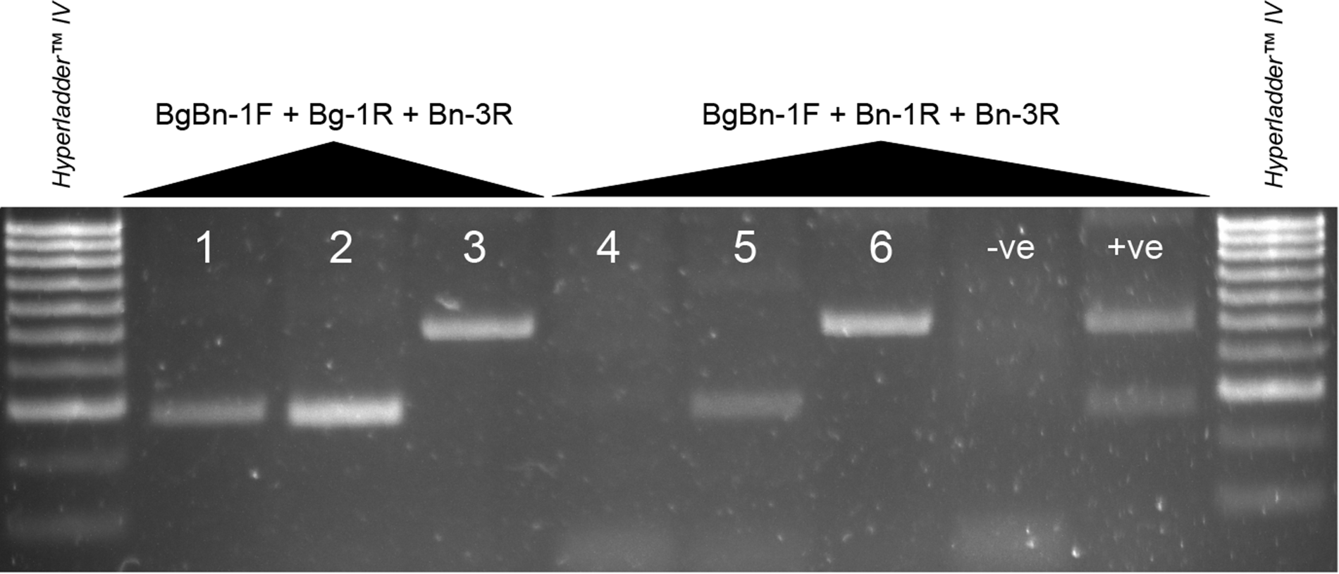
Gel electrophoresis showing PCR amplicon products of partial 5.8S and ITS2 genes of B. globosus from Tanzania (lanes 1 and 4) and Pemba (lanes 2 and 5) and B. nasutus productus from Tanzania (lanes 3 and 6). −ve = RNA-free water. +ve = B. nasutus from Pemba. See Supplementary Table 1 for full details of each sample per lane.

A third duplex reaction was therefore trialled using Bg-1R, which originally amplified a 307 bp fragment for both B. globosus and B. nasutus and Bn-3R that amplified an expected 553 bp fragment of B. nasutus and produced non-specific banding for B. globosus (Table 1 and Figure 2). Although initially Bg-1R was deemed not specific for B. globosus due to amplification of B. nasutus DNA (see Table 1 and Figure 2), it was predicted that in the presence of 2 primers, preferential amplification of the most suitable primer pair for each species would take place. This duplex reaction (BgBn-1F + Bg-1R + Bn-3R) was first trialled using samples of B. globosus from Pemba and mainland Tanzania and 1 B. productus with a S. haematobium infection from Tanzania (Supplementary Table 1), which demonstrated that the B. productus produced a banding pattern (single amplicon ∼553 bp) the same as observed from Pemba B. nasutus (Figure 3).
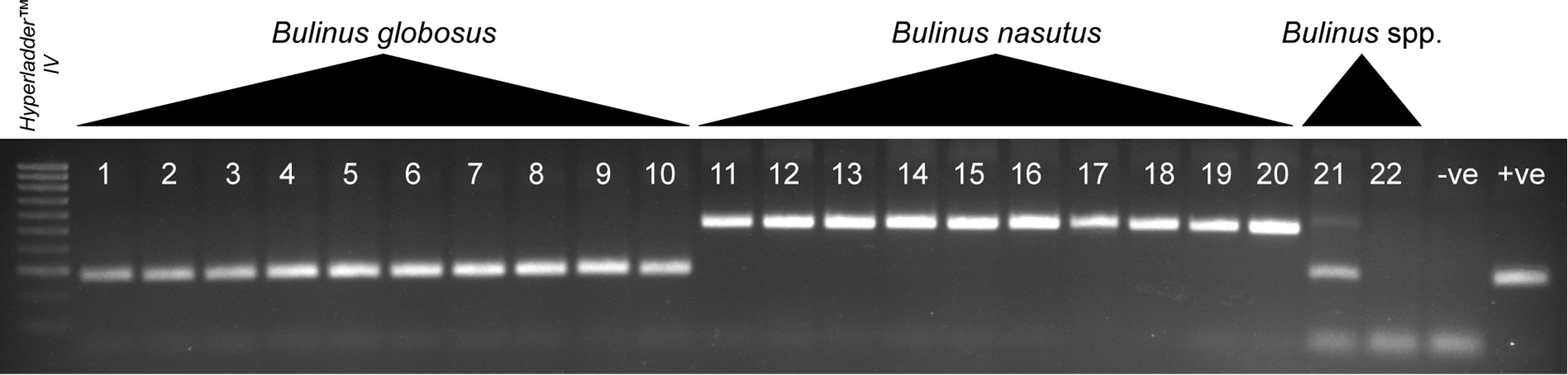
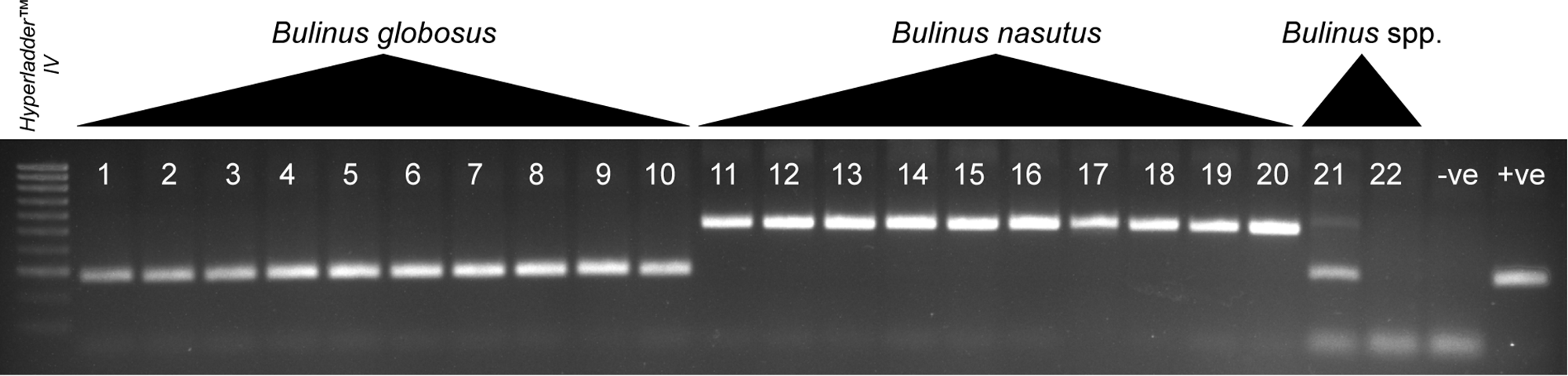
The tested duplex assay (BgBn-1F + Bg-1R + Bn-3R) was therefore performed on multiple B. globosus and B. nasutus from 3 shehias on Pemba (Pujini, Wambaa, Matale; see Pennance et al., Reference Pennance, Ame, Amour, Suleiman, Muhsin, Kabole, Ali, Archer, Allan, Emery, Rabone, Knopp, Rollinson, Cable and Webster2022), Bulinus sp. (africanus group) and B. globosus from Tanzania and B. forskalii from Niger (Figure 4). Expected banding patterns were observed from all B. globosus and B. nasutus. A B. globosus-specific amplicon, in addition to a faint secondary band of the B. nasutus-specific amplicon, was observed for the Bulinus sp. (africanus group) from Tanzania, and no amplification was observed from B. forskalii. As predicted, the additional amplicon produced in the singleplex PCR of BgBn-1F + Bg-1R with B. nasutus DNA observed in initial primer tests (see Figure 2) was not observed when reactions were performed in duplex with the B. nasutus Bn-3R primer.
Gel electrophoresis showing PCR amplicon products of partial 5.8S and ITS2 genes using primer combination BgBn-1F + Bg-1R + Bn-3R of B. globosus from Pemba (lanes 1–10), B. nasutus from Pemba (lanes 11–20), an undescribed Bulinus sp. (africanus group) from Tanzania (lane 21) and a B. forskalii isolate from Niger (lane 22). Legend: −ve RNA-free water. +ve = B. globosus from Tanzania. See Supplementary Table 1 for full details of each sample per lane.
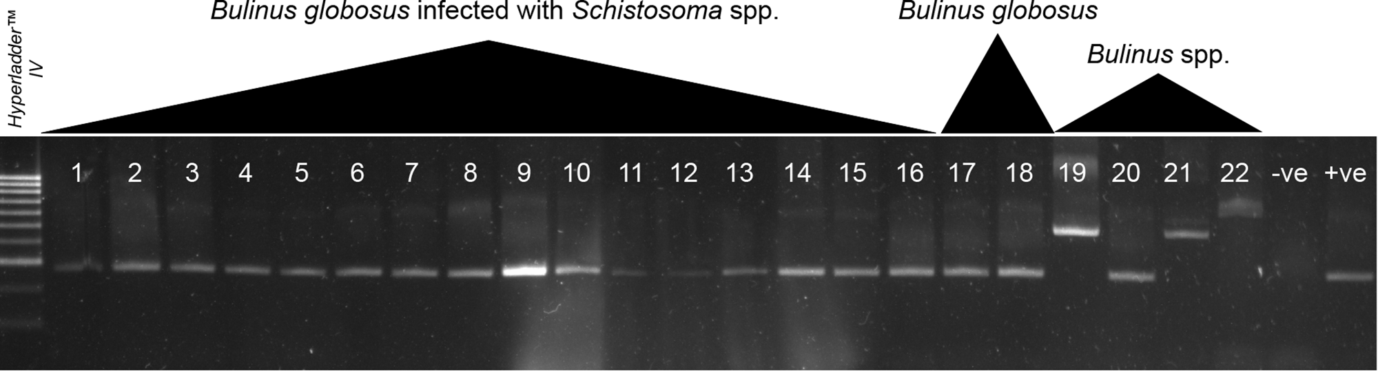
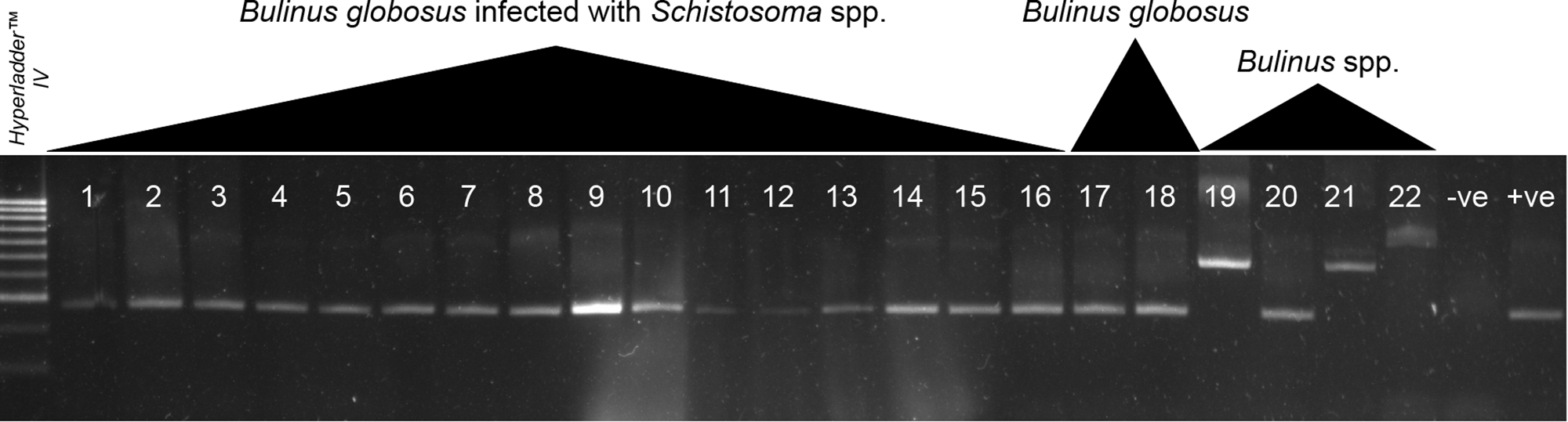
This duplex primer combination (BgBn-1F + Bg-1R + Bn-3R) showed no adverse amplifications with B. globosus with patent S. haematobium and S. bovis infections (see lanes 1–16, Figure 5); a single band was again observed for the B. productus with an S. haematobium infection (as observed in Figure 3) and weak non-specific banding for non-target B. truncatus species infected with S. haematobium (Figure 5).
Gel electrophoresis showing PCR amplicon products of partial 5.8S and ITS2 genes using primer combination BgBn-1F + Bg-1R + Bn-3R of B. globosus with patent S. haematobium (lanes 1 and 7–16) and S. bovis (lanes 2–6) infections, B. productus with a patent S. haematobium infection (lane 21), a non-target species B. truncatus with a patent S. haematobium (lane 22) and also uninfected B. globosus (lanes 17, 18 and 20) and uninfected B. nasutus (lane 19). Legend: −ve RNA-free water. +ve = B. globosus from Niger with a patent S. bovis infection. See Supplementary Table 1 for full details of each sample per lane.
Discussion
To employ effective malacological surveys in an endemic setting such as Pemba and Unguja, it is essential to fully understand schistosomiasis transmission dynamics in relation to the Bulinus spp. (B. globosus and B. nasutus) that act as vectors for the parasite. Here, we have developed a duplex PCR assay using the primers BgBn-1F + Bg-1R + Bn-3R targeting both the conserved (5.8S gene) and variable (ITS2) rDNA regions to accurately differentiate B. globosus and B. nasutus from Unguja and Pemba. Although there was no cross-reactivity with distantly related Bulinus spp. also present in Sub-Saharan Africa (B. truncatus and B. forskalii), we demonstrate that the assay may have limited applicability in distinguishing species/sub-species closely related to B. nasutus and B. globosus. For example, the banding patterns for B. nasutus and its closely related B. productus from Tanzania were the same. In addition, an undescribed Bulinus sp. from mainland Tanzania that is closely related to B. globosus from Pemba based on cox1 divergence produced a very similar, and not clearly distinct, amplicon banding pattern to B. globosus. Therefore, the assay could differentiate between the B. nasutus and B. globosus tested from Pemba (and likely Unguja given their similarity where these other closely related species are not present (Pennance et al., Reference Pennance, Ame, Amour, Suleiman, Muhsin, Kabole, Ali, Archer, Allan, Emery, Rabone, Knopp, Rollinson, Cable and Webster2022)), but does not yet support unambiguous species identification across mainland Africa where, as discussed further below, closely related species overlap in their distribution.
Other than circumventing unreliable morphological identifications, there are several advantages to using this diagnostic assay. The protocol is simple and involves a few steps, allowing high numbers of samples to be processed. The full protocol (including snail tissue digestion and DNA extraction) and interpretation of results (by visualizing amplicons using gel electrophoresis) only uses basic laboratory equipment (incubator, centrifuge, PCR machine, agarose gel electrophoresis) and does not require a cold chain for reagents or any additional steps requiring enzymes, such as for restriction digest (Stothard and Rollinson, Reference Stothard and Rollinson1997a, Reference Stothard and Rollinson1997b; Stothard et al., Reference Stothard, Mgeni, Alawi, Savioli and Rollinson1997; Tchami Mbagnia et al., Reference Tchami Mbagnia, Melachio Tanekou, Kengne Fokam, Nguiffo Nguete, Wondji and Njiokou2020). The assay involves just a single PCR for species determination, halving the number of reactions required in the previously described taxon-specific diagnostic assay (Stothard et al., Reference Stothard, Mgeni, Khamis, Seto, Ramsan, Hubbard, Kristensen and Rollinson2002c). Species identification can therefore be performed locally by researchers and technicians in Zanzibar using basic molecular laboratories. The assay is also not impacted by the presence of S. haematobium and S. bovis DNA in infected snails, an important consideration when considering active transmission regions such as in Unguja and Pemba. This all contributes towards a cost-effective and easy-to-implement protocol for mapping snail distribution and potential transmission risk areas in Zanzibar.
During field-based malacological surveys investigating urogenital schistosomiasis transmission, collected bulinids are often identified based on shell morphology alone, with schistosome identification taking precedence due to this being the etiological agent (Labbo et al., Reference Labbo, Djibrilla, Zamanka, Garba and Chippaux2007). The absence of basic and low-cost (i.e. without sequencing costs and sequence analysis) protocols for snail species delimitation, as is available for certain S. haematobium group schistosomes (Webster et al., Reference Webster, Rollinson, Stothard and Huyse2010), may be a reason for this. The accessibility and simplicity of this diagnostic marker for S. haematobium parasites enabled the discovery of introgressed S. haematobium group parasites from snails in West Africa (Tian-Bi et al., Reference Tian-Bi, Webster, Konan, Allan, Diakité, Ouattara, Salia, Koné, Kakou and Rabone2019; Pennance et al., Reference Pennance, Allan, Emery, Rabone, Cable, Garba, Hamidou, Webster, Rollinson and Webster2020b) and led to the discovery of S. bovis on Zanzibar (Pennance et al., Reference Pennance, Ame, Amour, Suleiman, Allan, Rollinson and Webster2018). The Bulinus spp. diagnostic assay reported here offers a tool for future intervention planning, being particularly pertinent considering the updated WHO recommendations for snail control and monitoring to be integrated as a complementary measure (World Health Organization (WHO), 2022; Garba Djirmay et al., Reference Garba Djirmay, Yadav, Guo, Rollinson and Madsen2024).
This duplex assay showed no cross-reactivity for distantly related non-target Bulinus species, B. truncatus and B. forskalii. To aid in interpreting these negative results (i.e. no PCR amplicon observed), the assay could be improved by including an internal control capable of amplifying a universal region of all Bulinus spp. DNA. Also, considering that misidentification of other Bulinus spp. is commonplace (e.g. B. globosus misidentified as B. truncatus prior to molecular analysis; Pennance et al., Reference Pennance, Allan, Emery, Rabone, Cable, Garba, Hamidou, Webster, Rollinson and Webster2020b), it would be useful to expand this duplex assay into a multiplex assay to also include B. truncatus group species control. Although not necessary on the Zanzibar archipelago, this would be particularly useful in West Africa, where B. globosus and B. truncatus are widespread and can inhabit the same freshwater body (Brown, Reference Brown1994), as shown in Niger (Rabone et al., Reference Rabone, Allan, Gouvras, Pennance, Hamidou, Webster, Labbo, Emery, Garba and Rollinson2019; Pennance et al., Reference Pennance, Allan, Emery, Rabone, Cable, Garba, Hamidou, Webster, Rollinson and Webster2020b) and Senegal (Ndione et al., Reference Ndione, Diop, Riveau, Ba and Jouanard2018, Reference Ndione, Bakhoum, Haggerty, Jouanard, Senghor, Ndao, Riveau and Ba2019). Although to a trained malacologist these 2 species are distinct in their shell morphology, misidentification by less experienced snail collectors is possible, particularly when dealing with juvenile snails. To produce another species-specific marker for B. truncatus, the available assembled and annotated rDNA data for B. truncatus can be used to explore further sites of variance and to design B. truncatus-specific primers (Young et al., Reference Young, Stroehlein, Wang, Korhonen, Mentink-kane, Stothard, Rollinson and Gasser2022; Zhang et al., Reference Zhang, Bu, Lu, Babbitt, Adema and Loker2022). Future iterations of snail vector identification assays may also help distinguish Bulinus spp. from morphologically similar freshwater snail species, namely those with conchological features (e.g. sinistral chirality) that overlap in geographical range, namely Physa and Lanistes spp. (although the latter can be distinguished by the presence of an operculum; Global Schistosomiasis Alliance, 2026).
Unlike species of the B. truncatus/tropicus complex, species belonging to the B. forskalii group have very distinct shell morphology (having a slender shell and a spire higher than the aperture) compared to snails of the B. africanus group (B. globosus and B. nasutus) that have a broader shell with a spire height less than that of the aperture. Because of this, it was deemed unnecessary to distinguish the B. forskalii group species present on Pemba and Unguja in this assay from those of the B. africanus group. However, significant morphological overlap does exist between species within the B. forskalii group, such as B. forskalii and B. senegalensis, that are host to S. haematobium group parasites, particularly in West Africa (Frandsen, Reference Frandsen1979; Ngonseu et al., Reference Ngonseu, Greer and Mimpfoundi1992; Labbo et al., Reference Labbo, Ernould, Djibrilla, Sidiki and Chippaux2003, Reference Labbo, Djibrilla, Zamanka, Garba and Chippaux2007), therefore a separate diagnostic assay to separate species within this group, improving on those previously developed (Jones et al., Reference Jones, Noble, Lockyer, Brown and Rollinson1997), may offer a useful solution for mapping freshwater snails in this region.
The assay was performed on 2 isolates of B. productus and 1 currently undescribed Bulinus sp. from Tanzania, which are closely related to B. nasutus and B. globosus, respectively (Kane et al., Reference Kane, Stothard, Emery and Rollinson2008). Both B. nasutus and B. productus act as vectors of S. haematobium in regions of East Africa (Webbe, Reference Webbe1962; Kinoti, Reference Kinoti1964; McCullough et al., Reference McCullough, Eyakuze, Msinde and Nditi1968; Loker et al., Reference Loker, Moyo and Gardner1981; Sarda et al., Reference Sarda, Simonsen and Mahikwano1985; Lwambo, Reference Lwambo1988; Kariuki et al., Reference Kariuki, Clennon, Brady, Kitron, Sturrock, Ouma, Ndzovu, Mungai, Hoffman, Hamburger, Pellegrini, Muchiri and King2004; Angelo et al., Reference Angelo, Buza, Kinung’hi, Kariuki, Mwanga, Munisi and Wilson2018), and a ‘typical’ form of each can allow for distinguishing between species (Brown, Reference Brown1994). Therefore, differentiation of these 2 species using a molecular assay may be less important. However, the undescribed Bulinus sp. with a currently undetermined role in S. haematobium transmission present in Tanzania could be confused with B. globosus due to both its overlapping morphology and similar amplicon size (albeit with a secondary larger non-specific amplicon also produced, the consistency of which was not tested). Therefore, the diagnostic marker described here should be used with caution outside of the Zanzibar archipelago. It should also not be ignored that several other B. africanus group snails are present in East Africa, such as B. africanus and B. ugandae, which could produce similar results to B. globosus or B. nasutus with this marker. The diversity of species of these B. africanus group species across mainland Africa, and their role in the transmission of schistosomiasis, is less well understood than on Zanzibar, and therefore the necessity for such species diagnostics needs to be determined.
Although B. globosus and B. nasutus from Unguja were not trialled with this assay, the primers used in this assay were developed using sequence data from B. globosus and B. nasutus from isolates collected in Unguja. It is therefore almost certain that the assay will function with B. globosus and B. nasutus endemic to Pemba, given the high mitochondrial cox1 sequence similarity between isolates (Pennance et al., Reference Pennance, Ame, Amour, Suleiman, Muhsin, Kabole, Ali, Archer, Allan, Emery, Rabone, Knopp, Rollinson, Cable and Webster2022), but tests are needed to confirm this. Further tests on a broader sample range are particularly necessary due to the variable nature of the ITS loci of Bulinus, i.e. intra-genomic non-homologous rDNA sequences (Stothard and Rollinson, Reference Stothard and Rollinson1997b). The developed primers may therefore behave in an allele-specific manner if heterogeneous copies of the ITS2 are present in individuals that interfere with the selected primers, although this was not observed from the B. globosus and B. nasutus from Pemba tested here; this may occur in populations included from a wider sample range. However, if feasibility to use this marker across the Zanzibar archipelago is established through testing on more endemic snails, it may be deemed beneficial for future risk planning and interventions to produce a comprehensive Bulinus species distribution map to assess potential at-risk areas without the need for DNA sequencing and analysis (Pennance et al., Reference Pennance, Ame, Amour, Suleiman, Muhsin, Kabole, Ali, Archer, Allan, Emery, Rabone, Knopp, Rollinson, Cable and Webster2022).
To aid in targeting control interventions on Zanzibar to those areas with continuing S. haematobium transmission, it is important to improve surveillance methods used for the detection of S. haematobium. Diagnosing human infections of S. haematobium rapidly using sensitive diagnostics, at the point of care, will be vital in avoiding reintroduction and recrudescence in areas that may become free of transmission (Rosser et al., Reference Rosser, Rollinson, Forrest and Webster2015; Archer et al., Reference Archer, LaCourse, Webster and Stothard2020; Rostron et al., Reference Rostron, Pennance, Bakar, Rollinson, Knopp, Allan, Kabole, Ali, Ame and Webster2019). Snail xenomonitoring protocols to identify Schistosoma spp. infections within snails will allow for the identification of ‘active’ transmission sites (Abbasi et al., Reference Abbasi, Webster, King, Rollinson and Hamburger2017; Pennance et al., Reference Pennance, Ame, Amour, Suleiman, Allan, Rollinson and Webster2018; Schols et al., Reference Schols, Carolus, Hammoud, Mulero, Mudavanhu and Huyse2019), but accurately determining and assessing freshwater snail species will additionally allow for infection risk mapping in endemic regions for targeting schistosomiasis interventions (World Health Organization (WHO), 2022).
This study demonstrates the utility of a cost-effective and deployable diagnostic assay for the differentiation of B. globosus and B. nasutus in Pemba and Unguja, providing a valuable tool for accurately mapping the distribution of these endemic snail vectors and refining risk assessment maps. By integrating molecular tools such as this in surveillance programmes, precise intervention strategies can be planned and implemented confidently, supporting efforts to accomplish schistosomiasis elimination.
Supplementary material
The supplementary material for this article can be found at https://doi.org/10.1017/S0031182026102418.
Data availability statement
The datasets supporting the conclusions of this article are included within the article and its additional file.
Author contributions
T.P., J.C. and B.L.W. conceived and designed the study. T.P., S.M.Am., S.M.Al., A.K.A. and K.R.S. conducted the data gathering. T.P. performed the formal analysis and wrote the original article. All authors reviewed and edited the final manuscript.
Financial support
The study was partially funded by a Wellcome Trust Seed Award (https://wellcome.org) grant number 207728 (awarded to B.L.W.). T.P. was funded by the Natural Environmental Research Council GW4+ DTP (www.nercgw4plus.ac.uk), NE/L002434/1. The funders had no role in study design, data collection and analysis, decision to publish, or preparation of the manuscript.
Competing interests
The authors declare there are no conflicts of interest.
Ethical standards
Not applicable.


 Open access

