





Introduction
Coccidiosis leads to approximately $14 billion economic losses globally in the poultry industry each year (Blake et al, Reference Blake, Knox, Dehaeck, Huntington, Rathinam, Ravipati, Ayoade, Gilbert, Adebambo, Jatau, Raman, Parker, Rushton and Tomley2020). This disease is caused by protozoan parasites of the Eimeria genus, which invade and damage the intestines of birds, leading to impaired nutrient absorption, diarrhoea and mortality in severe cases (Teng et al., Reference Teng, Yadav, FLdS, Tompkins, Fuller and Kim2020a). Once these parasites appear on a farm, they are difficult to eliminate and can rapidly spread throughout the flock if not effectively managed by vaccination or coccidiostats.
Eimeria infection begins when young chickens ingest oocysts from the used litter. Within hours, sporozoites are released from the oocysts and invade the enterocytes of the host. The parasites undergo the schizogony phase (asexual reproduction), followed by the gametogony phase (sexual reproduction), and eventually produce a new generation of oocysts (López-Osorio et al., Reference López-Osorio, Chaparro-Gutiérrez and Gómez-Osorio2020). Due to the complexity of the endogenous life cycle of Eimeria spp., establishing ex vivo or in vitro culture systems has remained a significant challenge (Felici et al., Reference Felici, Tugnoli, Piva and Grilli2021). The majority of in vitro assays have relied on primary enterocytes or mammalian epithelial cell lines, which provide limited support for the gametogony phase. Only a few studies cultivated E. tenella to gametogony stage in vitro, primarily through co-culture with chicken embryo kidney or lung epithelial cells (Bussiere et al., Reference Bussiere, Niepceron, Sausset, Esnault, Silvestre, Walker, Smith, Quere and Laurent2018; Hofmann and Raether, Reference Hofmann and Raether1990; Zhang et al, Reference Zhang, Wilson, Yang and Healey1997). However, the lack of gastrointestinal specific factors in these cells likely contributed to low oocyst yields (Felici et al., Reference Felici, Tugnoli, Piva and Grilli2021). Additionally, Bussiere et al (Reference Bussiere, Niepceron, Sausset, Esnault, Silvestre, Walker, Smith, Quere and Laurent2018) reported that the wild strains of E. tenella only generated oocysts in the monolayer culture system from the second generation of merozoites. The sporozoites of the wild strain failed to complete the endogenous life cycle in the model (Bussiere et al., Reference Bussiere, Niepceron, Sausset, Esnault, Silvestre, Walker, Smith, Quere and Laurent2018). Another drawback of using primary kidney cells is the requirement for fresh isolation for each experiment. Alternatively, Madin–Darby Bovine Kidney (MDBK) cells are more easily cultured and maintained in the lab but only support the parasite’s life cycle up to the merozoite stage. (Dimier-Poisson et al., Reference Dimier-Poisson, Bout and Quere2004; Marugan-Hernandez et al., Reference Marugan-Hernandez, Jeremiah, Aguiar-Martins, Burrell, Vaughan, Xia, Randle and Tomley2020). Since the development of gametocytes and oocysts are limited in MDBK cells, previous research has speculated that the merozoites cultured in this specific in vitro model are unlikely to reinvade epithelial cells and progress to the gametogony stage (Dimier-Poisson et al., Reference Dimier-Poisson, Bout and Quere2004; Felici et al., Reference Felici, Tugnoli, Piva and Grilli2021; Marugan-Hernandez et al., Reference Marugan-Hernandez, Jeremiah, Aguiar-Martins, Burrell, Vaughan, Xia, Randle and Tomley2020). Lastly, several other cell lines have been successful in culturing E. tenella, including the macrophage-like Cellosaurus LSCC-HD11 (Aguiar-Martins et al., Reference Aguiar-Martins, Tomley, Blake and Marugan-Hernandez2024) and the chicken lung epithelial cell line, CLEC-213 (Bussière et al., Reference Bussiere, Niepceron, Sausset, Esnault, Silvestre, Walker, Smith, Quere and Laurent2018). However, these models enable varying degrees of parasite development and do not always utilize cells derived from the intestinal tissue of the host, which could result in the expression of protein or metabolic profiles that differ when Eimeria occupy their natural habitat, potentially adding another layer of complexity when studying parasite biology and host–parasite interactions (Feix et al., Reference Feix, Cruz-Bustos, Ruttkowski and Joachim2023). Therefore, developing a robust in vitro model capable of completing the endogenous life cycle of Eimeria spp. within chicken enterocytes would be essential in advancing research on host–pathogen interactions and accelerating drug development for coccidiosis control.
In recent years, organoid culture technology has advanced in vitro systems for studying organ-specific functions and diseases. Organoids, which are three-dimensional mini-organ structures derived from adult stem cells or induced pluripotent stem cells, have been widely applied in human medical research (Kim et al., Reference Kim, Koo and Knoblich2020). Among various organoid models, intestinal organoids, also known as enteroids, closely recapitulate the functions and cell types of the intestinal epithelium (Xiang et al., Reference Xiang, Wang and Li2024). These intestinal organoids exhibit remarkable self-renewal and differentiation capabilities. With appropriate cell culture supplements, the intestinal stem cells can further differentiate into Paneth cells, goblet cells, enterocytes and enteroendocrine cells (Yang et al., Reference Yang, Wang, Zhou, Chen, Song, Deng, Yao and Yin2025). Intestinal organoid models have become powerful tools for establishing new in vitro systems to investigate complex host-pathogen interactions in apicomplexan infections, including Cryptosporidium and Toxoplasma gondii (Heo et al, Reference Heo, Dutta, Schaefer, Iakobachvili, Artegiani, Sachs, Boonekamp, Bowden, Hendrickx, Willems, Peters, Riggs, O’Connor and Clevers2018; Seo et al, Reference Seo, Han, Lee, Hong, Cho, Kim, Koo and Kim2020).
This advanced cell culture technology has been increasingly applied to poultry research as well. Avian intestinal organoids can be derived from chickens at different ages for various applications (Zhao et al., Reference Zhao, Farnell, Kogut, Genovese, Chapkin, Davidson, Berghman and Farnell2022). Previous studies have used the intestinal organoids to evaluate novel feed additives for nutritional research (Wang et al., Reference Wang, Hou, Wu, Xu, Liu, Chen, Xu, Guo, Gao and Yuan2022), and to develop multiple bacterial infections and toxin challenge models. (Ghiselli et al., Reference Ghiselli, Yu, Piva, Grilli and Li2023b; Kang and Lee, Reference Kang and Lee2024; Mitchell et al., Reference Mitchell, Sutton, Elango, Borowska, Perry, Lahaye, Santin, Arsenault and Vervelde2024; Lacroix-Lamande et al., Reference Lacroix-Lamande, Bernardi, Pezier, Barilleau, Burlaud-Gaillard, Gagneux, Velge and Wiedemann2023). Additionally, Nash et al (Reference Nash, Morris, Mabbott and Vervelde2021) demonstrated the potential of developing chicken intestinal organoids as a novel platform for studying Eimeria infections. However, a complete endogenous life cycle of Eimeria within chicken intestinal organoids has not yet been reported. Therefore, the present study aims to develop mature organoids using two commercially available media and establish a more comprehensive in vitro infection model to support the full progression of schizogony and gametogony phases in the life cycle of E. tenella.
Materials and methods
Isolation and culture of chicken intestinal organoids
The methods for isolation and culture of chicken intestinal organoids were followed and adjusted based on previous literature (Li et al., Reference Li, Li, Zhang, Li, Lin, Mi and Zhang2018; Oost et al., Reference Oost, Ijaz, van Haarlem, van Summeren, Velkers, Kraneveld, Venema, Jansen, Pieters and Ten Klooster2022; Pierzchalska et al., Reference Pierzchalska, Grabacka, Michalik, Zyla and Pierzchalski2012, Reference Pierzchalska, Panek, Czyrnek and Grabacka2019; Powell and Behnke, Reference Powell and Behnke2017; Zhao et al., Reference Zhao, Farnell, Kogut, Genovese, Chapkin, Davidson, Berghman and Farnell2022). In brief, a pool of intestines was collected from 1-day-old specific pathogen-free white leghorn chickens. The collected intestines were immediately stored in Dulbecco’s phosphate-buffered saline (DPBS) on ice for transportation to the lab. The ceca, colon, pancreas, yolk and connective tissue attached to the small intestine were carefully removed. Intestinal contents were gently flushed out from the lumen, and the entire small intestine was rinsed twice with DPBS. The tissue was then cut into 0.2 cm sections and transferred to 50 mL conical tubes. The tissue fragments were digested in Advanced DMEM/F12 (Thermo Fisher Scientific, Grand Island, NY, USA) formulated with 0.2 mg/mL collagenase I (Sigma Aldrich, St. Louis, MI, USA) and 6 µM ROCK inhibitor Y27632 (StemCell Technologies, Vancouver, Canada) at 37°C for 30 min, with intermittent mixing. After the first digestion, the suspension was filtered through a 100 µm strainer (Thermo Fisher Scientific, Grand Island, NY, USA) and the retained tissue fragments were transferred to a new conical tube for a second-round digestion under the same conditions for 30 min. The digested tissue was again filtered, and the fragments of tissue were discarded. The filtrate was collected and centrifuged at 300 × g for 3 min. The pellet was resuspended in 30 mL of 2% sorbitol (Spectrum Chemical Mfg. Corp., Gardena, CA, USA) in DMEM/F12 and centrifuged at 50 × g for 3 min. The supernatant was discarded, and the cell pellets were washed four additional times with the sorbitol solution to remove debris and single cells.
For developing apical-out organoids, the cell pellets were suspended directly in the culture media and transferred to a 96-well ultra-low attachment round bottom plate (Costar, Kennebunk, ME, USA). For developing basolateral-out organoids, the cell pellets were suspended in a mixture of 60% Matrigel® (Corning, Bedford, MA, USA) and 40% culture media, embedded in the Matrigel domes in a 24-well plate and cultured at 41°C. Two media formulations, L-WRN conditioned medium and IntestiCult Organoid Growth Medium (OGM, StemCell Technologies, Vancouver, Canada), were used for developing intestinal organoids in this study. The L-WRN conditioned medium was formulated by mixing the commercial 3dGRO™ L-WRN Conditioned Media (Sigma Aldrich, St. Louis, MI, USA) with the recommended supplements following the user guideline, N-2 (Thermo Fisher Scientific, Grand Island, NY, USA), B-27 (Thermo Fisher Scientific, Grand Island, NY, USA), L-Glutamine (Thermo Fisher Scientific, Grand Island, NY, USA), HEPES (Thermo Fisher Scientific, Grand Island, NY, USA), Niacinamide (Sigma Aldrich, St. Louis, MI), N-Acetyl-l-Cysteine (Sigma Aldrich, St. Louis, MI, USA), EGF (EMD Millipore Corp, Darmstadt, Germany), Gastrin I (Sigma Aldrich, St. Louis, MI, USA), Prostaglandin E2 (StemCell Technologies, Vancouver, Canada), A-83-01 (Tocris Bioscience, Bristol, UK) and SB202190 (StemCell Technologies, Vancouver, Canada). Both media were supplemented with Primocin (InvivoGen, San Diago, CA, USA) as an antibiotic. The media formulations used in this study are listed in Table 1.
Formulations of intesticult organoid growth medium (OGM) and L-WRN medium

a StemCell Technologies, Vancouver, Canada.
b InvivoGen, San Diago, California.
c Sigma Aldrich, St. Louis, Missouri.
d Thermo Fisher Scientific, Grand Island, New York.
e EMD Millipore Corp, Darmstadt, Germany.
f Tocris Bioscience, Bristol, United Kingdom.
The intestinal organoids were examined under a microscope (Nikon Eclipse Ti2, Nikon Instruments Inc., Melville, NY, USA) and images were captured by the NIS Elements software (Nikon Instruments Inc., Melville, NY, USA). Organoid size was measured 3 days after isolation and again at the second passage following thawing from cryopreservation. Fifteen organoids in focus within the image were randomly selected, and the length of the widest part of each organoid was measured using Image J (National Institutes of Health, Bethesda, MD, USA). The statistical analysis was conducted using Mann–Whitney test by GraphPad Prism 8 (GraphPad, Boston, MA).
Immunofluorescent staining
Immunofluorescent staining was performed on organoids cultured under different condition for approximately 1 week. For basolateral-out organoids embedded in Matrigel, the culture medium was gently aspirated first. The Matrigel domes were then rinsed once with DPBS and dissolved using Cell Recovery Solution (Corning, Bedford, MA, USA). The organoid suspension was transferred to 15 mL conical tubes and incubated on ice for 20 min. After incubation, the organoids were fixed with 4% PFA at 4°C for 20 min. For the apical-out organoids, approximately 150 organoids were collected from 16 wells. The pooled organoids were centrifuged at 300 × g at 4°C for 3 min. After the supernatant was removed, the organoid pellet was washed once with DPBS. The pellet was then resuspended and fixed in 4% paraformaldehyde (PFA, Thermo Fisher Scientific, Grand Island, NY, USA) at 4°C for 20 min, followed by permeabilization at 4°C for 1 h on a rotating platform. The organoids were further washed three times and incubated with the primary antibody overnight at 4°C. The fixed organoids were divided into different staining groups. Following the primary antibody incubation, the organoids were washed and incubated with the corresponding secondary antibody for at least 4 h. The organoids were washed with DPBS, resuspended in VectaShield Medium with DAPI (Vector Laboratories Inc, Newark, CA, USA), and mounted on a microscope slide with a spacer attached (Thermo Fisher Scientific, Grand Island, NY, USA). The antibodies used for immunofluorescent staining are listed in Table 2.
Primary and secondary antibodies for immunofluorescent staining assay

a Abcam, Waltham, Massachusetts.
b InvivoGen, San Diego, California.
c Immunostar, Hudson, Wisconsin.
d Santa Cruz Biotechnology, Dallas, Texas.
e Vector Laboratories Inc, Newark, California.
Parasite infection
The apical-out organoid was selected for developing the Eimeria infection model. After isolation from the chicken intestine, the organoids were initially cultured in L-WRN medium for two passage to accelerate expansion, followed by two additional passages in OGM to enhance enterocyte differentiation. Organoids were passaged approximately every 7 days. For the first 3 passages, organoids were collected and mechanically disrupted by passing through a 24 G needle 3 to 4 times (Seidler et al., Reference Seidler, Donath, Gentemann, Buettner, Heisterkamp and Kalies2024) before being plated at 10 to 15 organoids per well. In the final passage prior to Eimeria infection, the organoids were diluted to a single organoid per well.
Eimeria tenella oocysts from a field isolate were obtained from the University of Georgia. The oocysts were washed with 0.8 % sodium hypochlorite at room temperature for 5 min, followed by 3 washes with PBS to remove the sodium hypochlorite solution. Sporocysts were released from the oocysts by vortexing for 40–60 s in 50 mL conical tubes containing 3 mm glass beads (Thermo Fisher Scientific, Grand Island, NY, USA). The sporocysts were then incubated with 0.5% trypsin (Sigma Aldrich, St. Louis, MI, USA) and 4% taurocholic acid (Sigma Aldrich, St. Louis, MI, USA) at 37°C for 90 min. The excysted sporozoites were separated from debris and oocysts with a 5 µm strainer (PluriSelect, El Cajon, CA, USA), and further labelled with the PKH67 Green Fluorescent Cell Linker Midi kit (Sigma Aldrich, St. Louis, MI, USA) following the user guideline and a protocol described by Fuller and McDougald (Reference Fuller and McDougald2001). Finally, the PKH67-labeled sporozoites were washed with DPBS and resuspended in culture medium.
The sporozoites were co-cultured with chicken apical intestinal organoids at 41°C in a 96-well ultra-low attachment plate (1,500 sporozoites/organoid/well) with a total of 40 wells. At 3 days post-infection, extracellular sporozoites and merozoites in the medium were removed. Four wells of infected organoids were pooled into 1 well after centrifugation. Additionally, 4 wells of fresh parallel non-infected organoids were added to each infected well at 3 days post-infection, and 8 wells of fresh organoids were added at 7 days post-infection, following an adjusted protocol adapted from Nash et al (Reference Nash, Morris, Mabbott and Vervelde2021). Infected organoids were pooled from 8 wells for RNA extraction at 7 – and 11-days post-infection. RNA extraction and cDNA synthesis were performed using the RNeasy Micro Kit (Qiagen, Germantown, MD, USA) and the High Capacity cDNA Reverse Transcription Kit (Applied Biosystems, Foster City, CA, USA), respectively. The PCR amplification was performed using gametocyte-specific primers, EtGAM56, following a PCR program described by Bussiere et al (Reference Bussiere, Niepceron, Sausset, Esnault, Silvestre, Walker, Smith, Quere and Laurent2018). The infection area of the organoids was measured using an Incucyte SX5 (Sartorius, Göttingen, Germany) from 3- to 7-days post-infection which quantified the fluorescent signal area per field in real time. In addition to Incucyte analysis, chicken organoids infected with E. tenella were fixed with 4% PFA, and mounted on microscope slides on day 3-, 5-, 7- and 11-days post-infection. Images of organoids infected with E. tenella were captured by a confocal microscope (Leica DM6, Leica Microsystems, Wetzlar, Germany), and the 3D images were generated by the Leica Application Suit X software (Leica Microsystems, Wetzlar, Germany). The Eimeria infection experiments were conducted in two independent replicates with separated material preparations and infections, producing consistent results.
Results
L-WRN conditioned medium accelerated expansion of organoids
The size of organoids was measured only for those cultured in Matrigel domes, as organoids in floating conditions tend to cluster in the center of the well and potentially merge, which complicates the differentiation between organoid size increases due to cell proliferation and merging of multiple organoids. The results showed that chicken intestinal organoids grown in the L-WRN conditioned medium had significantly larger sizes on day 3 compared to those cultured in the OGM (P < 0.001, Figure 1C).
Chicken intestinal organoids embedded in Matrigel domes and cultured in IntestiCult Organoid Growth Medium or L-WRN conditioned medium. (A) Organoids cultured in the IntestiCult Organoid Growth Medium (OGM, StemCell Technologies, Vancouver, Canada) on day 3. Bar is 500 µm. (a) Magnified image highlights the smaller size of organoids cultured in the OGM. (B) Organoids cultured in the L-WRN conditioned medium (Merck KGaA, Darmstadt, Germany) supplemented with N-2, B-27, L-Glutamine, HEPES, niacinamide, N-acetyl-L-Cysteine, epidermal growth factor, gastrin I, prostaglandin E2, A-83-01, SB202190 on day 3. Bar is 500 µm. (b) Magnified image highlights the larger size of organoids cultured in the L-WRN conditioned medium. Figures A and B were captured by an inverted microscope (Nikon Eclipse Ti2). (C) Comparison of organoid size increase over 72 h between two different culture media after initial isolation from the intestine or the second passage post-thaw from cryopreservation.

Culture conditions determined organoid structure and cell population
The organoids were able to grow larger than 350 µm in diameter in both culturing conditions within 8 days, but apical-out organoids appeared darker than basolateral-out organoids under a phase contrast microscope (Figure 2A and 2B). F-actin staining (Figure 2C and 2D) revealed that organoids could develop into two opposite structures depending on the culture materials. Organoids that were cultured in the ultra-low attachment plates floated in the medium, facilitating the development of an apical-out structure. F-actin staining at the exterior of these organoids indicated the presence of microvilli on the surface. On the other hand, basolateral-out organoids cultured in Matrigel domes developed in an opposite way, where the F-actin staining localized at the centre of the spherical structure. These basolateral-out organoids also formed a bumpy brush border surface inward, resembling the intestinal structure shown in Figure 2C.
Chicken intestinal apical-out and basolateral-out organoids. (A) Phase contrast image of a basolateral-out organoid developed in a Matrigel dome. Bar is 100 µm. (B) Phase contrast image of an apical-out organoid developed in an ultra-low attachment plate. Bar is 100 µm. (C) 3D image of a basolateral-out organoid developed in a Matrigel dome. (D) 3D image of an apical-out organoid developed in an ultra-low attachment plate. Organoids were fixed with 4% PFA. Cell nuclei were stained by DAPI (blue), and brush borders (F-actin) were marked by Phalloidin (red). The X, Y and Z axis intervals in µm of figures are as follows, C (50, 50 and 5) and D (50, 50 and 5). (E) Different sections of confocal images of basolateral-out organoids developed in a Matrigel dome. (F) Different sections of confocal images of apical-out organoids developed in a ultra-low attachment plate. Cell nuclei were stained by DAPI (Blue) and brush borders (F-actin) were stained by Phalloidin. Figures A and B were captured by an inverted microscope (Nikon Eclipse Ti2). Figures C and D were captured by a confocal microscope (Leica DM6), and the 3D images were generated by the Leica Application Suit X software (Leica Microsystems). Figures E and F were captured by a confocal microscope (Leica DM6).

Besides the culture materials, the medium formulation also plays a crucial role in shaping different types of organoids. In this study, more Paneth cells (Figure 3B) and enteroendocrine cells (Figure 3D) were present in the apical-out organoids cultured with L-WRN-conditioned medium, whereas the OGM group had developed more microvilli covering the surface of the organoids (Figure 3E).
Characterization of apical-out organoids cultured in IntestiCult Organoid Growth Medium or L-WRN conditioned medium. (A), (C) and (E) were organoids cultured in the IntestiCult Organoid Growth Medium (StemCell Technologies, Vancouver, Canada). (B), (D) and (F), were organoids cultured in the L-WRN conditioned medium (Merck KGaA, Darmstadt, Germany) supplemented with N-2, B-27, L-Glutamine, HEPES, niacinamide, N-acetyl-l-Cysteine, epidermal growth factor, gastrin I, prostaglandin E2, A-83-01 and SB202190. Organoids were fixed with 4% PFA and stained with anti-lysozyme (orange, Paneth cells, A and B), anti-chromogranin A (yellow, enteroendocrine cells, C and D), antivillin (green, microvilli, E and F) and DAPI (blue, nuclei). Organoids in Figures 3 were captured by a confocal microscope (Leica DM6), and the 3D images were generated by the Leica Application Suit X software (Leica Microsystems). The X, Y and Z axis intervals in µm of figures are as follows, A (50, 50, and 5), B (50, 50 and 10), C (50, 50, 10), D (50, 50 and 10), E (20, 20 and 10), and F (50, 50 and 10).

Developing endogenous life cycle of Eimeria tenella in chicken intestinal organoids
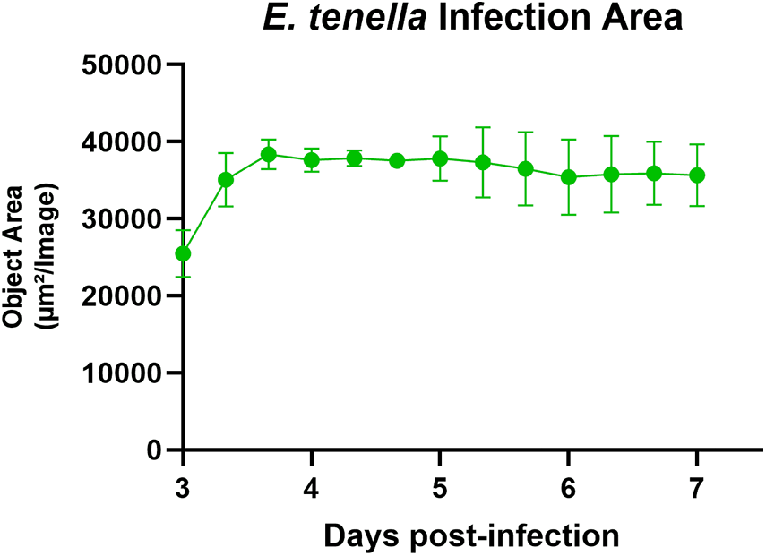
The infected area of organoids by E. tenella increased approximately 1.5-fold over 4 days, from 22.4 to 35.6 thousand µm2/image (Figure 4). The increased fluorescent signals rapidly spread throughout the organoids and even appeared in fresh organoids added on 3-days post-infection (Supplementary video A). Different stages of the endogenous life cycle of E. tenella were identified in the chicken organoids. By 3 days post-infection, sporozoites had developed into schizonts (Figure 5). Small DAPI-stained blue dots, representing parasite nuclei, were observed near the nuclei of chicken enterocytes (Figure 5A). The presence of fluorescent PKH67-labeled schizonts (Figures 5B and 5C) indicates that sporozoites successfully invaded the apical-out organoids and initiated their asexual reproduction cycle, generating merozoites in the in vitro model. The merozoites might undergo 2 to 3 asexual replication cycles before progressing to the gametogony stage. The gametocytes were found in the organoids fixed on 7- and 11-days post-infection (Figures 6C and 6D), as both DAPI and PKH67 staining marked the outline of the spheroid structure of the gametocytes (Figures 6E, 6F, and 6G). Additionally, a macrogamete marker, EtGAM56, was detected in the infected organoids by PCR (Supplementary Figure 1). A significant number of parasites were observed in the chicken organoids fixed on day 11 (Figure 6D), and oocysts were excreted from the organoids and observed in the culture medium (Figure 7A). The new generation oocysts exhibited fluorescent signals (Figure 7B and 7D), and some developed into sporulated oocysts within 24 h at room temperature (Figure 7C). As demonstrated in Figures 6 and 7, intestinal organoids derived from 1-day-old chickens have successfully supported E. tenella to complete their endogenous life cycle in vitro.
Real-time measurement of E. tenella infection area. The infection area in chicken intestinal apical-out organoids was measured by an Incucyte from 3- to 7-days post-infection. Organoids were infected with 1,500 PKH67-labeled sporozoites, and infection area was measured after the removal of extracellular parasites. Uninfected organoids were added in the well at 3-days post-infection.

Schizonts of E. tenella in a chicken intestinal apical-out organoid. Chicken intestinal organoids were infected with 1,500 PKH67-labeled sporozoites of E. tenella per organoid. The infected organoids were fixed with PFA and stained with DAPI at 3-days post-infection. (A) Chicken and parasites’ nuclei (blue, DAPI). (a) Magnified image highlights the nuclei of E. tenella. (B) PKH67 labeled schizont (Green, PKH67 fluorescent signal). (C) Merged image of DAPI and PKH67 fluorescent staining. Organoids in Figures 3 were captured by a confocal microscope (Leica DM6).

PKH67-labeled E. tenella colonized in chicken intestinal apical-out organoids. Chicken intestinal organoids were infected with 1,500 PKH67-labeled sporozoites of E. tenella per organoid. The infected organoids were fixed with PFA and stained with DAPI. (A) Infected organoids at 3-days post-infection. (B) Infected organoids at 5-days postinfection. (C) Infected organoids at 7-days post-infection. (D) Infected organoids at 11-days post-infection. (E) Gametocytes at 11-days post-infection (green, PKH67 fluorescent signal). (F) Nuclei of chicken enterocytes and E. tenella (blue, DAPI). (f) Magnified image of gametocytes at 11-days post-infection. (G) Merged image of DAPI and PKH67 fluorescent staining. Figures A, B, C and D were captured by a confocal microscope (Leica DM6), and the 3D images were generated by the Leica Application Suit X software (Leica Microsystems). The X, Y and Z axis intervals in µm of figures are as follows, A (20, 20 and 5), B (20, 20 and 5), C (20, 20 and 5) and D (20, 20 and 15). Figures E, F and G were captured by a confocal microscope (Leica DM6).

New generation of oocysts excreted from chicken intestinal organoids. Chicken intestinal organoids were infected with 1,500 pkh67-labeled sporozoites of E. tenella per organoid. The images were captured at 9-days post-infection (A) phase contrast image of an E. tenella-infected chicken intestinal organoid and an excreted oocyst highlighted in the square. Bar is 50 µm. (B) image of pkh67-labeled oocysts captured in the same field as Figure 7A. Bar is 50 µm. (C) phase contrast image of sporulated oocysts in the medium. Bars is 50 µm. (C) magnified image of a sporulated oocyst that contains sporocysts after 24 h incubation at room temperaturE. (D) Eimeria tenella-infected chicken intestinal organoids and excreted oocysts. Bar is 100 µm. Figures A, B, C and D were captured by an inverted microscope (Nikon eclipse ti2).

Discussion
Two commercially available media, IntestiCult OGM and L-WRN conditioned medium, were used to develop chicken intestinal organoids in this study. The organoids expanded faster in L-WRN medium, whereas OGM facilitated enterocyte maturation, particularly enhancing microvilli development, as shown in Figure 3E. The L-WRN-conditioned medium contains Wnt3a, R-spondin-3 and Noggin, derived from the Mus musculus cell line, and was supplied with other substrates to support stem cell maintenance. This specific medium has been widely used in human and murine studies and has also demonstrated efficacy in culturing organoids isolated from various livestock and companion animals (Powell and Behnke, Reference Powell and Behnke2017; Zhao et al., Reference Zhao, Farnell, Kogut, Genovese, Chapkin, Davidson, Berghman and Farnell2022). It is noteworthy that chicken organoids can be cultured and expanded in the L-WRN conditioned medium for up to 35 passages, as reported by Powell and Behnke (Reference Powell and Behnke2017).
In the intestinal epithelium, Wnt3a produced by Paneth cells and mesenchymal cells is highly concentrated within the crypt area, where it activates the Wnt signalling pathway in intestinal stem cells. The levels of Wnt3a gradually reduce toward the villus tip, as the signal is communicated through direct cell-to-cell interactions between Paneth cells and stem cells. In contrast, bone morphogenetic protein (BMP) signalling, which induces epithelial cell maturation, has an opposing gradient that increases toward the villus from the crypt. To mimic the crypt microenvironment, a BMP inhibitor, Noggin, is supplemented in the stem cell culture media along with Wnt3a to limit enterocyte differentiation and preserve the stemness of organoids (Yang et al., Reference Yang, Wang, Zhou, Chen, Song, Deng, Yao and Yin2025). Additionally, the activation of the Wnt pathway is dependent on R-spondins, which are ligands for Leucine-rich repeat-containing G-protein coupled receptors (LGR) that initiate Wnt signalling (Gehart and Clevers, Reference Gehart and Clevers2019; Koo and Clevers, Reference Koo and Clevers2014). Therefore, R-spondin is also included in L-WRN media to strengthen the Wnt pathway. Furthermore, Oost et al (Reference Oost, Ijaz, van Haarlem, van Summeren, Velkers, Kraneveld, Venema, Jansen, Pieters and Ten Klooster2022) reported chicken organoids can be maintained for multiple passages with chicken-derived Wnt3a compared to mammalian Wnt3a.
With the three WRN factors supporting the maintenance of stemness in stem cells, previous studies have reported that intestinal organoids cultured in L-WRN medium highly expressed the stem cell marker LGR-5, but showed low expression of differentiation markers such as Car1 (VanDussen et al., Reference VanDussen, Sonnek and Stappenbeck2019). Similarly, in the present study, the presence of microvilli was limited in the organoids cultured with L-WRN-conditioned medium (Figure 3F). In contrast, organoids cultured in OGM exhibited a greater abundance of microvilli on the surface (Figure 3E), suggesting that high concentration of WRN factors might suppress enterocyte differentiation in the chicken organoids.
In addition to mature enterocytes, both Paneth cells and enteroendocrine cells were observed in organoids cultured in L-WRN medium (Figures 3B and 3D). Previous studies also identified enteroendocrine cells in organoids cultured in DMEM/F12-based medium; however, these cells were not clustered together as observed in the present study (Nash et al., Reference Nash, Morris, Mabbott and Vervelde2021; Oost et al., Reference Oost, Ijaz, van Haarlem, van Summeren, Velkers, Kraneveld, Venema, Jansen, Pieters and Ten Klooster2022). Although the current research did not focus on developing specific intestinal cell types within the organoids, the results suggested the composition of the culture medium significantly influenced cell differentiation. Future studies could direct the development of intestinal cell types for diverse applications, as Paneth cells, goblet cells, enteroendocrine cells, and even M cells can be selectively differentiated by formulating the medium with various supplements (Yang et al., Reference Yang, Wang, Zhou, Chen, Song, Deng, Yao and Yin2025).
Most murine and human intestinal organoids models were initially cultured within extracellular matrix like Matrigel, which develop basolateral-out organoids with the brush border structures facing into the centre of the lumen (Xiang et al., Reference Xiang, Wang and Li2024). In contrast, when the isolated intestinal tissue fragments float in the culture media without extracellular matrix support, they tend to develop into apical-out organoids with brush border facing outward. The F-actin staining (Figure 2) highlights the opposite structures of basolateral-out organoids cultured in Matrigel domes and apical-out organoids grown in low-attachment plates. The observed F-actin staining was similar to that noted in both chicken organoid and human organoid studies (Co et al., Reference Co, Margalef-Catala, Monack and Amieva2021; Nash et al., Reference Nash, Morris, Mabbott and Vervelde2021). The chicken intestinal apical-out organoids have been used to establish various pathogen infection models including bacteria, viruses, and parasites (Nash et al., Reference Nash, Morris, Mabbott and Vervelde2021). The current study also selected the apical-out organoids to establish the Eimeria-infection model due to their externally accessible apical side (Figures 2D and 3E), which allows pathogens to be directly introduced into the culture medium rather than requiring microinjection into the lumen of basolateral-out organoids (Aguilar et al., Reference Aguilar, Alves da Silva, Saraiva, Neyazi, Olsson and Bartfeld2021).
The apical-out organoids were cultured in L-WRN media to enhance expansion of organoids, followed by OGM for differentiating more mature enterocytes prior to commence of Eimeria infection. In addition to using two commercial media for chicken organoid culture, the current study modified the protocol published by Nash et al (Reference Nash, Morris, Mabbott and Vervelde2021) by adding fresh organoids at 3- and 7-days post-infection. Furthermore, excess zoites were removed from the media at 3 days post-infection, a step not reported in previous studies. This modification may help maintain more viable cells for the parasites to complete both asexual and sexual reproduction phases in the chicken organoids.
At 3-days post-infection, the schizonts were found in multiple colonies spreading across the intestinal organoids (Figure 6A). The DAPI stained the nuclei of developing merozoites located close to the nuclei of host cells, while the fluorescence from PKH67 label, which initially stained the membrane of sporozoites, overlapped with but slightly larger than the DAPI signal. In the chicken intestine, once the schizonts were mature, the merozoites were released into the lumen, which further invaded adjacent cells. Although the current study did not differentiate the second or third cycles of schizogony, the schizonts were observed not only on 3-days but also 5-, 7- and 11-days post infection (Figure 6), suggesting the potential occurrence of multiple rounds of schizogony development similar to the in vivo circumstance.
The E. tenella infection had rapidly expanded in organoids from 3- to 11-days post-infection shown in Figures 6A-D. The infection area of the organoids was also captured in real time by an Incucyte. The recording started after removing extracellular parasites from the medium. This process not only reduced the ratio of parasites to host cells in each well but also prevented difficulties in distinguishing fluorescent signals between intracellular and extracellular zoites. It is noteworthy that the infected area increased dramatically from 3- to 4-days post-infection, immediately after fresh organoids were added to the well. This finding indicates that the parasites might proliferate and reinvade the cells too fast, resulting in overcrowded conditions that compelled the parasites to seek new host cells to continue their life cycle.
The gametocytes of E. tenella appear distinctly different from the schizonts in Figure 6. The mature gametocytes (approximately 20 µm) observed at 11-days post-infection were about two-fold larger than the schizonts (approximately 10 µm) observed at 3-days post-infection. Additionally, gametocytes exhibited a spheroid shape (Figure 6E) whereas mature schizonts showed a pleomorphic shape (Figure 5B). The membrane of schizonts is derived from the parasitophorous vacuole, which originates from the host cells (Burrell et al., Reference Burrell, Tomley, Vaughan and Marugan-Hernandez2020). It is speculated that PKH67 labels were transferred directly from the membrane of sporozoites to the merozoites, but not integrated with the parasitophorous vacuole. Thus, the fluorescent marker did not highlight the surface of schizonts; instead, it marked the pleomorphic-shaped merozoites inside.
A previous study showed that the microgametocytes can be marked by DAPI (Walker et al., Reference Walker, Sharman, Miller, Lippuner, Okoniewski, Eichenberger, Ramakrishnan, Brossier, Deplazes, Hehl and Smith2015), which aligns with the findings in the present study (Figure 6F). Alongside the visualization of microgametocyte structures using PKH67 and DAPI via confocal microscopy, pooled RNA samples extracted from infected organoids on 7- and 11-days post-infection (mixed in a 1:1 ratio) were used for PCR detection of the EtGAM56 marker. This marker is associated with oocyst wall formation and is specifically expressed by macrogametes (Bussiere et al., Reference Bussiere, Niepceron, Sausset, Esnault, Silvestre, Walker, Smith, Quere and Laurent2018). Supplementary Figure 1 shows a band at 178 bp, confirming the development of parasites to the gametogony stage. Furthermore, excreted oocysts were observed in the culture medium with a subtle fluorescent signal (Figure 7B). This confirms that a second generation of oocysts had been developed from the PKH67-labeled sporozoites in the chicken intestinal organoids. Additionally, some oocysts were sporulated after being incubated at room temperature for 24 h, as evidenced by the observation of four sporocysts within an oocyst in Figure 7C.
Various Eimeria in vitro infection models have been developed over the past decades as platforms for studying host-parasite interactions, as well as for the development of new anticoccidials and vaccines (Felici et al., Reference Felici, Tugnoli, Piva and Grilli2021; Teng et al., Reference Teng, Fuller and Kim2020b). However, cultivating Eimeria spp. and completing their endogenous life cycle in primary chicken intestinal epithelial cells has not been shown in previous studies. Early research has used primary kidney epithelial cells as an alternative approach to culture E. tenella and was able to generate new oocysts in vitro (Hofmann and Raether, Reference Hofmann and Raether1990; Zhang et al., Reference Zhang, Wilson, Yang and Healey1997). Beyond kidney cells, Bussiere et al (Reference Bussiere, Niepceron, Sausset, Esnault, Silvestre, Walker, Smith, Quere and Laurent2018) observed the developing oocysts in a chicken lung epithelial cell line, but only when they were infected with the second-generation merozoites or a precocious strain. The study also reported inconsistent sexual development when using wild-type parasites, highlighting the limitations of lung epithelial cells in supporting the complete endogenous life cycle of E. tenella (Bussiere et al, Reference Bussiere, Niepceron, Sausset, Esnault, Silvestre, Walker, Smith, Quere and Laurent2018). The development of gametogony in Eimeria spp. has not been replicated in either primary or immortalized intestinal epithelial cells (Dimier-Poisson et al., Reference Dimier-Poisson, Bout and Quere2004; Ghiselli et al., Reference Ghiselli, Felici, Piva and Grilli2023a), until Nash et al (Reference Nash, Morris, Mabbott and Vervelde2021) demonstrated E. tenella schizonts in their in vitro intestinal organoid model, with some parasites undergoing gametogony phase with detection of EtGAM56 gene transcription by PCR; however, the presence of oocysts was not reported.
To our knowledge, the present study is the first to successfully obtain a new generation of oocysts developed from wild-type E. tenella sporozoites in chicken intestinal organoids. However, the number of oocysts produced was lower than expected. It is speculated that a ‘crowding effect’ occurred. Despite fresh organoids being provided on 3- and 7-days post-infection, the available host cells might still be insufficient to support both sexual and asexual reproduction of the parasites, consequently leading to a small number of oocysts yielded in this study. The crowding effect was originally observed in chickens infected with an excessively high dose of oocysts. Williams (Reference Williams2001) reported that birds inoculated with 3,850 oocysts of E. tenella produced more oocysts in the intestine and faeces than chickens dosed with 16,300 oocysts. Moreover, if birds were infected with more than a million oocysts, no next generation of oocysts would be excreted, indicating that excessive parasite load can significantly limit oocyst production. Given the relatively fewer available host cells in an organoid compared to the chicken intestine, infecting each organoid with 1,500 sporozoites may still be too high causing the crowding effect. The infection dose in this in vitro model can be further optimized to mitigate this negative impact and increase oocyst production.
In conclusion, this study evaluated two commercial media for culturing chicken intestinal organoids and identified key differences in their effects on organoid development. The L-WRN conditioned medium promoted rapid expansion, while OGM facilitated enterocyte maturation in the organoids. A novel in vitro Eimeria infection model was established by expanding the apical-out organoids in L-WRN and maturing them in OGM before co-culturing with E. tenella sporozoites. To optimize the co-culture system, extracellular zoites were removed at 3-days post-infection, and fresh organoids were continuously supplemented to support the development of endogenous life cycle of E. tenella. The parasites successfully completed the schizogony and gametogony phases in the chicken intestinal organoids, eventually producing new generation of oocysts that could be sporulated at room temperature. Confirming that the observed sporulated oocytes could be infective would help establish whether this model could propagate E. tenella from schizogony, gametogony, to even sporogony phases. This in vitro infection model shows great potential for various applications. Future research may develop new assays based on this system for in vitro anticoccidial drug sensitivity testing, investigation of host–pathogen interactions across different life stages, and screening of novel therapeutic strategies to strengthen coccidiosis control in the poultry industry.
Supplementary material
The supplementary material for this article can be found at https://doi.org/10.1017/S0031182025100619.
Acknowledgements
This study was supported by the Greenhouse Innovation Initiative project at VMRD, Zoetis. We thank Dr Angela Hartman and George Sobell for providing chickens to develop intestinal organoids in this study, and Dr Lorraine Fuller at The University of Georgia for assistance with acquiring the E. tenella.
Author contributions
P.T. designed the study; P.T. and M.C. developed chicken intestinal organoids and conducted the immunofluorescent staining and parasite infection. P.T. wrote the manuscript. T.C. and T.F. supervised the study and reviewed the manuscript.
Financial support
This study was funded by Zoetis Inc.
Competing interests
P.T., T.C. and T.F. are current employees of Zoetis, M.C. was an intern at Zoetis. There were no conflicting interests that have influenced the model development and reporting of the study.
Ethical standards
Bird procedures were reviewed and approved by the Institutional Animal Care and Use Committee at Zoetis, ensuring that all animal welfare considerations were maintained to the highest standards.




 Open access

