





Introduction
Snail-borne diseases, such as trematodiases, are among the most neglected diseases globally. These infections, including schistosomiasis, fascioliasis and amphistomiasis, depend on freshwater snails as intermediate hosts and affect hundreds of millions of people and animals (Gaye et al., Reference Gaye, Doucouré, Sow, Sokhna and Ranque2024; Nyagura et al., Reference Nyagura, Malatji, Mukaratirwa and Gherman2024). Although many wildlife species are in imminent decline (Craigie et al., Reference Craigie, Baillie, Balmford, Carbone, Collen, Green and Hutton2010), little is known about the parasites affecting their physical condition and survival (Wells et al., Reference Wells, Gibson, Clark, Ribas, Morand and McCallum2018) despite clear evidence on the potential impact of such diseases (Tompkins & Begon, Reference Tompkins and Begon1999; Obanda et al., Reference Obanda, Mutinda, Gakuya and Iwaki2011; Pybus et al., Reference Pybus, Butterworth and Woods2015).
The common hippopotamus, Hippopotamus amphibius, is an ecosystem-engineering megaherbivore, strongly influencing freshwater habitats across sub-Saharan Africa (Voysey et al., Reference Voysey, de Bruyn and Davies2023). Despite this ecological importance, the species is currently classified as Vulnerable due to habitat fragmentation and human–wildlife conflict (Lawer and Ishaq, Reference Lawer and Ishaq2024). The potential impact of parasitic diseases, particularly snail-borne trematodes, on hippopotamus health and consequently African freshwater ecosystems has received little scientific attention (Adlard et al., Reference Adlard, Miller and Smit2015; Gilbert and Avenant-Oldewage, Reference Gilbert and Avenant-Oldewage2017; Schols et al., Reference Schols, Carolus, Hammoud, Muzarabani, Barson and Huyse2021). Because trematodes depend on freshwater snails as intermediate hosts, understanding infections in hippopotamuses requires simultaneous investigation of local freshwater snail communities.
This lack of integrated knowledge across parasites’ life cycles is particularly concerning in biodiversity hotspots (Gómez and Nichols, Reference Gómez and Nichols2013; Jorge and Poulin, Reference Jorge and Poulin2018; Carlson et al., Reference Carlson, Hopkins, Bell, Doña, Godfrey, Kwak, Lafferty, Moir, Speer, Strona, Torchin and Wood2020) such as the Greater Kruger region, where exotic and invasive freshwater snails have the potential to reduce local biodiversity and alter trematode transmission (De Kock and Wolmarans, Reference De Kock and Wolmarans2008). Although hippopotamuses are known hosts of a unique diversity of trematode taxa, the genetic signatures, life cycles and identity in general remain poorly documented (Leiper, Reference Leiper1910; Sey, Reference Sey1991).
This study aimed to address gaps in knowledge of trematode parasites in the Greater Kruger region, with a particular focus on those infecting H. amphibius, by integrating morphological and molecular approaches. Specifically, we sought to identify trematode species, determine their intermediate hosts and generate genetic reference data for previously poorly characterized taxa. Ultimately, this information provides a contemporary parasitological and malacological update that can help guide future conservation efforts in the Greater Kruger region.
Materials and methods
Sampling sites
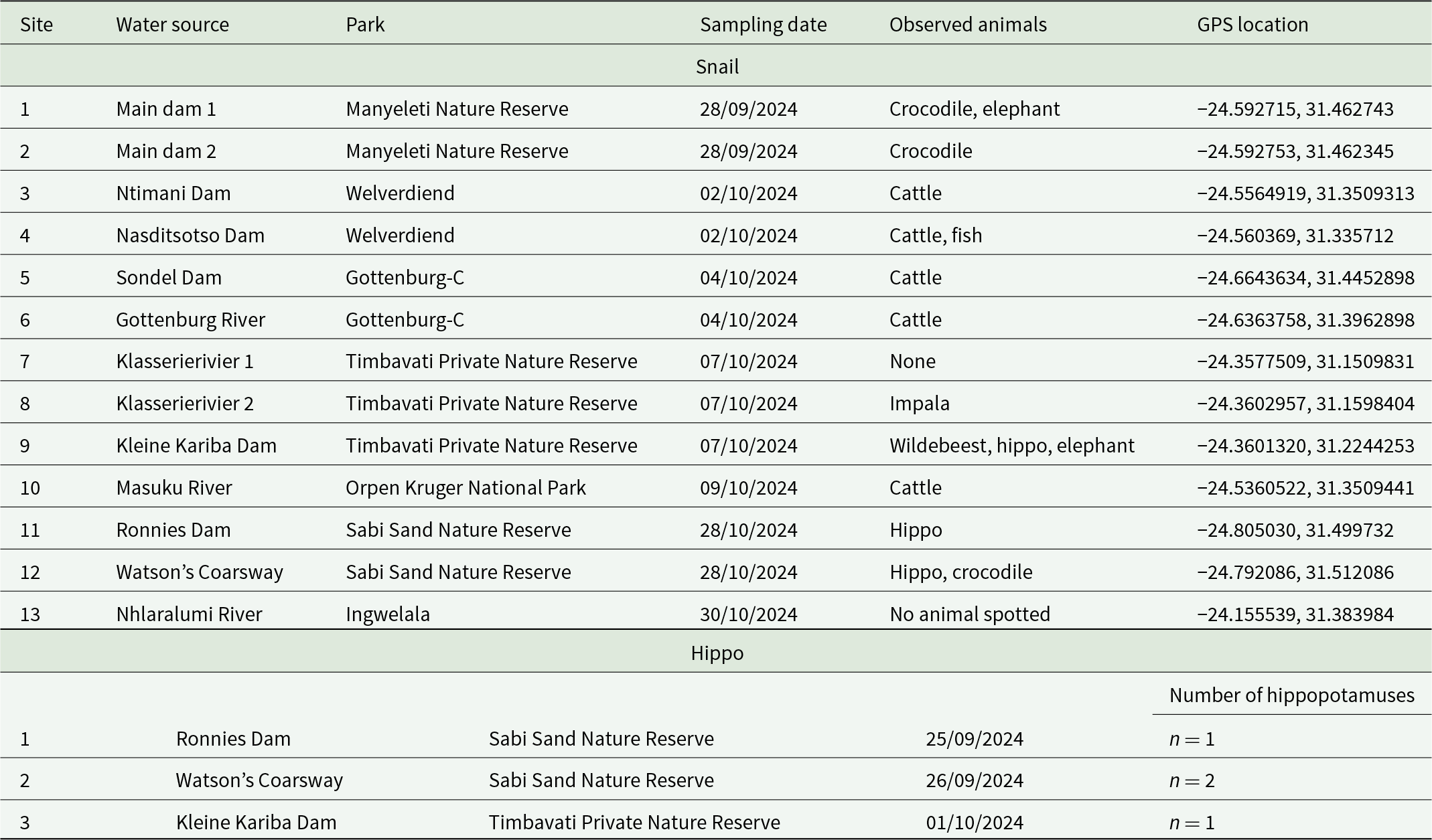
Freshwater snails were collected from 13 sites across the Greater Kruger region between 25 September and 30 October 2024. The sites were distributed across five protected areas and two neighbouring villages and comprised a range of aquatic habitats, including rivers, artificial lakes and livestock watering reservoirs. Of the sampled sites, seven were artificial lakes (sites 1–5, 9 and 11), while six were riverine habitats. Among the river sites, three were flowing systems (sites 7, 8 and 12) and three consisted of non-flowing rest pools (sites 6, 10 and 13) at the time of sampling. An overview of all sampling locations, including geographic coordinates and observed animal presence, is provided in Table 1.
Snail and hippopotamus sampling locations in the Greater Kruger region, including water body name, park name, sampling date, observed animals and GPS coordinates

Table 1 Long description
Sampling locations are listed for two datasets: snail collection sites and separate hippopotamus dissection observations, each with water source name, park, date, animals seen, and GPS coordinates. Snails were collected at 13 sites across Manyeleti, Welverdiend, Gottenburg-C, Timbavati, Orpen in Kruger National Park, Sabi Sand, and Ingwelala, with coordinates provided for each site. Observed animals at snail sites most often include cattle, with other sightings such as crocodile, elephant, impala, wildebeest, hippo, fish, or none reported. Hippopotamus dissections were done at three water bodies: Ronnies Dam in Sabi Sand had one hippopotamus, Watson’s Coarsway in Sabi Sand had two hippopotamuses, and Kleine Kariba Dam in Timbavati had one hippopotamus, all on late September to early October dates. The highest hippopotamus count in the table is at Watson’s Coarsway. . Only locations where snails were collected are included in the snail section, and some records may reflect shell-only recovery rather than live animals.
Snail collection and cercarial shedding
At each sampling site, snails were collected using scooping nets and manual collection from vegetation, rocks and substrates where possible. Sampling was done by two people for 30 min for sites 1 and 2, while all other sites were sampled by one person only.
Collected snails were transported in containers filled with site water and kept shaded and cooled to reduce mortality before transfer to the Hans Hoheisen Wildlife Research Station, South Africa. In the laboratory, snails were sorted per morphotype and identified through external morphological characteristics using established keys (Mandahl-Barth, Reference Mandahl-Barth1962; Frandsen, Reference Frandsen1980; Brown, Reference Brown1994). Individual snails were placed in multiwell plates (ThermoFisher Scientific, Waltham, USA) containing water from their site of origin at 20°C overnight. Where sample numbers exceeded plate capacity (only site 13), individuals were pooled by size to standardize biomass per well and optimize survival (i.e. smaller and larger specimens were pooled up to ten and five individuals, respectively). All containers were inspected using a dissecting microscope for cercarial shedding – release of larval trematodes – at sunrise (5.30 am) whereafter all snails were exposed to indirect natural and artificial light followed by a final inspection at 10 am.
After this experiment, all specimens were fixed in ethanol using a stepwise preservation protocol (70% ethanol for 24 h, 90% ethanol for 72 h and final fixation in 90% ethanol) and stored at 4°C until further processing and transport. Transportation from South Africa to Belgium occurred at ambient temperatures.
Hippopotamus sampling
Four adult H. amphibius (>4 years) culled at three locations – Sabi Sand’s Ronnie’s Dam (n = 1), Watson’s Coarsway (n = 2) and Timbavati’s Kleine Kariba Dam (n = 1) – were examined post-mortem for endo- and ectoparasites (Table 1). Internal organs were examined for flukes: the rumen for amphistomes, the liver for liver flukes and mesenteric veins for blood flukes. All specimens were stored per morphotype per host individual in 70–96% ethanol and kept at 4°C. Additionally, two schistosome specimens collected from two culled hippopotamuses in 2016 were included.
Morphological identification of amphistomes
Amphistomes were further analysed via scanning electron microscopy (SEM) and median sagittal sections as described in Schols et al. (Reference Schols, Henrard, Brecko, Mudavanhu, Goossens, Steffanie, Clegg, Vanhove and Huyse2025). Samples were dehydrated through graded ethanol, butanol and paraffin embedding, then sectioned at 10 μm using a Leica SM 2000R microtome. Sections were deparaffinized, rehydrated and stained with haematoxylin–erythrosin before dehydration, clearing in xylene and mounting in Depex. Internal morphology was analysed following Sey (Reference Sey1991), and SEM images of the genital pore area were used to aid identification (Sey, Reference Sey1991; Eduardo, Reference Eduardo2005).
Specimen photography
Up to 15 snails per morphotype per site were photographed using a high-quality focus stacking setup at the Royal Museum for Central Africa, following Brecko et al. (Reference Brecko, Mathys, Dekoninck, Leponce, VandenSpiegel and Semal2014) and adapted by Schols et al. (Reference Schols, Mudavanhu, Carolus, Hammoud, Muzarabani, Barson and Huyse2020). A Canon DSLR with EOS Utility 3 captured anterior, posterior and, when relevant, frontal views, which were combined into high-resolution composites using Zerene Stacker® (v. 1.04) and post-processed in GIMP (v. 2.10.32) to unify the background, merge orientations and add a scale bar. Adult flukes were photographed fully submerged in ethanol using the same protocol.
DNA extraction of snail tissues and trematodes
Snail soft tissue was separated from the shell, homogenized using a scalpel and residual ethanol was allowed to evaporate prior to DNA extraction. Due to the strong shells of T. granifera, these specimens required crushing with sterilized pliers to access soft tissue. To avoid cross contamination, all utensils and work surfaces were sterilized before every sample using RNase AWAY™ Surface Decontaminant (Thermo Scientific™) and a flame, while the sterile absorbent paper was replaced after each sample. DNA was extracted from all snails and 15 T. granifera using the E.Z.N.A.® Mollusc DNA Kit and eluted in 150 µL. An additional 500 T. granifera were extracted using 200 μL of 5% Chelex® (Biorad™) (Carolus et al., Reference Carolus, Muzarabani, Hammoud, Schols, Volckaert, Barson and Huyse2019). DNA extracts were aliquoted for storage at – 20°C (1/4), dried on Gentegra™ tubes (2/4), or 1:10 diluted for downstream analyses (1/4).
Adult trematodes were processed using the same sterilization protocols as snails. A subsection of most worms was used for DNA extraction: a portion of the acetabulum for stomach flukes, a small triangle from the middle of liver flukes and whole bodies for blood flukes. DNA was extracted using the E.Z.N.A.® Tissue DNA Kit (OMEGA bio-tek, Inc.), eluted in 150 µL, and aliquoted according to snail DNA extracts.
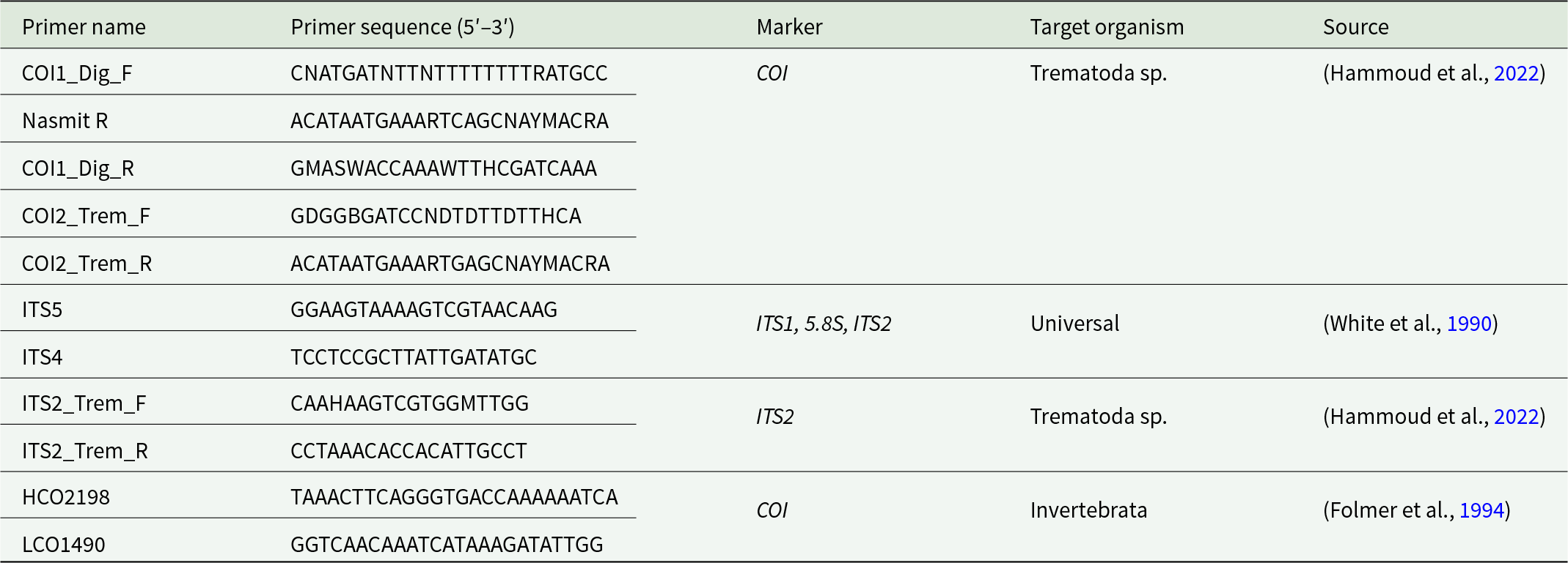
Snail identification was confirmed through amplification of partial COI gene using HCO2198 and LCO1490 primers, producing a 710 bp fragment (Folmer et al., Reference Folmer, Black, Hoeh, Lutz and Vrijenhoek1994; Table 3). Trematode DNA amplification required up to 9 primers (Table 3); ITS4/ITS5 and COI1_DIG_F/NASMIT_R were initially used to amplify the full ITS1–5.8S–ITS2 region and an 871 bp COI fragment (White et al., Reference White, Bruns, Lee, Taylor, White, Lee, Innis, Gelfand and Sninsky1990; Hammoud et al., Reference Hammoud, Mulero, Boissier, Van Bocxlaer, Verschuren, Albrecht, Huyse, Boissier, Verschuren, Albrecht and Huyse2022). If unsuccessful, alternative primers targeting shorter ITS2 and COI fragments (ITS2_Trem_F/R, and COI1_DIG_F/R, COI2_Trem_F/R, resp.) were utilized (Hammoud et al., Reference Hammoud, Mulero, Boissier, Van Bocxlaer, Verschuren, Albrecht, Huyse, Boissier, Verschuren, Albrecht and Huyse2022).
Detection of snail infections by PCR
The infection status of collected snails was molecularly determined using the infection Rapid Diagnostic PCR (infection RD-PCR) for trematodes and Schistosoma spp. as described in Schols et al. (Reference Schols, Carolus, Hammoud, Mulero, Mudavanhu and Huyse2019) (Table 2). PCRs (15 μL) contained 1 μL snail DNA, 7.5 μL QIAGEN™ Multiplex PCR Master Mix, 1.5 μL primer mix, and 5 μL dH₂O, with final primer concentrations of 0.1 μM 18S snail, 0.2 μM 18S trematode and 0.6 μM ITS2 Schistosoma spp. primers. Cycling consisted of 95°C for 5 min; 39 cycles of 94°C for 30 s, 62°C for 45 s, 72°C for 45 s; and a final extension at 72°C for 10 min. All snails were tested individually, except T. granifera, which were pooled in groups of eight for the infection RD-PCR; positive pools were retested individually, and detected infections were processed separately. DNA samples showing a strong band of the expected length were selected for Sanger sequencing, which was done at Macrogen™ (Netherlands). The resulting sequences were curated in Geneious Prime (v2025.1.2) before being compared to representatives on the BLAST (https://blast.ncbi.nlm.nih.gov/Blast.cgi) and BOLD (https://id.boldsystems.org/) databases. Thresholds for trematode species separation were set at divergence values of >5% for COI and >1% for ITS sequences (Vilas et al., Reference Vilas, Criscione and Blouin2005). For snail species a threshold of >2% for COI was set (Christian Albrecht pers. comm.).
Primer sets used to amplify the partial COI gene and ITS region for sequencing

Table 2 Long description
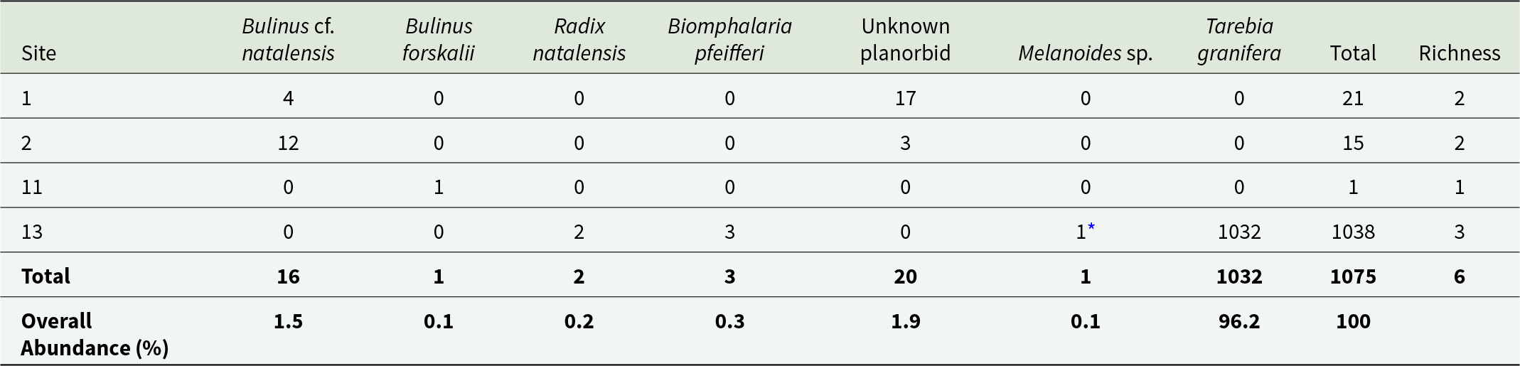
Counts of freshwater snails are listed by sampling site for seven taxa, with a total count and species richness per site. Across all sites, 1075 snails were recorded and Tarebia granifera accounts for 1032, representing 96.2 percent of overall abundance. Site 13 contains nearly all snails, with 1038 total and richness of 3, including 1032 Tarebia granifera plus small numbers of Radix natalensis (2), Biomphalaria pfeifferi (3), and Melanoides sp. (1). Sites 1 and 2 have much lower totals, 21 and 15 respectively, and both have richness of 2 driven by Bulinus cf. natalensis (4 at site 1; 12 at site 2) and the unknown planorbid (17 at site 1; 3 at site 2). Site 11 has only 1 snail, Bulinus forskalii, and richness of 1. Totals by species other than Tarebia granifera are low overall: Bulinus cf. natalensis 16, the unknown planorbid 20, and each of Bulinus forskalii, Melanoides sp., Radix natalensis, and Biomphalaria pfeifferi at 3 or fewer.
Results
Snail abundance
A total of 1074 snails were collected during this study. The number of collected snails per site was highly variable, with most sites (n = 9) having no snails. The highest snail count (n = 1037) was observed at site 13, while the lowest non-zero count (n = 1) occurred at site 11. The number of species found per site ranged from one to three, while six species were collected across all sites. Additionally, one Melanoides sp. shell was identified from site 13, indicating the presence of this species in the past (Table 3).
Abundance of freshwater snails per sampling site and species, including species richness and overall abundance of snail presence

Table 3 Long description
The table lists primer names alongside their nucleotide sequences written from 5-prime to 3-prime, the genetic marker amplified, the intended target organism group, and the literature source.
* Only sites where snails were collected are shown. An asterisk (*) indicates that only an empty shell of the species was recovered.
Snail species and diversity
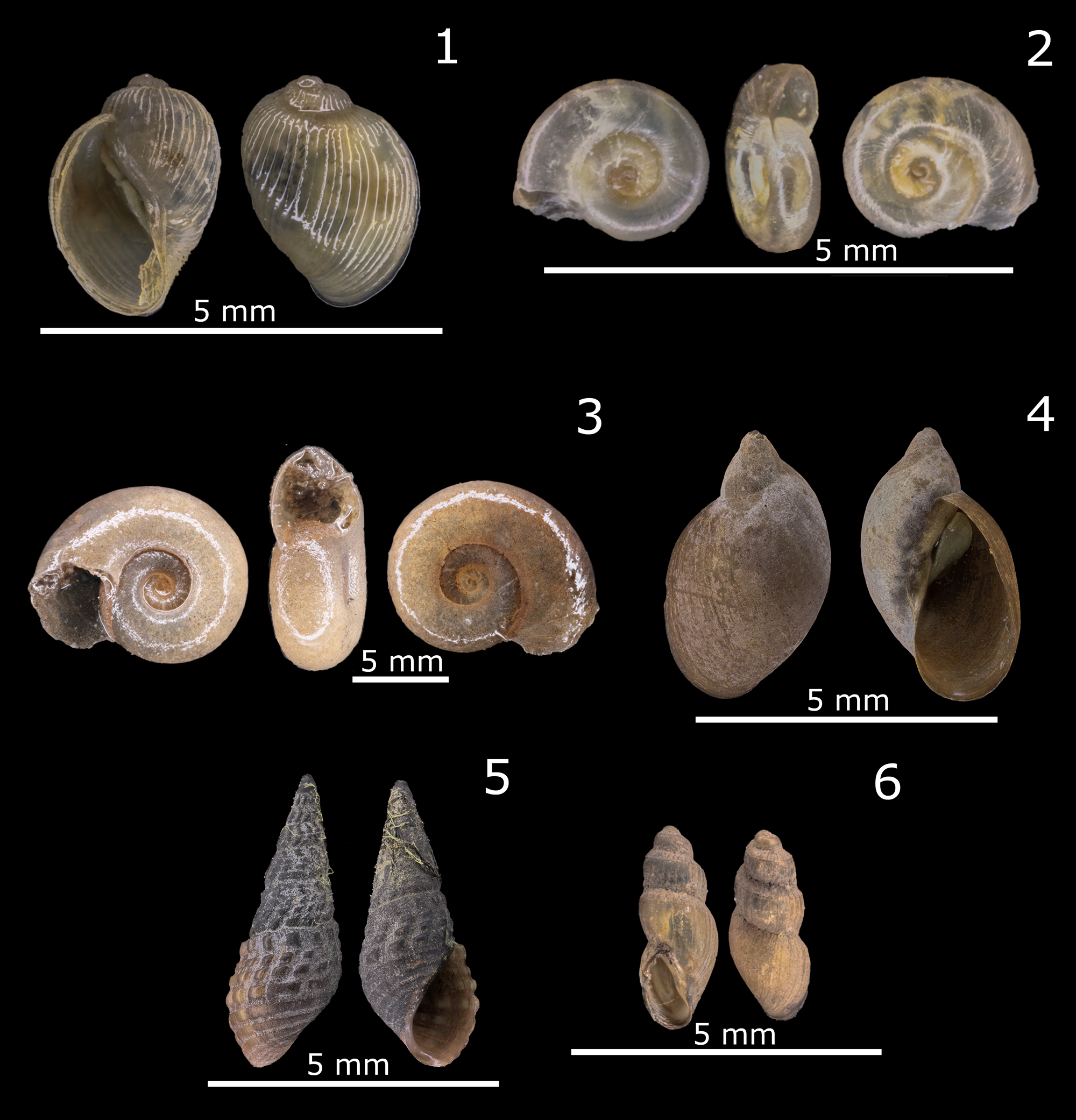
Key-based identification in South Africa revealed six snail morphotypes: a Bulinus sp. belonging to the truncatus/tropicus species complex, an unknown species belonging to the Planorbidae, Biomphalaria pfeifferi, Radix natalensis, Tarebia granifera and Bulinus forskalii (Figure 1). COI amplification of one snail per morphotype per site (n = 7), along with all trematode-infected individuals (n = 18, three morphotypes) detected by RD-PCR, yielded 20 high-quality sequences (>400 bp, HQ > 80) suitable for BLAST analysis.
Photographs of representative individuals from each collected snail species. DNA extraction and molecular analysis were performed on all individuals shown. Bulinus cf. natalensis (1), unknown planorbid (2), Biomphalaria pfeifferi (3), Radix natalensis (4), Tarebia granifera (5) and Bulinus forskalii (6). Scale bars of 5 mm are shown in every image.

All seven Bulinus sp. specimens showed ∼0.2% internal divergence (1 nucleotide difference in 572 positions of the partial COI gene, GenBank: PZ392945 & 47–52). All sequences showed affinity to the B. truncatus/tropicus group with the smallest p-distance to B. natalensis (0.5–0.7%, GenBank: AM286311, South Africa). Therefore, the morphotype is identified as Bulinus cf. natalensis, requiring a more detailed taxonomic account of the radula combined with karyotyping to allow a definitive identification (DBL-WHO, 1977, 1987). All three partial COI gene sequences linked to the unknown planorbid represented a single, yet undescribed species, with 0% internal divergence (across 564 nucleotides, GenBank: PZ392941-3) and 1.6% divergence from the nearest sequence on GenBank also identified as an unknown planorbid from Lake Victoria (GenBank: LC491295, Kenya). All other BLAST results exceeded 12% divergence. Such divergence levels indicate a different genus within the Planorbidae (Christian Albrecht pers. comm.). Therefore, the identification of this morphotype remains inconclusive. Both B. pfeifferi specimens were identical to each other (0% internal divergence across 446 nucleotides, GenBank: PZ392944 & 46) and to GenBank records from Zimbabwe (GenBank: MG780186) and Malawi (GenBank: OR880348). Both R. natalensis specimens were identical to each other (0% internal divergence across 525 nucleotides, GenBank: PZ392939-40) and showed 1.3% divergence from the nearest sequence on GenBank classified as R. natalensis (GenBank: PP228864, South Africa). Both T. granifera specimens exhibited 0% internal divergence (GenBank: PZ392937-8) and 0% divergence from reference sequences from South Africa, China and Thailand (resp., BOLD:ADE4657, MZ662113, MK000368). The single B. forskalii specimen (GenBank: PZ392967) closely matched reference sequences of B. forskalii with 0.4% divergence (GenBank: OP233130, Kenya).
Snail infection prevalence and parasite diversity
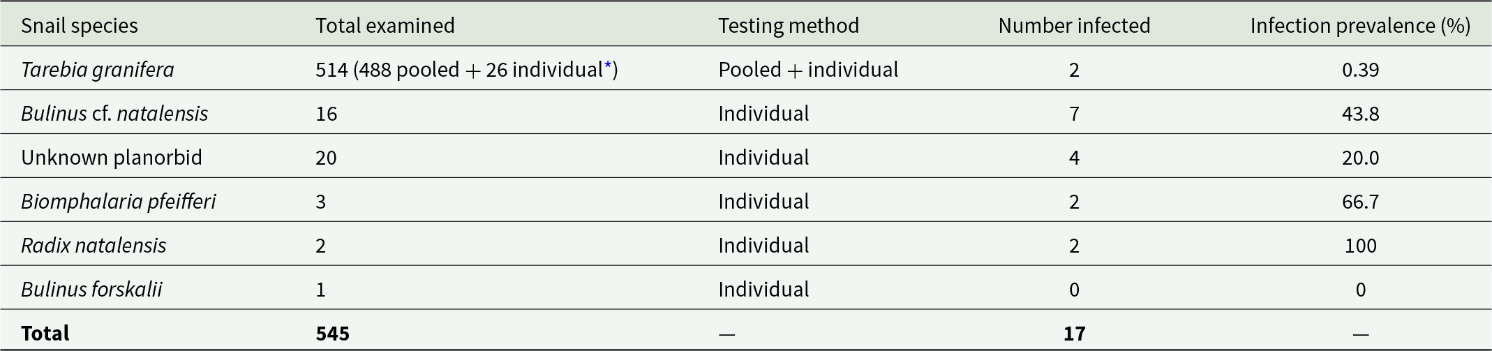
The shedding experiment revealed no patent infections in the 1074 collected freshwater snails. Of these, 545 specimens were screened molecularly for trematode infections. Due to the high abundance of T. granifera at site 13, a randomly selected subset of individuals was analysed. Trematode infections were detected in all sampled snail taxa except B. forskalii, which only consisted of a single specimen (Table 4). Like snail counts, infection prevalence varied widely among species, ranging from 0.39% in T. granifera (2/545) to 100% in R. natalensis (2/2), with intermediate prevalence observed in B. cf. natalensis (43.8%, 7/16), the unknown planorbid (20%, 4/20) and B. pfeifferi (66.7%, 2/3).
Molecular screening through RD-PCR to detect trematode infections in freshwater snail samples

Table 4 Long description
The table reports RD–PCR screening results for trematode infections in freshwater snails, listing how many were examined, whether testing was pooled or individual, how many were infected, and the infection prevalence. Across all species, 545 snails were examined and 17 were infected. Radix natalensis showed the highest prevalence, with two infected out of two examined, reported as 100 percent. Biomphalaria pfeifferi also had high prevalence, with two infected out of three, reported as 66.7 percent, and Bulinus cf. natalensis had seven infected out of 16, reported as 43.8 percent. The unknown planorbid had four infected out of 20, reported as 20.0 percent, while Bulinus forskalii had 0 infected out of one, reported as 0 percent. Tarebia granifera had the largest sample size, 514 examined using pooled plus individual testing, but only two infections, reported as 0.39 percent. Comparisons across species should be interpreted cautiously because several species have very small sample sizes.
* Photographed samples and positive pools were (re)tested individually.
Molecular identification revealed diverse trematode taxa: Haplorchis taichui in T. granifera; Nudacotyle sp. in B. cf. natalensis; Orientocreadium sp. in R. natalensis; a Ribeiroia sp. in B. pfeifferi; and 2 Echinostomatidae species in the unknown planorbid. One of the two infections from T. granifera was successfully amplified for ITS2 (212 nucleotides, GenBank: PZ390619), 28S (154 nucleotides, GenBank: PZ395368) and COI (294 nucleotides, GenBank: PZ397341). The ITS2-28S fragments showed 0% divergence (GenBank: KX815126), while the COI fragment showed 1% divergence (GenBank: MG972809), from adult Haplorchis taichui infecting humans in Vietnam. The ITS2 fragment was successfully sequenced for all seven B. cf. natalensis infections (GenBank: PZ395383-7 & PZ395374-5), showing no internal divergence and no divergence with Nudacotyle sp. isolated from B. pfeifferi in Kenya (GenBank: MN745950, 393 nucleotides). Of these, two specimens were successfully sequenced for the partial COI marker, showing 0.8% divergence (three mutations across 375 overlapping nucleotides, GenBank: PZ392968-9). No significant BLAST results emerged from these sequences (>17% divergence). Both infections in R. natalensis showed 0% internal divergence for the ITS2 marker across 285 nucleotides (GenBank: PZ395388-9) and 0% divergence from Orientocreadium sp. from R. natalensis in Zimbabwe (GenBank: MZ600135). Similarly, the partial COI sequences obtained for these infections showed 0% internal divergence (308 nucleotides, GenBank: PZ392966 & PZ392979). No significant BLAST results emerged from these sequences (>15% divergence). Only one of two infected B. pfeifferi specimens resulted in an ITS2 sequence of sufficient quality yet was still unqualified to deliver high-resolution information (no consensus sequence, 170 bp, GenBank: PZ395376). It overlapped 100% with Ribeiroia marini (GenBank: AY761147, 0% divergence) and Ribeiroia sp. (GenBank: KF525784, 0% divergence), both reported from Biomphalaria. As no COI information was obtained for these infections, a tentative identification as Ribeiroia sp. is made. Two out of four infections in the unknown planorbid were successfully amplified for the ITS2 fragment and showed 12.7% divergence (GenBank: PZ395390-1). Type 1 (GenBank: PZ395390) showed the lowest divergence (1.9%) from Echinoparyphium sp. from Uganda (GenBank: OQ548298). Type 2 (GenBank: PZ395391) showed the lowest divergence (5%) from Cathaemasia longivitellata (GenBank: PP177542, Czechia). Therefore, both trematode infections in the unknown planorbid appear to belong to the Echinostomatidae yet no further identification is possible with these divergence levels.
Trematode assemblages in hippopotamuses
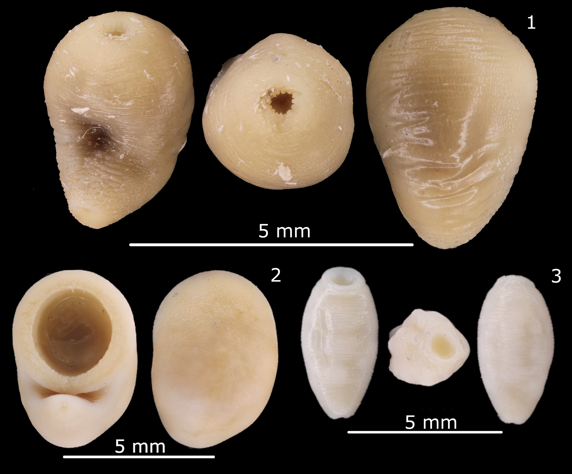
The dissections of 4 hippopotamuses yielded blood flukes, liver flukes and amphistomes (Figure 2). Molecular analyses identified the presence of two Schistosoma edwardiense specimens from the post-mortem examinations in this study both based on the ITS-region (766 nucleotides, 0% internal divergence, GenBank: PZ395415-6) and partial COI (792 nucleotides, 0% internal divergence, GenBank: PZ392985-6). The ITS BLAST results indicated a 0.1% divergence from an S. edwardiense infection in Biomphalaria sudanica in Uganda (GenBank: AY197344). Notably, this divergence corresponds to a Single Nucleotide Polymorphism (SNP) across all 766 nucleotides. This SNP is represented by a faint Thymine signal (matching the SNP of AY197344) underlying the strong Guanine signal within our consensus sequence. The COI BLAST results indicated 2% divergence from an infection in Biomphalaria sudanica in Uganda (GenBank: AY197347) and 0.3% divergence from an infection in B. pfeifferi in Zimbabwe (GenBank: MT886702). COI amplification was not successful for the 2016 samples, whereas only amplification of the ITS region was successful for these specimens (GenBank: PZ395417-8). Molecular analyses identified the presence of two Schistosoma hippopotami specimens (867 nucleotides, 0% internal divergence). The ITS BLAST results indicated 0% divergence from an S. hippopotami infection in Bulinus truncatus in Uganda (GenBank: AY197343).
Images of one individual representing each of the different amphistome species collected: Carmyerius aff. schoutedeni (1), Gigantocotyle gigantocotyle (2) and Nilocotyle cf. praesphinctris (3). Scale bars of 5 mm are shown in every image.

The ITS-region was successfully amplified for all six F. nyanzae specimens, revealing no internal divergence across 943 nucleotides (GenBank: PZ395377-82). The ITS BLAST revealed no divergence with reference sequences of F. nyanzae from both Zimbabwe and South Africa (GenBank: MT909820-1 and ON661092, resp.). The COI fragment was successfully amplified for five out of six F. nyanzae specimens with a single SNP across 748 nucleotides (0.1% internal divergence, GenBank: PZ392980-4). One to two SNPs are noted compared to F. nyanzae sequences from South Africa (GenBank: ON661094-100) and 3–8 SNPs to those from Zimbabwe (GenBank: MT909542-3).
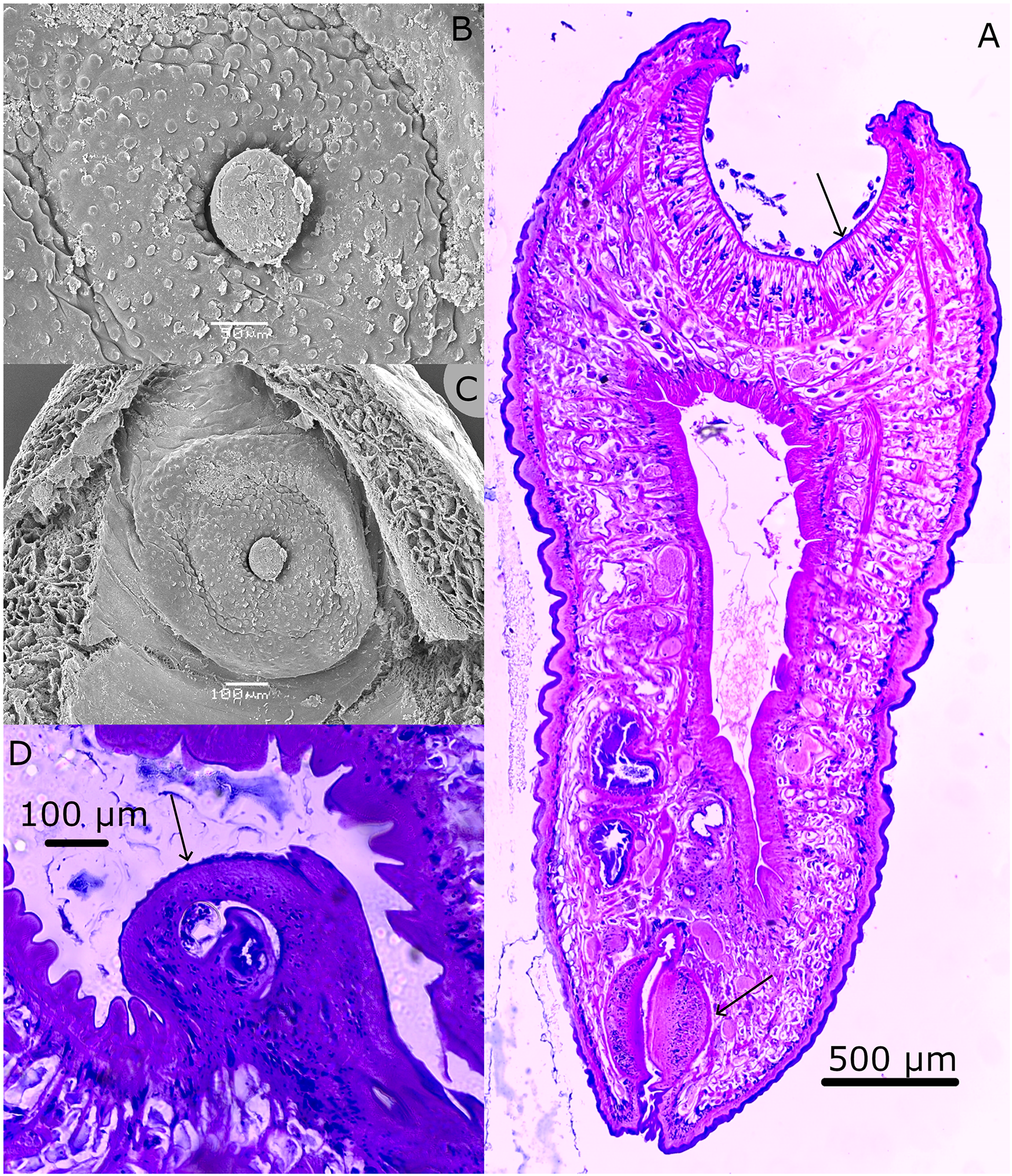
The ITS-region and partial COI gene were successfully sequenced for 28 out of the 33 collected amphistome specimens. Molecular analysis revealed the presence of three species: Gigantocotyle gigantocotyle, Nilocotyle cf. praesphinctris and Carmyerius sp. (represented by 1, 2 and 25 specimens, resp.). For G. gigantocotyle, the ITS-region (GenBank: PZ395393) showed no divergence from G. gigantocotyle from Zimbabwe and South Africa (244 nucleotides, GenBank: PV655540 & LC660654, resp.), while the COI region (GenBank: PZ392978) showed 0.3% divergence from a specimen from Zimbabwe (730 nucleotides, GenBank: PV639488). For N. cf. praesphinctris, the ITS-region (GenBank: PZ395395-6) showed no divergence from N. praesphinctris from South Africa (393 nucleotides, GenBank: LC660653), while the COI region (GenBank: PZ392964-5) showed 14.1% divergence from Carmyerius aff. chabaudi from Zimbabwe (700 nucleotides, GenBank: PV639486). Currently, no COI reference sequence exists for N. praesphinctris. For Carmyerius sp., the ITS-region (GenBank: PZ395392, PZ395394 and PZ395397-414) showed 1.5% divergence from C. aff. chabaudi from Zimbabwe (390 nucleotides, GenBank: PV655542), while the COI region (GenBank: PZ392953-63 & PZ392970-7) showed 9.5% divergence from the same specimen (722 nucleotides, GenBank: MT909560). This Carmyerius sp. was further examined using SEM and median sagittal sectioning: The caeca terminating at the anterior level of the testes; esophagus without bulb; genital papilla and genital fold well developed, papillae present along the exterior surface of the genital fold, no ventral fold present; terminal genitalium of the Schoutedeni type (sensu Sey, Reference Sey1983) in SEM image; combined with measurements from 1 specimen (genetically identical based on ITS2 to all other 29 specimens of this morphotype) most closely resemble, but still differ from, Carmyerius schoutedeni (Figure 3). Notably, the acetabulum diameter described in Sey (Reference Sey1991) for C. schoutedeni did not match the illustration they provided, which leaned more to 1 mm aligning with our specimen. Combined, the close, yet non-perfect, resemblance to C. schoutedeni justifies an identification as Carmyerius aff. schoutedeni.
Various visualizations of Carmyerius aff. schoutedeni. (A) Median sagittal section. The upper arrow points to the acetabulum, and the lower arrow points to the pharynx. (B) and (C) Scanning electron microscope images at different zoom levels of the terminal genitalium. Papillae can be seen on the genital fold (large circular structure), the smaller inner circular structure is the genital tube, containing the genital pore. Separating both of these structures is the genital atrium. (D) Sectional view of the genital pore with papillae visible, marked by a black arrow. All of the images are accompanied by an individual scale bar.

Discussion
The malacological component of our survey in the Greater Kruger revealed low abundances of freshwater snails, a highly uneven spatial distribution and dominance by an invasive species. The concurrent parasitological survey detected six trematode species within the hippopotamuses and six additional trematode species across all but one sampled snail taxon. These findings contribute to the limited knowledge on freshwater snail and trematode diversity in the Greater Kruger region. However, none of the trematode infections identified in snails could be linked to adult parasites recovered from the examined hippopotamuses. Notably, the Haplorchis taichui infection in Tarebia granifera represents both the first record in Southern Africa of a trematode infection in this snail species, as well as the first detection of this exotic parasite in the region.
Overall, the invasive thiarid snail T. granifera dominated numerically, with 1032 individuals (96.2% of all snails collected), yet was only present in one site. This pattern aligns with previous reports describing T. granifera as a highly successful invader in southern African freshwater systems due to its parthenogenetic reproduction and broad ecological tolerance (Appleton et al., Reference Appleton, Forbes and Demetriades2009; Miranda et al., Reference Miranda, Perissinotto and Appleton2011). In our study, collections were made in late spring, which may explain the observed abundance pattern. Previous studies have shown that T. granifera exhibits heterogeneous distribution across seasons, with peak littoral densities in summer corresponding to increases in water temperature and rainfall (Makherana et al., Reference Makherana, Cuthbert, Dondofema, Wasserman, Chauke, Munyai and Dalu2022). The high density observed in late spring in our study system could represent the onset of this seasonal increase, as water and sediment conditions become increasingly favourable for population growth, supporting the species’ invasive success.
The unknown planorbid remained unresolved due to limitations in available reference sequences and general neglect of small planorbids throughout Africa (pers. comm. Christian Albrecht). An urgent malacological effort targeting this group is required to fill this void in African biodiversity, which would benefit the mapping of general and malacological biodiversity. These in turn will benefit future parasitological efforts and help elucidate parasite transmission and life cycle information.
Trematode infections were detected in all snail species except B. forskalii (only one specimen collected), demonstrating active parasite transmission in local aquatic habitats. Infection prevalences were particularly high in B. pfeifferi, B. cf. natalensis and R. natalensis, although small sample sizes may have inflated these estimates. Importantly, none of the larval trematodes detected in snails matched the adult trematodes recovered from hippopotamuses, preventing direct reconstruction of parasite life cycles. Nevertheless, the presence of established intermediate hosts for various trematodes indicates that environmental conditions support the maintenance of complex trematode transmission cycles (Rollinson et al., Reference Rollinson, Knopp, Levitz, Stothard, Tchuem Tchuenté, Garba, Mohammed, Schur, Person, Colley and Utzinger2013).
A notable finding of this study was the detection of H. taichui in T. granifera, representing the first record of a trematode infection in this invasive snail and the first record of this parasite in Southern Africa. Previous surveys in Zimbabwe and South Africa consistently reported T. granifera as uninfected (Pearson, Reference Pearson2022; Mudavanhu et al., Reference Mudavanhu, Schols, Goossens, Nhiwatiwa, Manyangadze, Brendonck and Huyse2024) and suggested that its invasion could reduce trematode transmission through competitive displacement of native intermediate hosts (Appleton et al., Reference Appleton, Forbes and Demetriades2009; Pointier et al., Reference Pointier, David, Jarne, Toledo and Fried2011; Mudavanhu et al., Reference Mudavanhu, Schols, Goossens, Nhiwatiwa, Manyangadze, Brendonck and Huyse2024). For now, this argument still holds for native parasites such as schistosomes and fasciolids, yet it also illustrates how precarious the limited benefit offered by these invasive snails – if any – truly is. Moreover, these invaders have the potential to drastically increase parasite transmission ( Kelly et al., Reference Kelly, Paterson, Townsend, Poulin and Tompkins2009; Mastitsky & Veres, Reference Mastitsky and Veres2010; Grabner et al., Reference Grabner, Mohamed, Nachev, Me, Sabry, Sures, Méabed, Sabry and Sures2014; Schols et al., Reference Schols, Carolus, Hammoud, Muzarabani, Barson and Huyse2021), while the opposite can also be true (Thieltges et al., Reference Thieltges, Reise, Prinz and Jensen2009). Clearly, the role of T. granifera in trematode transmission on the African continent requires more attention.
Interestingly, H. taichui is one of three species known to cause haplorchiasis in humans across Northern Africa, Eurasia and Australia (Murrell and Fried, Reference Murrell and Fried2007; Van Van et al., Reference Van Van, Dalsgaard, Blair and Le2009). To the best of our knowledge, this is the first record of this species in Southern Africa (Pearson, Reference Pearson1964; Murrell and Fried, Reference Murrell and Fried2007). Based on the limited consumption of raw fish in Southern Africa, only a limited zoonotic risk from the presence of H. taichui is expected. However, drying or salting fish has proven ineffective at preventing the transmission of food-borne trematodes (Abdussalam et al., Reference Abdussalam, Käferstein and Mott1995), and some kapenta (Limnothrissa miodon), a frequently consumed freshwater fish, is typically consumed raw on the market to assess product quality (pers. comm. Aspire Mudavanhu). Therefore, the presence of H. taichui is of potential zoonotic concern for the important kapenta industry in Southern Africa. For example, over 90% of fish production in Lake Kariba, Zimbabwe originates from L. miodon (Mudzengi et al., Reference Mudzengi, Dahwa, Matanga, Taderera and Kapembeza2021). Currently, evidence of metacercariae in kapenta is limited to unpublished observations (pers. comm. Nikol Kmentova). The abovementioned risks – zoonotic, broad intermediate host range, uncooked fish consumption and the presence of unidentified metacercariae in kapenta – warrant further examination into the possible transmission routes in H. taichui in Southern Africa.
Post-mortem examination of hippopotamuses revealed a diverse trematode assemblage, including S. edwardiense, S. hippopotami, F. nyanzae and three amphistome species. The presence of both schistosome species confirms their continued circulation in hippopotamus populations of the Greater Kruger region decades after their initial description (Pitchford and Visser, Reference Pitchford and Visser1981). Although the examined animals showed no overt clinical signs, schistosome infections in hippopotamuses have previously been associated with vascular lesions and chronic pathology, suggesting potential subclinical impacts on host fitness (McCully et al., Reference McCully, van Niekerk and Kruger1967) and a potential importance for conservation efforts.
Genetic analyses of F. nyanzae revealed close similarity between South African and Zimbabwean specimens, as shown earlier in Bargues et al. (Reference Bargues, Halajian, Artigas, Luus-Powell, Valero and Mas-coma2022), indicating potential gene flow across the Greater Limpopo Transfrontier Conservation Area. Such connectivity is consistent with the extensive movement of hippopotamuses along river systems and floodplains (Eltringham, Reference Eltringham1999) and the broad ecological tolerance of its known intermediate hosts: the native R. natalensis and the invasive Pseudosuccinea columella (Brown, Reference Brown1994; Bargues et al., Reference Brown1994; Schols and Huyse, Reference Schols and Huyse2024). The latter was somewhat surprisingly absent from the current dataset as it is the third-most-widespread freshwater snail species in South Africa with earlier records in the Kruger National Park (De Kock and Wolmarans, Reference De Kock and Wolmarans2008). Notably, the other known intermediate host, R. natalensis, was only found uninfected. This is consistent with the typically low, or at least highly heterogeneous, infection prevalence of Fasciola spp. infections in intermediate snail hosts (Ngcamphalala et al., Reference Ngcamphalala, Nyagura, Malatji and Mukaratirwa2025) and specifically so for F. nyanzae infections in R. natalensis (Dinnik and Dinnik, Reference Dinnik and Dinnik1961; Schols et al., Reference Schols, Carolus, Hammoud, Muzarabani, Barson and Huyse2021).
Amphistomes were the most abundant trematodes recovered from hippopotamuses and consisted three species within the Paramphistomidae and Gastrothylacidae families. Molecular identification was successful for G. gigantocotyle and tentatively for Nilocotyle cf. praesphinctris, while the dominant species required morphological identification and was classified as Carmyerius aff. schoutedeni. The difficulty in molecular identification reflects the limited representation of amphistomes in public sequence databases (Laidemitt et al., Reference Laidemitt, Zawadzki, Brant, Mutuku, Mkoji and Loker2016), a challenge widely reported in trematode barcoding studies (Brant et al., Reference Brant, Morgan, Mkoji, Snyder, Rajapakse and Loker2006; Soldánová et al., Reference Soldánová, Georgieva, Roháčová, Knudsen, Kuhn, Henriksen, Siwertsson, Shaw, Kuris, Amundsen, Scholz, Lafferty and Kostadinova2017; Mudavanhu et al., Reference Mudavanhu, Schols, Goossens, Nhiwatiwa, Manyangadze, Brendonck and Huyse2024).
Similarly, several trematodes detected in snails not linked to hippopotamus infections could not be identified to species level. Nevertheless, they represent important faunal records, including Nudacotyle, Orientocreadium, Ribeiroia and two Echinostomatidae species. These genera and families are associated with diverse definitive hosts such as birds, fish and amphibians, reflecting the ecological complexity in the Greater Kruger region and the role of multi-host transmission networks in maintaining trematode diversity (Poulin and Cribb, Reference Poulin and Cribb2002). The sampling of definitive hosts, followed by morphological study of collected trematodes should enable the generation of suitable reference sequences that will enable the reconstruction of these parasites’ life cycles.
Overall, this study demonstrates that freshwater ecosystems within the Greater Kruger region support a relatively diverse community of snails and trematodes. It also demonstrates that this large conservation area is under pressure, as illustrated by the presence of invasive species and parasites with a zoonotic potential. The genetic sequences generated in this work contribute to the catalogue of molecular data of parasites of large African herbivores while laying the foundation to fully resolve life cycles of hippo-infecting parasites through experimental and field studies.
Data availability statement
All 102 generated sequences are freely available on GenBank: ‘PZ390619, PZ392937-86 and PZ395368-418’.
Acknowledgements
We would like to thank Christian Albrecht for his insights into identification methods for the planorbid snails. We would like to thank Kato Dillen and Benjamin André for their help in sequencing snail specimens. Chris Goodman from Londolozi is thanked for his assistance with our research. Our gratitude goes out to the rest of the field team, especially Monique Bester and Piet King, making this research possible. We want to thank Nikol Kmentova for providing insights into possible metacercariae in kapenta and Aspire Mudavanhu for insights into kapenta consumption in Southern Africa. The SEM imagery was made possible thanks to Dr Rudy Jocqué. Median sagittal sections were made by Natascha Steffanie.
Author contributions
Conceptualization: R.S., T.H., L.B., J.W. and W.J.L.-P.; data collection: R.S., K.L., F.R., J.W. and W.J.L.-P.; formal analysis: R.S. and K.L.; investigation: R.S. and K.L.; resources: F.R., J.W. and W.J.L.-P.; funding acquisition: R.S., K.L., T.H., F.R., J.W. and W.J.L.-P.; data curation: R.S.; writing – original draft: R.S. and Z.T.H.M.; writing – review and editing: R.S., Z.T.H.M., K.L., L.B., T.H., F.R., J.W. and W.J.L.-P.; supervision: T.H., L.B. and W.J.L.-P. All authors read and approved the final manuscript.
Financial support
Schols R. was supported through BRAIN-be 2.0 under the MicroResist project (B2/191/P1/MicroResist) during fieldwork, and by KU Leuven (Grant No. PDMT2/24/045) and ETH Zürich (25-2 FEL-082) during the data analysis and writing process. Lambaerts K. was supported by VLIR-UOS during his fieldwork. Luus-Powell W. J. was supported through the DSTI-NRF SARChI Chair funding (no 101054).
Competing interests
None.
Ethical standards
Section 20 permit of the Animal Diseases Act (Act No.35 of 1984) was approved by the Directorate Animal Health, Department of Agriculture, Land Reform and Rural Development [12/11/1/8/6668(HP)] and permit MPB 9422 F. Roux, for culling. Permits to move snails through the Kruger National Park and export of specimens from South Africa were obtained from the state veterinarian Dr Monique Bester of the Departement Landbou [20241009-ORM-VMMP-01, 20241001-ORM-VMMP-0]. The transport of specimens from South Africa to Belgium was further approved by the Federal Agency for the Safety of the Food Chain (FASFC) [2024000671]. Finally, a permit from the Federale Overheidsdienst FINANCIEN [2024/BO/vt/027-S/10] was approved for the import of specimens into Belgium.

 Open access

