





Introduction
Behavioural modification of the hosts caused by parasites is a diffuse phenomenon that is described in a broad range of host–parasite relations across many taxa (Moore, Reference Moore2013; Miroliubov et al., Reference Miroliubov, Lianguzova and Libersat2025). Such alterations of behaviour can affect the survival of the host, its reproduction and its relations with predators and preys. Considerable research effort has been carried out to investigate the proximate mechanisms that are responsible for behavioural changes (Miroliubov et al., Reference Miroliubov, Lianguzova and Libersat2025). Among the parasites most extensively studied in this framework, there is Toxoplasma gondii an obligate intracellular apicomplexan parasite with a global distribution and a complex heteroxenous life cycle, which alternates between definitive felid hosts, in which sexual reproduction occurs, and a broad range of endotherm vertebrates that can act as intermediate hosts (Dubey, Reference Dubey2021). Substantial evidence indicates that latent T. gondii infection is associated with behavioural changes in infected hosts (reviewed by Webster, Reference Webster2007; Vyas and Sapolsky, Reference Vyas and Sapolsky2010). These changes include impaired motor performance, deficits in spatial awareness and sensory learning and memory, lack of neophobia, reduced anxiety, display of neurological signs and reduced avoidance of feline, but not other, predators (Berdoy et al., Reference Berdoy, Webster and Macdonald2000; Lamberton et al., Reference Lamberton, Donnelly and Webster2008; Kannan et al., Reference Kannan, Moldovan, Xiao, Yolken, Jones-brando and Pletnikov2010; Webster and McConkey, Reference Webster and McConkey2010; Berenreiterová et al., Reference Berenreiterová, Flegr, Kuběna and Němec2011).
The influence of T. gondii on host behaviour has been documented across a broad range of species and ecological contexts. Aside from experimental studies on rodents (Witting, Reference Witting1979; Webster, Reference Webster1994; Berdoy et al., Reference Berdoy, Webster and Macdonald1995, Reference Berdoy, Webster and Macdonald2000) and observational studies in humans (Torrey and Yolken, Reference Torrey and Yolken2003), associations between aberrant behaviours and infection with T. gondii have also been reported in wild species such as spotted hyenas (Crocuta crocuta Erxleben, 1777) from Kenya (Gering et al., Reference Gering, Laubach, Weber, Hussey, Turner, Lehmann, Montgomery, Holekamp and Getty2020), wolves (Canis lupus Linnaeus, 1758) from North America (Meyer et al., Reference Meyer, Cassidy, Stahler, Brandell, Anton, Stahler and Smith2022), chimpanzees (Pan troglodytes troglodytes Blumenbach, 1775) from Gabon (Poirotte et al., Reference Poirotte, Kappeler, Ngoubangoye, Bourgeois, Moussodji and Charpentier2016) and red deer (Cervus elaphus Linnaeus, 1758) from northern Italy (Nava et al., Reference Nava, Corlatti, Formenti, Trogu, Pedrotti, Gugiatti, Lanfranchi, Luzzago and Ferrari2023). A neuropathological study carried out by Milne et al. (Reference Milne, Fujimoto, Bean, Peters, Hemmington, Taylor, Fowkes, Martineau, Hamilton, Walker, Mitchell, Léger, Priestnall and Webster2020) also demonstrated behavioural impacts of infection with T. gondii and other neurotropic pathogens, such as fox-specific circovirus in red foxes (Vulpes vulpes Linnaeus, 1758) from England. This presentation, named Dopey Fox Syndrome (DFS), is characterized by neurological signs and abnormal behaviours such as reduced fear, increased sociability, repetitive pacing, facial twitching and anorexia (Milne et al., Reference Milne, Fujimoto, Bean, Peters, Hemmington, Taylor, Fowkes, Martineau, Hamilton, Walker, Mitchell, Léger, Priestnall and Webster2020).
In spite of the growing evidence, linking toxoplasmosis with behavioural alterations, the biological mechanisms responsible for these changes remain poorly understood. Several studies have attempted to elucidate the proximate mechanisms behind behavioural changes associated with latent toxoplasmosis; however, no consensus has yet been reached regarding the biological processes involved (Berenreiterová et al., Reference Berenreiterová, Flegr, Kuběna and Němec2011; Adamo and Webster, Reference Adamo and Webster2013). Latent toxoplasmosis has been shown to cause cellular damage and focal necrosis, potentially mediated by toxic molecules that induce neuron death (Hermes et al., Reference Hermes, Ajioka, Kelly, Mui, Roberts, Kasza, Mayr, Kirisits, Wollmann, Ferguson, Roberts, Hwang, Trendler, Kennan, Suzuki, Reardon, Hickey, Chen and McLeod2008). Furthermore, the pro-inflammatory cytokines required to maintain dormancy of T. gondii might affect turnover and efficiency of many neuromodulators, such as dopamine, glutamate and serotonin (Webster and McConkey, Reference Webster and McConkey2010). Since the mid-1980s, dopamine dysregulation has been postulated to be the primary mechanism of parasite-induced behavioural changes (Stibbs, Reference Stibbs1985). This hypothesis was supported by findings showing increased dopamine levels in infected cells (Prandovszky et al., Reference Prandovszky, Gaskell, Martin, Dubey, Webster and McConkey2011), and by the identification of 2 parasite-related genes (TgAAH1 and TgAAH2) encoding for tyrosine hydrolase-like enzymes involved in dopamine synthesis (Gaskell et al., Reference Gaskell, Smith, Pinney, Westhead and McConkey2009; Prandovszky et al., Reference Prandovszky, Gaskell, Martin, Dubey, Webster and McConkey2011; McConkey et al., Reference McConkey, Martin, Bristow and Webster2013). However, later studies using knock-out strains of T. gondii for these genes did not detect significant changes in dopamine levels (Wang et al., Reference Wang, Harmon, O’malley and Sibley2015), despite detecting impairment in other biological functions unrelated to host manipulation (Wang et al., Reference Wang, Verma, Dubey and Sibley2017). Collectively, these findings suggest that increased dopamine production alone is unlikely to explain the effects of latent toxoplasmosis on host behaviour, pointing towards a more complex and likely multifactorial mechanism.
While the biochemical mechanisms behind the phenomenon of host behaviour manipulation have been extensively investigated, fewer studies have been carried out to map the distribution of parasitic elements in the host brain, with the majority of them carried out in experimental infections with known strains of T. gondii using murine models (Berenreiterová et al., Reference Berenreiterová, Flegr, Kuběna and Němec2011; McConkey et al., Reference McConkey, Martin, Bristow and Webster2013; Schneider et al., Reference Schneider, Figueroa Velez, Orchanian, Shallberg, Agalliu, Hunter, Gandhi, Lodoen and Weiss2022; Ramírez-Flores and Mondragón-Flores, Reference Ramírez-Flores and Mondragón-Flores2025). It has been speculated that the manipulation of host behaviour could stem from neuroinflammation of specific brain regions associated with fear processing such as the amygdala (Misslin, Reference Misslin2003; House et al., Reference House, Vyas and Sapolsky2011; McConkey et al., Reference McConkey, Martin, Bristow and Webster2013). An experimental study by Blanchard and Blanchard (Reference Blanchard and Blanchard1972), carried out on rats with lesions affecting the amygdala, showed similar behavioural disorders to those observed in T. gondii-infected rats (Berdoy et al., Reference Berdoy, Webster and Macdonald2000; Vyas et al., Reference Vyas, Kim, Giacomini, Boothroyd and Sapolsky2007), suggesting a potential role of this brain region which is responsible for classical fear responses such as fight-or-flight and freezing behaviours (McConkey et al., Reference McConkey, Martin, Bristow and Webster2013). However, whether T. gondii or its associated neuropathological changes preferentially target specific brain regions in naturally infected hosts remains unclear.
The main question addressed in this study is whether the topographical distribution of T. gondii and its associated lesions within the brain is functionally linked to regions involved in regulation of behaviour. Since it is well established that behavioural manipulation is mediated through specific neural circuits (Prandovszky et al., Reference Prandovszky, Gaskell, Martin, Dubey, Webster and McConkey2011; McConkey et al., Reference McConkey, Martin, Bristow and Webster2013), parasitic elements and associated lesions should be disproportionately associated with the brain regions responsible for fear processing and behavioural control. The aim of the present study was to map the distribution of T. gondii and its associated lesions in the brain of red foxes from Italy and Australia, to assess the potential role of T. gondii in the onset of behavioural alterations.
Materials and methods
Study area and sample collection
Red fox carcasses were opportunistically collected from November 2023 to May 2025 from central Italy and from the state of Victoria, Australia. Foxes from Italy were provided by the WildUmbria wildlife rescue centre, which works alongside the Veterinary Teaching Hospital of Perugia University in the rescue and rehabilitation of wildlife in Umbria, Central Italy. Foxes from Australia were sourced from professional trappers and shooters, legally operating in the state of Victoria under pest control regulations. All animals were already dead at the time of sampling and no animal was killed for the specific purposes of this study; therefore, no animal ethics approval was required from either country. To minimize artefacts caused by autolysis of brain tissue, only animals that were sampled within 48 h of death were included in the analysis. The exact time of death was obtained directly from carcass providers and confirmed according to carcass condition scores (McAloose et al., Reference McAloose, Colegrove, Newton, Terio, McAloose and Leger2018).
All carcasses were refrigerated at 4 °C within hours from death and processed within 48 h post-mortem. Body mass and sex were recorded and age class was estimated based on incisor teeth eruption and wear examination (Harris, Reference Harris1978). Serum samples were obtained by collecting and centrifuging (5000 rpm for 10 min) either cardiac blood clots or thoracic blood-tinged fluid. The resulting serum samples were collected and stored at −20 °C until further use in serological analysis. The entire brain was removed from each fox following standard neuropathological procedures and fixed by immersion in 10% buffered formalin for a minimum of 7 days (Vandevelde et al., Reference Vandevelde, Higgins and Oevermann2012).
Serological analysis
The presence of anti-T. gondii antibodies (IgG) was assayed by means of Immunofluorescence Antibody Assay (Morganti et al., Reference Morganti, Rigamonti, Marchesi, Maggi, Angeli, Moretta, Brustenga, Diaferia and Veronesi2024) for all Italian foxes, and a modified agglutination test (Liyanage et al., Reference Liyanage, Amery-gale, Uboldi, Adriaanse, Firestone, Tonkin, Jabbar and Hufschmid2024) for all Australian foxes, following established protocols that were in use in the laboratories of the 2 universities. Two-fold serial dilutions of sera were performed in Phosphate-buffered saline and used to identify the antibody titre of samples in each serological assay, according to protocols already described (Brustenga et al., Reference Brustenga, Scarcelli, Rigamonti, Moretta, Diaferia, Morganti, D’avino, Gobbi, Ranucci, Sgroi, Passamonti and Veronesi2025a, Reference Brustenga, Liyanage, Loukopoulos, Fisher, Haining, Gauci, Lucentini, Uboldi, Tonkin, Morganti, Rigamonti, Passamonti, Veronesi, Jabbar and Hufschmid2025b). Samples were considered seropositive when positive reactions were detected at titres equal to or higher than 1:20 (Verin et al., Reference Verin, Mugnaini, Nardoni, Papini, Ariti, Poli and Mancianti2013; Dubey et al., Reference Dubey, Whitesell, Culp and Daye2014). End-point titres were defined as the highest dilution yielding a positive reaction.
Histological and immunohistochemical analyses
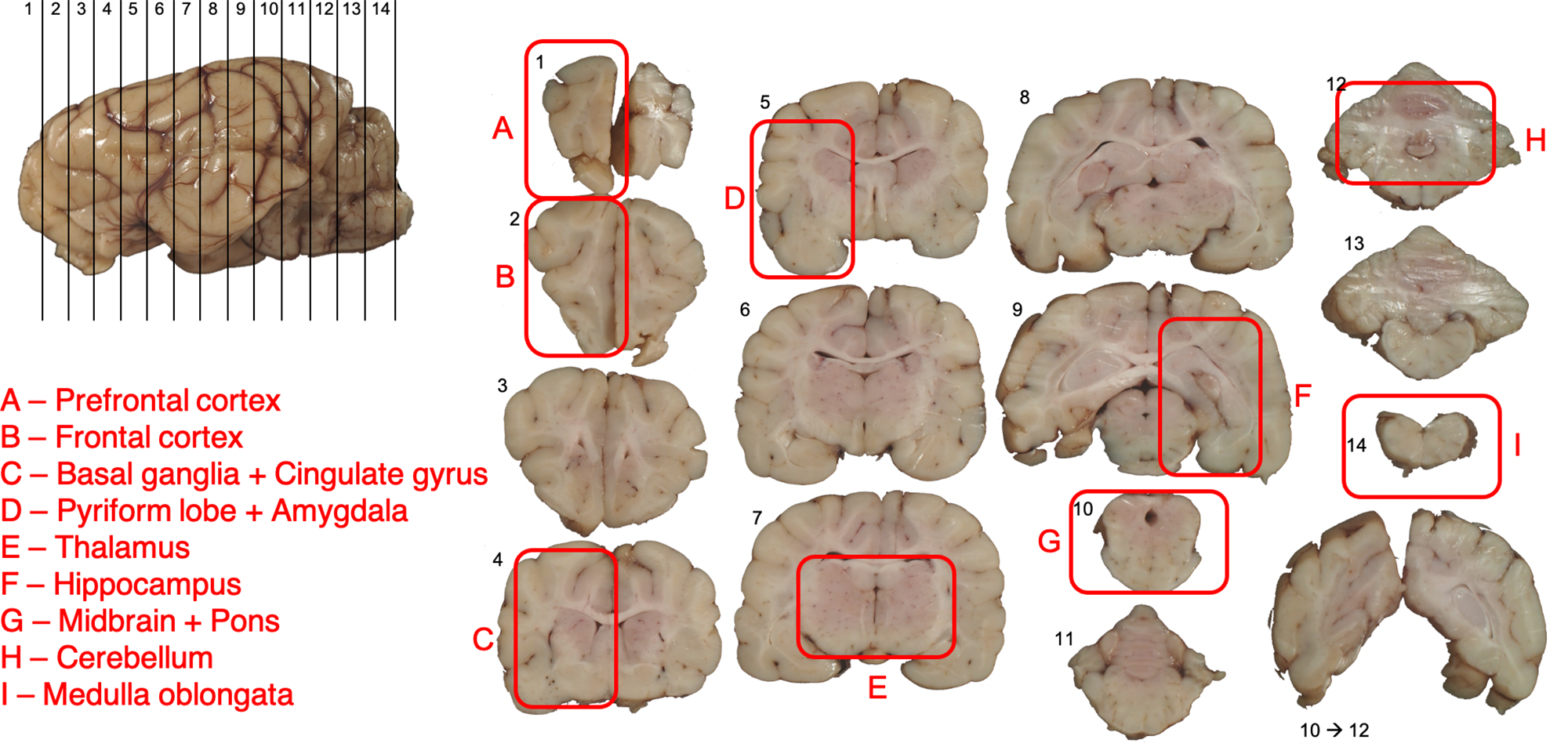
After fixation, gross examination was performed and serial 0.5 cm thick coronal sections were cut in a rostro-caudal direction across the entire brain. Trimming of brain sections was carried out only for T. gondii seropositive foxes. Using the fox brain atlas by Rogers Flattery et al. (Reference Rogers Flattery, Abdulla, Barton, Michlich, Trut, Kukekova and Hecht2023), 12 functionally representative areas of the brain were selected, and samples for each fox were placed into nine histocassettes (A–I) (Figure 1). Samples were routinely processed to obtain 5 µm sections stained with hematoxylin and eosin (H&E). Additional unstained paraffin-embedded 5 µm sections from blocks with inflammatory lesions were submitted for molecular (formalin-fixed and paraffin-embedded [FFPE]-PCR) and immunohistochemical (IHC) assessment. IHC staining was carried out with polyclonal anti-T. gondii rabbit antibody (LSBio, LS C312239, 1:200) using a standard avidin-biotin complex protocol (Morell et al., Reference Morell, Ijsseldijk, Berends, Gröne, Siebert, Raverty, Shadwick and Kik2021) and suitable controls.
Trimming protocol for the study of the anatomical distribution of Toxoplasma gondii in the brain of red foxes (Vulpes vulpes). Black lines crossing the whole brain represent the cutting planes operated to obtain coronal sections (1–14). Red rectangles denote the regions (A–I) that were sampled. Each region was sampled to contain one (A, B, E, F, H, I) or 2 (C, D, G) of the 12 functionally representative areas selected for histological, immunohistochemical and biomolecular analyses.

Molecular analysis
FFPE tissue blocks of all the brain areas with inflammatory lesions were subjected to molecular analyses. Five 8 µm-thick sections from each FFPE block were deparaffinized at room temperature by washing for 10 min in 1200 µL of xylene at 56 °C, spinning the tube to pellet the tissue, pipetting out the xylene and rinsing the pellet in 1200 µL of absolute ethanol at room temperature vortex mixing to remove xylene residues. Samples were centrifuged at 10 000 × g for 5 min to form a pellet and the fluid was decanted. The deparaffinization step was repeated twice.
Total genomic DNA was extracted with the ExgeneTM FFPE Tissue DNA Kit (GeneAll, Seoul, Korea), according to the manufacturer’s protocol. Extracted DNA was used to amplify a 130 bp fragment of the B1 gene widely used for T. gondii diagnostics according to a validated semi-nested PCR protocol (Lin et al., Reference Lin, Chen, Kuo, Tseng and Tseng2000; Veronesi et al., Reference Veronesi, Santoro, Milardi, Diaferia, Morganti, Ranucci and Gabrielli2017). Both the first and semi nested amplifications were carried out in a total volume of 25 µL containing 2.5 µL 10 × CoralLoad PCR Buffer (Qiagen, Hilden, Germany), 2 µL of 10 mM dNTP Mix (ThermoFisher Scientific, Waltham, Massachusetts, USA), 1 µL of each 10 µM primers, 0.15 µL of TaqDNA Polymerase (Qiagen, Hilden, Germany), 2 µL of template and nuclease free water (Qiagen, Hilden, Germany) to reach final volume. A positive control, DNA extracted from a FFPE lung of a cat with a high tissue cyst burden, and a negative control, nuclease free water, were included in each PCR run. Samples were checked on a 1.5% agarose gel stained with EuroSafe (Euro Clone, Milan, Italy).
Results
Overall, 25 and 75 fox brains were collected from Italian and Australian foxes, respectively. Serological analyses showed seropositivity to Toxoplasma gondii in 13 (52%, 95% confidence interval [CI] 0.32–0.72) Italian foxes and 28 (37.3%, 95% CI 0.26–0.48) Australian foxes (Table 1). Specific data on T. gondii seroprevalence in Italian and Australian red fox populations derived from this dataset have previously been published (Brustenga et al., Reference Brustenga, Scarcelli, Rigamonti, Moretta, Diaferia, Morganti, D’avino, Gobbi, Ranucci, Sgroi, Passamonti and Veronesi2025a,Reference Brustenga, Liyanage, Loukopoulos, Fisher, Haining, Gauci, Lucentini, Uboldi, Tonkin, Morganti, Rigamonti, Passamonti, Veronesi, Jabbar and Hufschmidb, respectively) and contextualized through comparison with seroprevalence estimates reported at both regional and global scales (Wei et al., Reference Wei, Gao, Lv, Wang, Chen, Zhao, Gong and Zhang2021).
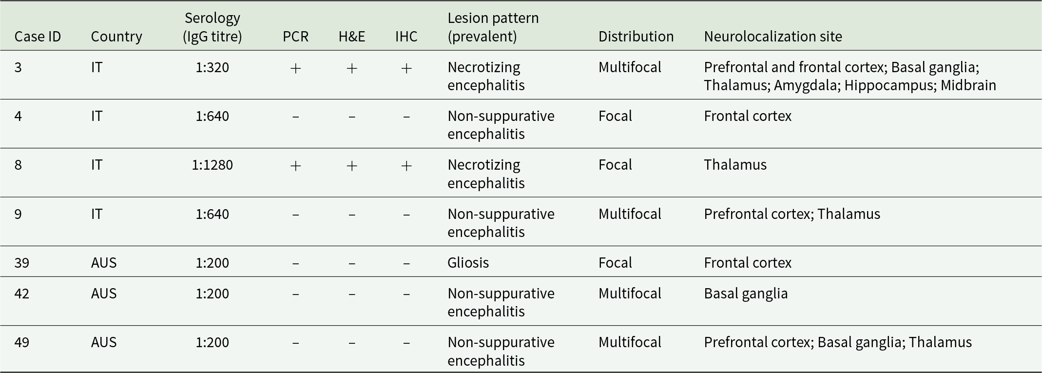
Overview of test results for Toxoplasma gondii in 7 (17.1%; 7/41) seropositive red foxes (Vulpes vulpes), 4 (30.8%; 4/13) from Italy and 3 (10.7%; 3/28) from Australia, that presented brain lesions

Table 1 Long description
Seven seropositive red fox cases with brain lesions are summarized by country, IgG titer, PCR result, histology, immunohistochemistry, lesion pattern, distribution, and neuroanatomic sites. Two Italy cases (IDs 3 and 8) were positive on PCR, histology, and immunohistochemistry and both had necrotizing encephalitis, with multifocal involvement in case 3 and focal thalamic involvement in case 8. The other two Italy cases (IDs 4 and 9) were negative on PCR, histology, and immunohistochemistry and showed non-suppurative encephalitis, either focal in the frontal cortex or multifocal including prefrontal cortex and thalamus. All three Australia cases (IDs 39, 42, 49) were negative on PCR, histology, and immunohistochemistry; lesions were gliosis in one case and non-suppurative encephalitis in two cases, mostly multifocal and involving basal ganglia, thalamus, and prefrontal or frontal cortex. IgG titers were higher in the Italy cases than in the Australia cases, but positive tissue-based tests occurred only in the necrotizing encephalitis cases from Italy. Interpretation is limited because the table includes only seropositive animals with lesions and does not indicate timing of infection or other causes of similar neuropathology.
H&E, hematoxylin and eosin stain; IHC, immunohistochemistry; IT, Italy; AUS, Australia; + = positive; – = negative.
Gross pathological lesions were not observed in any of the examined cases. Inflammatory lesions were detected in 7 seropositive animals, 4 Italian and 3 Australian, all adults, showing a small number of mild to moderate necrotizing or non-suppurative foci of encephalitis (Table 1). Histological changes mainly consisted of small foci of necrosis, associated with gliosis and perivascular cuffs around small calibre vessels, prevalently characterized by lymphocytes, and by a less prominent population of macrophages with rare neutrophils. In some areas, macrophages were the predominant cell population infiltrating the neuroparenchyma. In all affected cases, lesions were localized in the prosencephalon, predominantly involving the telencephalon and/or diencephalon. The basal ganglia were involved in 3 cases, whereas the midbrain was only affected in 1 case. No lesions were detected in the brainstem and cerebellum in any of the cases.
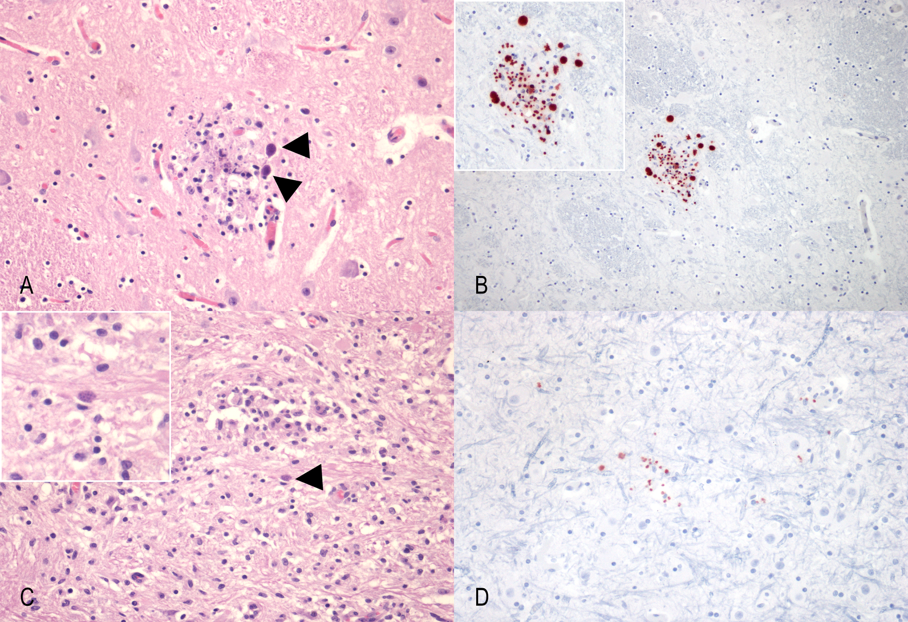
Apicomplexan parasitic elements (tachyzoites and tissue cysts) were histologically detected in 2 out of the 7 cases (cases 3 and 8; Table 2), both within and outside areas with tissue lesions. In both cases the thalamus was involved (Figure 2A, C). Tissue cysts presented as round to oval structures approximately 20 µm in diameter, with a thin cyst wall enclosing numerous 1–2 µm long oval bradyzoites. Tachyzoites were observed as extracellular, oval to crescent-shaped organisms measuring 1–2 µm within the neuroparenchyma. These parasitic elements showed strong immunoreactivity with a polyclonal anti-T. gondii antibody on immunohistochemistry (Figure 2B, D).
Histological and immunohistochemical findings in the brains of red fox (Vulpes vulpes) with lesions associated with Toxoplasma gondii elements. (A) Thalamus, focus of tissue damage characterized by cell debris (necrosis of the neuroparenchyma) and gliosis in association with tissue cysts (arrowheads) and extracellular tachyzoites (H&E; ×400); (B) Thalamus, same area as shown in (A), showing intralesional intensely immunolabelled T. Gondii cysts and extracellular tachyzoites (Anti-Toxoplasma IHC; Streptavidin and biotin; ×400); Inset, detail of the immunolabelled tissue cysts and extracellular tachyzoites (H&E; ×400); (C) Thalamus, large area of gliosis and mononuclear cell infiltration represented mainly by macrophages in association with a tissue cyst (arrowhead) (H&E; ×400); (D) Midbrain, immunolabelled tissue cysts and extracellular tachyzoites in an area of neuroparenchyma without inflammatory lesion (Anti-Toxoplasma IHC; ×400).

Details on the neurolocalization of Toxoplasma gondii parasitic elements in 2 red foxes (Vulpes vulpes) with confirmed T. Gondii-induced damage

Table 2 Long description
Neurolocalization results are listed for two red fox cases, indicating whether inflammatory lesions and parasite elements were detected across 11 brain regions. Case 3 is positive in prefrontal cortex, frontal cortex, basal ganglia, amygdala, hippocampus, midbrain, and thalamus, and negative in cingulate gyrus, pyriform lobe, cerebellum, and brainstem. Case 8 is negative in every region except the thalamus, which is positive. The thalamus is the only region positive in both cases. Overall, case 3 shows broader distribution across multiple forebrain and midbrain areas, whereas case 8 appears localized. Interpretation should note that the symbols distinguish inflammation from parasite detection methods, so a positive mark may reflect different underlying findings depending on the column notation.
+ = presence of the inflammatory lesion; – = absence of the inflammatory lesion.
* = presence of tachyzoites and/or tissue cysts in H&E.
§ = presence of tachyzoites and/or tissue cysts in IHC.
Abbreviations: Pc, Prefrontal cortex; Fc, Frontal cortex; Bg, Basal ganglia; Cg, Cingulate gyrus; T, Thalamus; A, Amygdala; Pl, Pyriform lobe; H, Hippocampus; M, Midbrain, C – cerebellum, B – Brainstem (Pons + Medulla oblongata).
Molecular testing of samples from all brain sections that displayed inflammatory changes was conducted using PCR, but only samples containing IHC-positive areas tested positive (n = 2).
Discussion
The study aimed to investigate the presence of Toxoplasma gondii and the anatomical distribution of T. gondii-associated lesions in the brain of naturally infected red foxes from central Italy and southeast Australia. Histopathological investigation of T. gondii-seropositive red foxes revealed mild to moderate, focal to multifocal inflammatory lesions, potentially consistent with toxoplasmic encephalitis, in 7 animals (17.1%, 7/41), with the detection of parasitic elements in 2 cases through both H&E staining and IHC.
The prevalent histological pattern of lesions associated with T. gondii elements was in agreement with what has been previously reported, with areas of tissue necrosis and macrophage infiltration as well as mononuclear perivascular cuffs (Mandara et al., Reference Mandara, Cantile, Baroni and Bernardini2011; Vandevelde et al., Reference Vandevelde, Higgins and Oevermann2012). Considering the limited severity of the observed lesions and the absence of extensive tissue destruction, it is safe to assume that acute toxoplasmosis was not fatal for any of the examined animals. Unfortunately, behavioural data from the collected animals were not available. However, neurolocalization of the lesions and parasitic elements was consistent with involvement of the limbic system, which is closely associated with the types of behavioural changes previously described as DFS by Milne et al. (Reference Milne, Fujimoto, Bean, Peters, Hemmington, Taylor, Fowkes, Martineau, Hamilton, Walker, Mitchell, Léger, Priestnall and Webster2020).
The heterogeneous distribution of lesions observed in this study mirrors findings from experimental murine models, in which tissue cysts were unevenly distributed throughout the brain and lacked a consistent neuroanatomical tropism, supporting the hypothesis that cerebral colonization by T. gondii is largely stochastic (Berenreiterová et al., Reference Berenreiterová, Flegr, Kuběna and Němec2011; Rodrigues Meurer et al., Reference Meurer, Brito, da Silva, Andade, Linhares, Pereira Junior, de Andrade-neto, de Sá and Oliveira2020). In contrast, other studies have proposed that the magnitude of neuroinflammation and overall cyst burden, rather than localization to specific brain regions, may be the principal drivers of parasite-associated behavioural alterations (Boillat et al., Reference Boillat, Hammoudi, Dogga, Pagès, Goubran, Rodriguez and Soldati-favre2020; Yin et al., Reference Yin, Xu, Zhao and Xie2022). Under this framework, behaviours previously interpreted as adaptive parasite manipulation aimed at facilitating trophic transmission (Berdoy et al., Reference Berdoy, Webster and Macdonald2000; Vyas et al., Reference Vyas, Kim, Giacomini, Boothroyd and Sapolsky2007) may instead reflect the broader consequences of cerebral inflammation affecting behaviourally relevant neural circuits. The lesions observed in the 7 cases were distributed across several functional brain areas, with specific involvement of the prefrontal and frontal cortex, the amygdala, the hippocampus, the basal ganglia and the thalamus. The rhombencephalon (including vestibulocerebellar system, cranial nerve nuclei and ascending reticular formation) seemed to be completely spared.
Although behavioural data was not available for the examined animals, the anatomical localization of the lesions observed in this study still allows for a functional interpretation of the potential neural systems affected by T. gondii-induced tissue damage. The prefrontal and/or frontal cortex were affected in at least 5 of the 7 animals examined. These regions are both part of the limbic system, which is associated with executive control, decision making and inhibitory behaviours (Noonan et al., Reference Noonan, Mars and Rushworth2011; Friedman and Robbins, Reference Friedman and Robbins2022). Tissue damage or inflammation in these areas can therefore be expected to influence behaviours associated with lack of self-control and inhibition, alter decision making patterns and potentially lead to the host taking riskier decisions and expressing bolder behaviours; this has been observed in another naturally infected canid species, the wolf (Canis lupus occidentalis) (Meyer et al., Reference Meyer, Cassidy, Stahler, Brandell, Anton, Stahler and Smith2022).
Equally noteworthy is the involvement of both the thalamus and basal ganglia in at least 2 of the foxes examined. These regions are responsible for sensory integration, emotional motivation, motor and postural control (Kropotov and Etlinger, Reference Kropotov and Etlinger1999; Utter and Basso, Reference Utter and Basso2008; Haber and Calzavara, Reference Haber and Calzavara2009). The basal ganglia are critically dependent on dopaminergic signalling (Utter and Basso, Reference Utter and Basso2008); therefore, infection of these nuclei by a pathogen capable of modulating dopamine synthesis and turnover (Gaskell et al., Reference Gaskell, Smith, Pinney, Westhead and McConkey2009; Prandovszky et al., Reference Prandovszky, Gaskell, Martin, Dubey, Webster and McConkey2011; McConkey et al., Reference McConkey, Martin, Bristow and Webster2013) may plausibly contribute to dysregulated dopaminergic neurotransmission. The thalamus, as the principal relay for sensory and limbic information to the cerebral cortex, regulates attention, consciousness and individual responses to emotions (Sherman and Guillery, Reference Sherman and Guillery2013; Halassa and Sherman, Reference Halassa and Sherman2019). Previous work on murine models (Berenreiterová et al., Reference Berenreiterová, Flegr, Kuběna and Němec2011; Boillat et al., Reference Boillat, Hammoudi, Dogga, Pagès, Goubran, Rodriguez and Soldati-favre2020) has shown that the thalamus and its associated centres are frequently areas of T. gondii tissue cyst accumulation. In the present study, the thalamus was the only region in which inflammatory lesions, immunohistochemically detectable parasites and PCR-confirmed T. gondii DNA co-occurred, suggesting that disruption of thalamic pathways may represent an important target of infection with potential functional consequences (Milne et al., Reference Milne, Fujimoto, Bean, Peters, Hemmington, Taylor, Fowkes, Martineau, Hamilton, Walker, Mitchell, Léger, Priestnall and Webster2020).
Inflammatory changes in the amygdala, hippocampus and midbrain were only observed in 1 animal. The amygdala is a crucial area involved in the processing of fear, threat evaluation and emotional learning, regulating behavioural responses to potential threats (LeDoux, Reference LeDoux2000; Janak and Tye, Reference Janak and Tye2015). Toxoplasma gondii tissue cysts have been repeatedly observed in the amygdala of experimentally infected rodents and their presence has been associated with the loss of innate aversion to feline odours (Vyas et al., Reference Vyas, Kim, Giacomini, Boothroyd and Sapolsky2007; Berenreiterová et al., Reference Berenreiterová, Flegr, Kuběna and Němec2011). Inflammation in the amygdala is known to project its influence to cortical and subcortical pathways amplifying emotional dysregulation and affecting decision-making (Tedford and McConkey, Reference Tedford and McConkey2017). The hippocampus, together with the amygdala, is central to learning, memory formation, spatial navigation and modulation of stress responses (O’Keefe and Nadel, Reference O’Keefe and Nadel1978; Fanselow and Dong, Reference Fanselow and Dong2010; Moser et al., Reference Moser, Rowland and Moser2015; Sheldon and Levine, Reference Sheldon and Levine2016). Experimental ablation studies in dogs have demonstrated that hippocampal damage impairs spatial memory in proportion to lesion extent (Kowalska, Reference Kowalska1995). It is therefore plausible that T. gondii–induced tissue damage in this region may impair spatial cognition and environmental awareness, potentially increasing the likelihood of risky behaviours and maladaptive responses to threats (Berenreiterová et al., Reference Berenreiterová, Flegr, Kuběna and Němec2011; Gering et al., Reference Gering, Laubach, Weber, Hussey, Turner, Lehmann, Montgomery, Holekamp and Getty2020).
The midbrain orients responses to both visual and auditory stimuli for innate defensive behaviours and for sensory and motor coordination (Redgrave et al., Reference Redgrave, Rodriguez, Smith, Rodriguez-oroz, Lehericy, Bergman, Agid, DeLong and Obeso2010; Hikosaka et al., Reference Hikosaka, Yasuda, Nakamura, Isoda, Kim, Terao, Amita and Maeda2019). Tissue damage disrupting midbrain circuits can impair the ability to respond to environmental threats or to modulate the responses to both fear and stress. Furthermore, T. gondii’s tropism for dopaminergic neurons was the focal point of the behavioural modification theory. By enhancing dopamine turnover, T. gondii could trigger fatal feline attraction in rodents and disinhibited behaviours in other animals, facilitating the connection of the parasite to its definitive host (Prandovszky et al., Reference Prandovszky, Gaskell, Martin, Dubey, Webster and McConkey2011; McConkey et al., Reference McConkey, Martin, Bristow and Webster2013; Milne et al., Reference Milne, Fujimoto, Bean, Peters, Hemmington, Taylor, Fowkes, Martineau, Hamilton, Walker, Mitchell, Léger, Priestnall and Webster2020).
No lesions or parasitic elements were detected in the cingulate gyrus, piriform lobe (with the exception of the amygdala), cerebellum, pons, or medulla oblongata. This distribution aligns with experimental findings in rodents, which reported lower cyst densities in the cerebellum, caudal brainstem and highly myelinated regions (Berenreiterová et al., Reference Berenreiterová, Flegr, Kuběna and Němec2011). These areas are characterized by dense white matter tracts, lower metabolic activity, reduced blood perfusion and limited immune cell trafficking, potentially restricting parasite invasion and persistence (Engelhardt and Sorokin, Reference Engelhardt and Sorokin2009; Owens et al., Reference Owens, Allen, Ondobaka and Friston2018). The relative sparing of the piriform lobe is less readily explained and may reflect species-specific neuroanatomical differences between carnivores and rodents (Miodoński, Reference Miodoński1974). However, interindividual variability and limited sample size must also be considered.
One of the examined foxes (Case ID: 3) presented with tissue damage but not parasitic elements in hippocampus and midbrain, another one (Case ID: 8) presented parasitic elements in IHC but not tissue damage in the same areas, highlighting the asynchronous dynamics of cerebral T. gondii infection. Similar scenarios were already reported (Ferguson and Hutchison, Reference Ferguson and Hutchison1987; Hunter and Sibley, Reference Hunter and Sibley2012) in relation to the acute and chronic stages of infection. Tissue necrosis is mostly caused by acute infection, in which tachyzoites replicate causing direct damage to the neuroparenchyma. An immune response with inflammatory infiltrates subsequently steps in to clear the necrotic debris and reacts to the tissue damage caused by the presence of parasitic elements. In contrast, the presence of tissue cysts with bradyzoites may reflect the chronic stage of infection, in which the parasite creates an immunologically silent environment in the host tissue and persists in its cystic stage without causing inflammation or necrosis (Dubey, Reference Dubey2021).
Despite the novel insights provided by this study, there are a number of limitations that warrant acknowledgement. First, although a relatively large number of fox brains was examined, only 7 animals exhibited histopathological lesions and only 2 showed direct evidence of parasitic elements, precluding statistical analysis and limiting inference regarding parasite tropism. This low detection rate may partly reflect sampling constraints inherent to large carnivore brains, in which lesions and cysts may be widely dispersed and therefore underrepresented in standard histological sections. Secondly, the cross-sectional and post-mortem nature of the study precluded behavioural assessment and limited interpretation to a single time point during infection. Thirdly, samples originated from 2 geographically distinct regions, and although circulating T. gondii genotypes are broadly comparable between Italy and Australia (Amouei et al., Reference Amouei, Sarvi, Sharif, Aghayan, Javidnia, Mizani, Moosazadeh, Shams, Hosseini, Hosseininejad, Nayeri Chegeni, Badali and Daryani2020; Hataminejad et al., Reference Hataminejad, Montazeri, Tanzifi and Galeh2025; Brustenga et al., Reference Brustenga, Liyanage, Loukopoulos, Fisher, Haining, Gauci, Lucentini, Uboldi, Tonkin, Morganti, Rigamonti, Passamonti, Veronesi, Jabbar and Hufschmid2025b), differences in exposure ecology and host–parasite interactions may have influenced lesion prevalence and distribution.
Future studies integrating larger sample sizes, longitudinal designs, behavioural observations and parasite genotyping will be essential to further elucidate the neuropathological basis of DFS and the broader behavioural consequences of T. gondii infection in wild carnivores.
Conclusions
The study provides the first spatial characterization of Toxoplasma gondii infection within the brain of naturally infected red foxes, offering an anatomical framework to evaluate existing hypotheses on parasite-induced behavioural changes. Inflammatory lesions, tissue cysts and free tachyzoites were detected in behaviourally relevant brain regions, including the prefrontal cortex, the thalamus, the hippocampus and the basal ganglia. No selective tropism for certain brain area was detected, corroborating the findings of experimental infections carried out on murine models. Nevertheless, the localization of T. gondii–associated lesions within these areas supports a potential link between parasite-induced neuroinflammation and the behavioural alterations described as DFS. While causality cannot be established, these findings advance understanding of the neuropathological substrates through which T. gondii may influence host behaviour in wild carnivores and generates testable predictions for future studies integrating neuropathology with ethological observations.
Data availability statement
All data generated and analysed in this study is provided in the manuscript.
Acknowledgements
The authors would like to express their most sincere acknowledgements to all the staff of WildUmbria wildlife rescue centre, the Veterinary Teaching Hospital of Perugia University and the Experimental Zooprophylactic Institute of Umbria and Marche. Furthermore, this work could have never been carried out without the extensive help of local trappers and shooters active in Victoria as well as Fields and Games Australia who were invaluable for the collection of Australian fox carcasses.
Author contributions
L.B.: conceptualization, sampling, lab work, data curation, manuscript preparation, manuscript review and editing; G.G.: conceptualization, sampling, lab work, data curation, manuscript review and editing; KLDTDL: sampling, lab work, data curation, manuscript review and editing; G.R.: sampling, lab work, data curation, manuscript review and editing; P.L.: sampling, manuscript review and editing; M.F.: sampling, manuscript review and editing; Je.Ha.: sampling, manuscript review and editing; L.L.: resources, validation, manuscript review and editing; G.M.: validation, manuscript review and editing; I.M.: lab work, manuscript review and editing; M.D.: validation, manuscript review and editing; N.D.: sampling, manuscript review and editing; M.G.: sampling, manuscript review and editing; A.R.: sampling, manuscript review and editing; F.P.: validation, supervision, manuscript review and editing; A.J.: resources, supervision, manuscript review and editing; Ja.Hu.: resources, supervision, manuscript review and editing; M.T.M.: conceptualization, resources, supervision, manuscript review and editing; F.V.: conceptualization, resources, supervision, manuscript review and editing.
Financial support
This research received no specific funding.
Competing interests
The authors declare no competing interests.
Ethical standards
All animals sampled were dead for causes independent to this study, therefore no ethical authorization was required to perform the analyses.


 Open access

