To save content items to your account,
please confirm that you agree to abide by our usage policies.
If this is the first time you use this feature, you will be asked to authorise Cambridge Core to connect with your account.
Find out more about saving content to .
To save content items to your Kindle, first ensure no-reply@cambridge.org
is added to your Approved Personal Document E-mail List under your Personal Document Settings
on the Manage Your Content and Devices page of your Amazon account. Then enter the ‘name’ part
of your Kindle email address below.
Find out more about saving to your Kindle.
Note you can select to save to either the @free.kindle.com or @kindle.com variations.
‘@free.kindle.com’ emails are free but can only be saved to your device when it is connected to wi-fi.
‘@kindle.com’ emails can be delivered even when you are not connected to wi-fi, but note that service fees apply.
The significance of the use of floral diagrams is discussed in this chapter, including different types as well as its advantages and limitations. Floral diagrams can catch the diversity of flowers, including accessory structures and floral heteromorphism. The importance of floral development, the drive of pollination and evolutionary developmental genetics is shown in the shaping of floral structures and as a reflection of floral evolution.
Floral diagrams are presented for fourteen families out of six orders of the basal groups of angiosperms. The floral diagrams of the basal angiosperms illustrate the early progression of flower diversification. Starting with the most basal angiosperms, different trends in flower evolution are shown through diagrams, illustrating the transition of spiral to whorled trimerous flowers, incipient transformations of floral organs and accessory structures, and the progressive fusion of organs. The major orders are presented with representatives of the most important families and their trends, including Laurales, Magnoliales and Piperales.
Floral diagrams are presented for twenty-eight families out of ten orders of monocots, covering the diversity and evolutionary trends within this largely homogeneous group. The monocots are basically defined by their trimerous Bauplan, regulating the structure of the flower as well as its evolution. The basalmost orders demonstrate greater variation with higher or lower stamen numbers, while higher groups have evolved within the constraint of a pentacyclic arrangement that has changed very little. Variations in flower morphology as adaptations to different pollination systems are regulated by hypanthial growth, reductions of organs or stamen increases, and monosymmetry.
The Pentapetalae have diversified very rapidly, leading to two major successful clades, the superrosidae and superasteridae. Although it is still uncertain how the Pentapetalae diverged from ancestral flowers, their regular floral pentacyclic and pentamerous Bauplan is almost unversal and firmly established in all evolutionary lines. The possibility is presented that intermediates such as Berberidopsis regulate the transition from a spiral to a pentamerous pentacyclic flower. The diversity and unique evolutionary trends of early diverging orders with unclear affinity, Dilleniales and Santalales, are presented.
Floral diagrams are presented for thirty-six families out of nine orders of SupperAsteridae. Asterids represent the pinacle in Pentapetalae floral evolution, expressing a strong synorganization between whorls and the stabilization of a tetracyclic Bauplan with five sepals, five petals, a single whorl of stamens (haplostemony) and two carpels. The lower Asterids share characters of basal Pentapetalae and rosids and present all characteristics of a transition to euasterids. Diferent trends are higlighted in lamiids and campanulids. The development of a stamen-petal tube emerges in the basal Asterids and is prominent in the lamiids in association with median monosymmetry. An important evolutionary trend representing the sterilisation of stamens in Lamiales is shown through floral diagrams. In Campanulids there is a shift towards the reduction of the stamen-petal tube as well as the reduction of the calyx linked with an inferior ovary and a lower number of ovules.
This chapter represents a summary of the major taxonomic groups and their floral evolution. The value of floral diagrams is highlighted as a means to stress similarities and differences between the major angiosperm groups.
Floral diagrams are presented for twenty-one families of the highly diverse orders caryophyllales and polygonales. The caryophyllids represent a small but important clade close to the asterids. The flowers show important evolutionary trends linked with a progressive loss of petals, and their reinvention of petal-like structures in derived groups, such as coloured sepals or staminodes. Stamen evolution also shows unique trends linked with spatial constraints and secondary multiplications. The ovary shows a general tendency to the development of central placentas and the reduction in the number of ovules. Floral diagrams are used to illustrate the major evolutionary trends within caryophyllales and specifically within the diverse family Polygonaceae, in addition to specific diagrams representative of families.
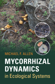
Mycorrhizae are mutualisms between plants and fungi that evolved over 400 million years ago. This symbiotic relationship commenced with land invasion, and as new groups evolved, new organisms developed with varying adaptations to changing conditions. Based on the author's 50 years of knowledge and research, this book characterizes mycorrhizae through the most rapid global environmental changes in human history. It applies that knowledge in many different scenarios, from restoring strip mines in Wyoming and shifting agriculture in the Yucatán, to integrating mutualisms into science policy in California and Washington, D.C. Toggling between ecological theory and natural history of a widespread and long-lived symbiotic relationship, this interdisciplinary volume scales from structure-function and biochemistry to ecosystem dynamics and global change. This remarkable study is of interest to a wide range of students, researchers, and land-use managers.
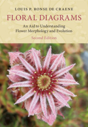
Floral morphology is key for understanding floral evolution and plant identification. Floral diagrams are two-dimensional representations of flowers that replace extensive descriptions or elaborate drawings to convey information in a clear and unbiased way. Following the same outline as the first edition, this comprehensive guide includes updated and relevant literature, represents the latest phylogeny, and features 28 new diagrams. Diagrams are presented in the context of the most recent classifications, covering a variety of families and illustrating the floral diversity of major groups of plants. A strong didactic tool for observing and understanding floral structures, these diagrams are the obvious counterpart to any genetic study in flowering plants and to the discussion of major adaptations and evolutionary trends of flowers. This book is invaluable for researchers and students working on plant structure, development and systematics, as well as being an important resource for plant ecologists, evolutionary botanists and horticulturists.
Third-generation long-read sequencing is transforming plant genomics. Oxford Nanopore Technologies and Pacific Biosciences are offering competing long-read sequencing technologies and enable plant scientists to investigate even large and complex plant genomes. Sequencing projects can be conducted by single research groups and sequences of smaller plant genomes can be completed within days. This also resulted in an increased investigation of genomes from multiple species in large scale to address fundamental questions associated with the origin and evolution of land plants. Increased accessibility of sequencing devices and user-friendly software allows more researchers to get involved in genomics. Current challenges are accurately resolving diploid or polyploid genome sequences and better accounting for the intra-specific diversity by switching from the use of single reference genome sequences to a pangenome graph.
Stomata are cellular pores on the leaf epidermis that allow plants to regulate carbon assimilation and water loss. Stomata integrate environmental signals to regulate pore apertures and adapt gas exchange to fluctuating conditions. Here, we quantified intraspecific plasticity of stomatal gas exchange and anatomy in response to seasonal variation in Brachypodium distachyon. Over the course of 2 years, we (a) used infrared gas analysis to assess light response kinetics of 120 Bd21-3 wild-type individuals in an environmentally fluctuating greenhouse and (b) microscopically determined the seasonal variability of stomatal anatomy in a subset of these plants. We observed systemic environmental effects on gas exchange measurements and remarkable intraspecific plasticity of stomatal anatomical traits. To reliably link anatomical variation to gas exchange, we adjusted anatomical gsmax calculations for grass stomatal morphology. We propose that systemic effects and variability in stomatal anatomy should be accounted for in long-term gas exchange studies.
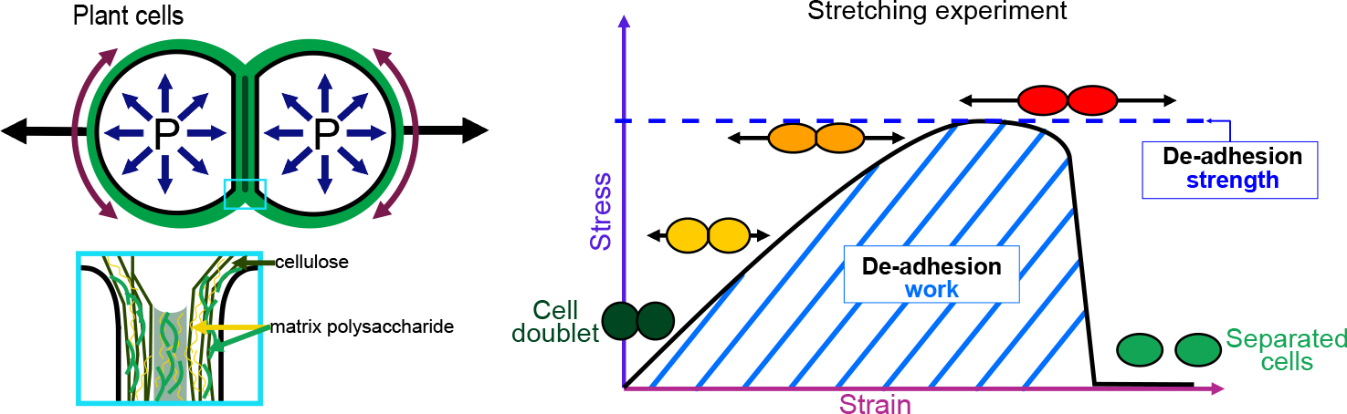
Cell–cell adhesion is a fundamental feature of multicellular organisms. To ensure multicellular integrity, adhesion needs to be tightly controlled and maintained. In plants, cell–cell adhesion remains poorly understood. Here, we argue that to be able to understand how cell–cell adhesion works in plants, we need to understand and quantitatively measure the mechanics behind it. We first introduce cell–cell adhesion in the context of multicellularity, briefly explain the notions of adhesion strength, work and energy and present the current knowledge concerning the mechanisms of cell–cell adhesion in plants. Because still relatively little is known in plants, we then turn to animals, but also algae, bacteria, yeast and fungi, and examine how adhesion works and how it can be quantitatively measured in these systems. From this, we explore how the mechanics of cell adhesion could be quantitatively characterised in plants, opening future perspectives for understanding plant multicellularity.
In, essentially, all species where meiotic crossovers (COs) have been studied, they occur preferentially in open chromatin, typically near gene promoters and to a lesser extent, at the end of genes. Here, in the case of Arabidopsis thaliana, we unveil further trends arising when one considers contextual information, namely summarised epigenetic status, gene or intergenic region size, and degree of divergence between homologs. For instance, we find that intergenic recombination rate is reduced if those regions are less than 1.5 kb in size. Furthermore, we propose that the presence of single nucleotide polymorphisms enhances the rate of CO formation compared to when homologous sequences are identical, in agreement with previous works comparing rates in adjacent homozygous and heterozygous blocks. Lastly, by integrating these different effects, we produce a quantitative and predictive model of the recombination landscape that reproduces much of the experimental variation.
Cell division is a tightly regulated mechanism, notably in tissues where malfunctions can lead to tumour formation or developmental defects. This is particularly true in land plants, where cells cannot relocate and therefore cytokinesis determines tissue topology. In plants, cell division is executed in radically different manners than in animals, with the appearance of new structures and the disappearance of ancestral mechanisms. Whilst F-actin and microtubules closely co-exist, recent studies mainly focused on the involvement of microtubules in this key process. Here, we used a root tracking system to image the spatio-temporal dynamics of both F-actin reporters and cell division markers in dividing cells embedded in their tissues. In addition to the F-actin accumulation at the phragmoplast, we observed and quantified a dynamic apico-basal enrichment of F-actin from the prophase/metaphase transition until the end of the cytokinesis.
Studies on the mechanics of plant cells usually focus on understanding the effects of turgor pressure and properties of the cell wall (CW). While the functional roles of the underlying cytoskeleton have been studied, the extent to which it contributes to the mechanical properties of cells is not elucidated. Here, we study the contributions of the CW, microtubules (MTs) and actin filaments (AFs), in the mechanical properties of Nicotiana tabacum cells. We use a multiscale biomechanical assay comprised of atomic force microscopy and micro-indentation in solutions that (i) remove MTs and AFs and (ii) alter osmotic pressures in the cells. To compare measurements obtained by the two mechanical tests, we develop two generative statistical models to describe the cell’s behaviour using one or both datasets. Our results illustrate that MTs and AFs contribute significantly to cell stiffness and dissipated energy, while confirming the dominant role of turgor pressure.