To save content items to your account,
please confirm that you agree to abide by our usage policies.
If this is the first time you use this feature, you will be asked to authorise Cambridge Core to connect with your account.
Find out more about saving content to .
To save content items to your Kindle, first ensure no-reply@cambridge.org
is added to your Approved Personal Document E-mail List under your Personal Document Settings
on the Manage Your Content and Devices page of your Amazon account. Then enter the ‘name’ part
of your Kindle email address below.
Find out more about saving to your Kindle.
Note you can select to save to either the @free.kindle.com or @kindle.com variations.
‘@free.kindle.com’ emails are free but can only be saved to your device when it is connected to wi-fi.
‘@kindle.com’ emails can be delivered even when you are not connected to wi-fi, but note that service fees apply.
Stem cell fates are spatio-temporally regulated during plant development. Time-lapse imaging of fluorescence reporters is the most widely used method for spatio-temporal analysis of biological processes. However, excitation light for imaging fluorescence reporters causes autofluorescence and photobleaching. Unlike fluorescence reporters, luminescence proteins do not require excitation light, and therefore offer an alternative reporter for long-term and quantitative spatio-temporal analysis. We established an imaging system for luciferase, which enabled monitoring cell fate marker dynamics during vascular development in a vascular cell induction system called VISUAL. Single cells expressing the cambium marker, proAtHB8:ELUC, had sharp luminescence peaks at different time points. Furthermore, dual-color luminescence imaging revealed spatio-temporal relationships between cells that differentiated into xylem or phloem, and cells that transitioned from procambium to cambium. This imaging system enables not only the detection of temporal gene expression, but also facilitates monitoring of spatio-temporal dynamics of cell identity transitions at the single cell level.
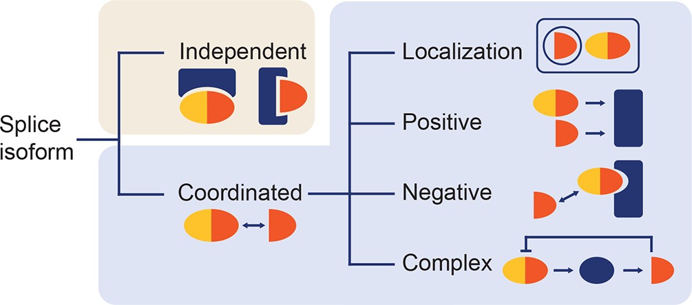
Most plant primary transcripts undergo alternative splicing (AS), and its impact on protein diversity is a subject of intensive investigation. Several studies have uncovered various mechanisms of how particular protein splice isoforms operate. However, the common principles behind the AS effects on protein function in plants have rarely been surveyed. Here, on the selected examples, we highlight diverse tissue expression patterns, subcellular localization, enzymatic activities, abilities to bind other molecules and other relevant features. We describe how the protein isoforms mutually interact to underline their intriguing roles in altering the functionality of protein complexes. Moreover, we also discuss the known cases when these interactions have been placed inside the autoregulatory loops. This review is particularly intended for plant cell and developmental biologists who would like to gain inspiration on how the splice variants encoded by their genes of interest may coordinately work.
Phyllotaxis, the regular arrangement of plant lateral organs, is an important aspect of quantitative plant biology. Some models relying on the geometric relationship of the shoot apex and organ primordia focus mainly on spiral phyllotaxis, a common phyllotaxis mode. While these models often predict the dependency of Fibonacci spirals on the Golden Angle, other models do not emphasise such a relation. Phyllotactic patterning in Asteraceae is one such example. Recently, it was revealed that auxin dynamics and the expansion and contraction of the active ring of the capitulum (head) are the key processes to guide Fibonacci spirals in gerbera (Gerbera hybrida). In this Insights paper, we discuss the importance of auxin dynamics, distinct phases of phyllotactic patterning, and the transition of phyllotaxis modes. These findings signify the local interaction among primordia in phyllotactic patterning and the notion that Fibonacci spirals may not need the Golden Angle.
Quantitative analysis of experimental metabolic data is frequently challenged by non-intuitive, complex patterns which emerge from regulatory networks. The complex output of metabolic regulation can be summarised by metabolic functions which comprise information about dynamics of metabolite concentrations. In a system of ordinary differential equations, metabolic functions reflect the sum of biochemical reactions which affect a metabolite concentration, and their integration over time reveals metabolite concentrations. Further, derivatives of metabolic functions provide essential information about system dynamics and elasticities. Here, invertase-driven sucrose hydrolysis was simulated in kinetic models on a cellular and subcellular level. Both Jacobian and Hessian matrices of metabolic functions were derived for quantitative analysis of kinetic regulation of sucrose metabolism. Model simulations suggest that transport of sucrose into the vacuole represents a central regulatory element in plant metabolism during cold acclimation which preserves control of metabolic functions and limits feedback-inhibition of cytosolic invertases by elevated hexose concentrations.
The ability of plants to absorb CO2 for photosynthesis and transport water from root to shoot depends on the reversible swelling of guard cells that open stomatal pores in the epidermis. Despite decades of experimental and theoretical work, the biomechanical drivers of stomatal opening and closure are still not clearly defined. We combined mechanical principles with a growing body of knowledge concerning water flux across the plant cell membrane and the biomechanical properties of plant cell walls to quantitatively test the long-standing hypothesis that increasing turgor pressure resulting from water uptake drives guard cell expansion during stomatal opening. To test the alternative hypothesis that water influx is the main motive force underlying guard cell expansion, we developed a system dynamics model accounting for water influx. This approach connects stomatal kinetics to whole plant physiology by including values for water flux arising from water status in the plant .
Biomechanical properties of the cell wall (CW) are important for many developmental and adaptive responses in plants. Expansins were shown to mediate pH-dependent CW enlargement via a process called CW loosening. Here, we provide a brief overview of expansin occurrence in plant and non-plant species, their structure and mode of action including the role of hormone-regulated CW acidification in the control of expansin activity. We depict the historical as well as recent CW models, discuss the role of expansins in the CW biomechanics and address the developmental importance of expansin-regulated CW loosening in cell elongation and new primordia formation. We summarise the data published so far on the role of expansins in the abiotic stress response as well as the rather scarce evidence and hypotheses on the possible mechanisms underlying expansin-mediated abiotic stress resistance. Finally, we wrap it up by highlighting possible future directions in expansin research.
The ability of plants to sense and orient their root growth towards gravity is studied in many laboratories. It is known that manual analysis of image data is subjected to human bias. Several semi-automated tools are available for analysing images from flatbed scanners, but there is no solution to automatically measure root bending angle over time for vertical-stage microscopy images. To address these problems, we developed ACORBA, which is an automated software that can measure root bending angle over time from vertical-stage microscope and flatbed scanner images. ACORBA also has a semi-automated mode for camera or stereomicroscope images. It represents a flexible approach based on both traditional image processing and deep machine learning segmentation to measure root angle progression over time. As the software is automated, it limits human interactions and is reproducible. ACORBA will support the plant biologist community by reducing labour and increasing reproducibility of image analysis of root gravitropism.
A community has a diverse suite of definitions, as many as there are scientific fields and sociological units. Among humans, there is a place-based variant such as a city defined by political boundaries, or an interest-based variant focused on a group of people defined by their interactions. The same issues plague the field of community ecology, especially when we address a topic such as a mycorrhiza, which is a functional relationship. Early measurements of communities looked for spatial boundaries, the edge of a meadow; a shift in forest type with a physical edge such as a shift in topography; an obvious shift in plant types such as a forest edge. Theophrastus (~44 BCE) noted that most mushrooms were found in forests (which included EM species) but not grasslands (which were almost exclusively AM). Alexander von Humbolt drove some of the earliest developments in community and ecosystem ecology by showing relationships between climate and vegetation both up elevation gradients, such as his beautiful and accurate 1807 Tableau Physique, drawings of vegetation up Mount Chimborazo in Ecuador, and his Geographical Distribution of Plants – climate consortia of plants – in relation to global climate patterns (371).
The definition of symbiosis is two organisms living intimately together, and this chapter examines the physiological basis of the interaction. A mycorrhiza is comprised of two distinctly different organisms, a plant and a fungus, that interface down to the molecular level. Because of this intimate physical closeness, the biochemistry, physiology, and ecology become highly intertwined. At the most basic definitional level, the fungus picks up nutrients and water in the soil, transfers those resources to the host, in exchange for carbon fixed by the plant from the atmosphere. This physical dimension means that resources available to one partner are less available to the other. But both sets of resources are essential to both organisms.
The study of population ecology in plants is as old as the field of ecology but is more complex for fungi. Due to their microscopic morphology, identifying individuals for measuring and modeling is challenging. We have examined the general morphology of fungi and plants comprising the mycorrhizal symbiosis, and we have looked at the larger-scale evolutionary patterns that resulted in the mycorrhizae that we observe and study today. However, selection acts on the individual organism (472). An organism survives to reproduce offspring that in turn reproduce, or it does not: a binary outcome. And, an organism is comprised of a complete genetic code that allows it to survive to reproduction (or not).
Probably no research topic in mycorrhizae has undergone as much change over the past few decades as the evolution of the symbiosis. The rapid development of techniques and reduction in costs of sequencing, increase in databases and new approaches to sequence database management, data mining, and sequencing analyses has generated a plethora of new phylogenic reorganization, molecular clocks, and theory. Newer sequencing concepts often readily integrate with the fossil record as the field of paleoecology itself rapidly evolves. But for understanding the mechanisms of evolution in a symbiosis, we need to go beyond phylogenetic relationships to understanding both the role of and the shifts in environments that determine how mycorrhizae develop, adapt, and diversify. Here I will summarize the key topic areas relating to mycorrhizal symbiosis, recognizing that there are likely many ideas that will change in the near future. Specifically, I address the hypotheses that: (1) mycorrhizae were crucial to the invasion of land and related to the regulation of atmospheric CO2, (2) mycorrhizal symbioses are fundamentally stable, and (3) there are both genetic and ecological underpinnings supporting the mycorrhizal symbiosis. Here I explore the four lines of evidence of how evolution has played a key role in the ecology of modern mycorrhizae (36), including (1) paleobiology evidence, (2) extant plant mycorrhizal status, (3) the molecular basis of interaction, and (4) models of mutualism.
Travel to your nearest nature reserve. It could be anywhere, within a human habitation or wildland, on continents or islands around the globe. It could be a forest, shrubland, grassland, forbland, or even desert. Across your view is a diverse array of plants, complete with a complex architecture with several levels from the ground surface to the highest leaf, be it a redwood tree extending hundreds of meters into the sky, or a diminutive, but still complex, structure only 10 cm high of mosses, liverworts, and cryptogams. As you separate the stems and look down, you see roots: tens of meters of roots or rhizoids for every square meter of the soil surface. Take your hand lens and pull up some of those roots. There will be soil hanging from those roots, held together by tens to hundreds of meters of threads of fungal hyphae per cubic centimeter. The vast majority of these hyphae form mycelia from many species of fungi, directly connecting fine roots to the larger soil matrix, serving as a living pathway interconnecting the plant, which is actively fixing carbon, within the micro patches of nutrients and water necessary to fix that carbon.
From the beginning of mycorrhizal research, understanding of functioning was based on the morphological structure of the plant–fungus interface. The structure of the fungal hyphae within the root, which regulates the exchange of resources between plant and fungus, and the extramatrical hyphae, the extent and locations of which dictate the ultimate flows of resources between soil, hyphae, and root, characterizes a mycorrhiza, determines the type of mycorrhiza, and, to a large extent, determines the functioning of that mycorrhiza (255). Overall, knowing these two structural components, internal root colonization and extramatrical hyphae, allows us to make a number of predictions about the mechanisms and quantity of resource exchange between the two symbionts.
During the nineteenth century, the Selkirk Settlement of Canada and the Homestead Act in the United States led to some of the most dramatic and widespread destruction of native ecosystems across a short time period in history. Soils across the Great Plains, from the Mississippi River to the Rocky Mountains, from the Chihuahuan desert of Mexico to the Boreal Forests of Canada, an area of around 4 million kilometers squared, were nearly all turned over for agriculture, from prairies with dense grasslands to riparian regions with extensive forest cover and deep roots, within the decades from approximately 1820 to 1890. By the 1890s, a protracted drought led to a collapse of agriculture in the United States, Canada, the Ukraine (the Selk’nam genocide), and elsewhere. The young field of ecology was just beginning, documenting the community ecology of recovery at lakeshores (182), glaciers (175), and from the abandonment of highly disturbed agricultural lands (170).