To save content items to your account,
please confirm that you agree to abide by our usage policies.
If this is the first time you use this feature, you will be asked to authorise Cambridge Core to connect with your account.
Find out more about saving content to .
To save content items to your Kindle, first ensure no-reply@cambridge.org
is added to your Approved Personal Document E-mail List under your Personal Document Settings
on the Manage Your Content and Devices page of your Amazon account. Then enter the ‘name’ part
of your Kindle email address below.
Find out more about saving to your Kindle.
Note you can select to save to either the @free.kindle.com or @kindle.com variations.
‘@free.kindle.com’ emails are free but can only be saved to your device when it is connected to wi-fi.
‘@kindle.com’ emails can be delivered even when you are not connected to wi-fi, but note that service fees apply.
This chapter reviews the types of shunt complications, their likelihood, and some clinical examples. Cerebrospinal fluid (CSF) shunting and particularly ventriculoperitoneal shunting is a common procedure used to treat a variety of CSF flow dynamic pathologies including obstructive, communicating, and normal pressure hydrocephalus (NPH) from a variety of etiologies. There are several types of specific long-term complications associated with shunting procedures in addition to those more likely seen in the NPH population in particular. These include underdrainage of CSF, overdrainage of CSF, mechanical failures of the hardware such as alterations in valve resistance over time, malposition of the tubing, and frank breakage, CSF leak, subdural hemorrhage or hygroma, seizure, and infection. Underdrainage conditions arise when shunt systems develop increased resistance across the valve over time, disconnection or kinking of the system components, or migration of the tubing out of an appropriate position for proper drainage and/or reabsorption.
This chapter describes many of the techniques and protocols that can be used to minimize the risk of shunt failure, focusing on the surgical procedure at each anatomic location for proximal and distal catheter placement. It includes the extended discussions of ventricular and lumbar proximal catheters, and peritoneal, atrial, pleural, and other distal catheters. Valves can be divided into four different categories: differential pressure valves, flow-regulated valves, antisiphon valves, and adjustable valves. Antisiphon valves are used to prevent the siphoning of cerebrospinal fluid (CSF) into the distal compartment. Antisiphon devices (ASDs) are primarily used in management of normal pressure hydrocephalus (NPH) patients in the adult population to prevent the formation of subdural hematomas and hygromas, and in the management of hydrocephalus in the pediatric population to prevent the slit ventricle syndrome and proximal catheter obstruction.
Genetic studies in animal models have started to open new ways for understanding the underlying molecular pathophysiology of hydrocephalus. Human hydrocephalus can be classified as syndromic versus non-syndromic, and congenital versus acquired. Comparative twin studies have been performed to analyze the genetic influences in congenital structural defects including hydrocephalus. Familial hydrocephalus has long been suggested as a heritable disease, with heterogeneous causes, which may result from distinct monogenic or multifactorial disorders. Congenital hydrocephalus (CHC) is usually the consequence of deficient brain development and perturbed cellular function, implicating the important roles that CHC genes play during brain development. The majority of identified hydrocephalus loci and genes are from genetic analysis in hydrocephalic animal models. The pathophysiology of hydrocephalus in the ventricular system has been extensively studied through either down- or up-regulation of certain targeted gene expression, followed by comparative morphological and molecular studies.
Cerebrospinal fluid (CSF) plays a role in homeostatic hormonal signaling, chemical buffering, circulations of nutrients, and neurodevelopment. The two lateral ventricles drain into the third ventricle through the foramina of Monro. The third ventricle subsequently drains into the fourth ventricle through the narrowest portion of the ventricular system called the cerebral aqueduct. The roof of the fourth ventricle is bounded by the vermis of the cerebellum and the fastigium. CSF production is to a degree dependent on blood perfusion. In instances of increased intracranial pressure (ICP) with subsequent decreased cerebral perfusion, there will be a decrease in CSF production. CSF flows through net bulk flow from the lateral ventricles through the foramen of Monro into the third ventricle, then on into the fourth ventricle through the cerebral aqueduct. The pathways of CSF reabsorption include the ependymal layer of the ventricular system, and endothelial layer of the brain parenchyma.
Structural and functional brain imaging have helped to elucidate the neural pathways involved in hydrocephalic cognitive impairment. In addition, studies of brain metabolism and blood flow, molecular imaging, and cerebrospinal fluid (CSF) physiology have provided novel windows into the pathogenesis of dementia in idiopathic normal pressure hydrocephalus (iNPH). A number of pathophysiologic mechanisms have been identified that are potentially relevant to the pathogenesis of the cognitive symptoms of iNPH, namely, mechanical distortion, pressure effects, and cerebrovascular compromise. A possible synthesis of these mechanisms would be that an imbalance of CSF production and clearance leads to progressive ventricular enlargement. The profile of cognitive impairments in iNPH is recognizably that of a subcortical pathological process. Deficiencies in attention, working memory, set shifting, response inhibition and other aspects of executive functioning are commonly observed in iNPH and can be seen early in the disease course.
This chapter first focuses on the ethics of animal models and then discusses the formal requirements general to any experimental model and the specific requirements for models of hydrocephalus. It also discusses the recent and current research areas in experimental hydrocephalus. Ethical standards for experimental studies involving animals are legally set by laws and regulations. Animal models resemble human disease by conditions which are genetically determined, naturally acquired, or induced by the investigator. Shunted animal models provide a unique potential for insight into questions concerning the destructive effects of hydrocephalus on the brain and its development, as it is rarely possible to obtain brain tissue from humans with hydrocephalus. Most of the hydrocephalus animal models are neonatal or juvenile animals and the majority of research is thus directed at congenital or pediatric hydrocephalus.
Lack of expertise in fixation of these fractures does not equate to the fracture being unreconstructable. Refer to a more experienced surgeon for fixation if necessary. The results of TEA are better than ORIF in the elderly. The results of revision of a failed fixation to a TEA are inferior to a primary TEA.
Contraindications
Infection.
Lack of soft tissue coverage.
Age under 70 years.
Non-compliant patient.
Neurological injury afecting the elbow lexors.
Preoperative planning
Clinical assessment
Full history and examination. Document status of radial, ulnar, median and posterior interosseous nerves individually.
Exclude associated injuries.
Radiological assessment
Plain radiographs of the elbow: anteroposterior and lateral views (Fig. 4.1.1).
A CT scan is oten very helpful to deine the pattern of fracture and the extent of intra-articular comminution.
Up to 2 cm of humeral shat bone loss can be addressed using standard implants. Greater than 2 cm of bone loss requires the use of implants with extra-long anterior flanges.
Determine the humeral and ulnar canal diameters to conirm that they can accommodate the implants.
Preoperative consent
Obtain informed consent from the patient, including but not limited to risks, beneits, alternatives, complications and potential outcome.
Associated injury to the brachial plexus and/or subclavian artery.
Floating shoulder.
Bilateral fractures.
Multiple-injured patient.
Soft tissue interposition between the fragments.
Impending skin necrosis or penetration from a prominent fragment.
Consideration for surgical fixation should be given to completely displaced, comminuted midshaft fractures that contain a transverse fragment (Z-shaped fractures). There is evidence in the current orthopaedic literature that these fractures have a high risk of non-union.
Operative management is indicated in displaced type III, IV and V distal clavicle fractures according to the Neer classification.
Clinical assessment
Mechanism of injury: motor vehicle collision, sports injury, fall on outstretched hand (FOOSH), direct trauma.
This chapter attempts to develop biomarkers specific for idiopathic normal pressure hydrocephalus (iNPH) to distinguish it from other neurodegenerative conditions. The case for emerging biomarkers in iNPH has arisen because of similar developments in other common causes of dementia and the increasing awareness of both the epidemiology of NPH and its impact on the quality of life of elderly patients. Changes in the neurochemical composition of cerebrospinal fluid (CSF) due to hydrocephalus have been widely documented. The problem with identifying diagnostic biomarkers between NPH and Alzheimer's disease (AD) is that NPH was used in those studies more as a comparative control group rather than as the primary studied group, apart from a few exceptions. The difficulty in the differential diagnosis of PD with patients with NPH lies in the features of the gait disorder. There have been much fewer studies developing prognosis biomarkers when compared with those assessing diagnostic biomarkers.
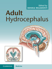
Adult hydrocephalus is an insidious yet treatable condition that develops slowly, with usual onset around 60 years of age. It is poorly recognized and many cases are not diagnosed until late in the course of disease, leading to poorer patient outcomes and a high financial cost to healthcare providers. The resulting neurological symptoms include gait/balance problems, loss of bladder control, and a cognitive decline leading to dementia, which is often mistaken for Alzheimer's disease. This book - the first published on this topic since 1993 - provides comprehensive guidelines to improve the speed and accuracy of diagnosis, and covers various neurosurgical techniques used to treat the disease, including the insertion of different types of shunts and endoscopic third ventriculostomy. This is essential reading for neurologists, neurosurgeons, family physicians, and radiologists who may well encounter adult patients with hydrocephalus more often than they realize.
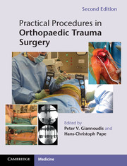
This highly illustrated textbook is an essential guide for surgeons in training, providing step-by-step approaches to performing surgical procedures. Practical guidance is given on patient positioning, approach and reduction techniques, which implant to insert, protocols for postoperative mobilisation, possible complications, when the patient should be seen in the outpatient clinic, and whether the implant should be removed. This second edition has been extensively expanded and revised. Additional chapters address fractures of the scapula, fractures around the elbow, around the foot and minimal invasive plate osteosynthesis procedures. All of the other chapters have been expanded and revised to comprehensively cover the range of common trauma procedures performed by surgeons in training. Written by experts in orthopaedics and trauma, the book includes numerous intraoperative colour photographs to help readers visualise the techniques described. This will be an invaluable resource for all surgical trainees in orthopaedics, trauma, and general disciplines.