Refine search
Actions for selected content:
106117 results in Materials Science
Introduction
-
- Journal:
- Journal of Materials Research / Volume 30 / Issue 1 / 14 January 2015
- Published online by Cambridge University Press:
- 15 January 2015, p. 1
- Print publication:
- 14 January 2015
-
- Article
- Export citation
JMR volume 30 issue 1 Cover and Back matter
-
- Journal:
- Journal of Materials Research / Volume 30 / Issue 1 / 14 January 2015
- Published online by Cambridge University Press:
- 15 January 2015, pp. b1-b4
- Print publication:
- 14 January 2015
-
- Article
-
- You have access
- Export citation
JMR volume 30 issue 1 Cover and Front matter
-
- Journal:
- Journal of Materials Research / Volume 30 / Issue 1 / 14 January 2015
- Published online by Cambridge University Press:
- 15 January 2015, pp. f1-f5
- Print publication:
- 14 January 2015
-
- Article
-
- You have access
- Export citation
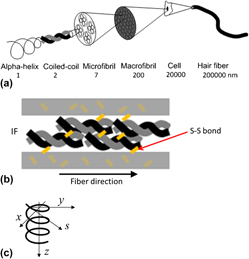
Mechanics of trichocyte alpha-keratin fibers: Experiment, theory, and simulation
-
- Journal:
- Journal of Materials Research / Volume 30 / Issue 1 / 14 January 2015
- Published online by Cambridge University Press:
- 15 January 2015, pp. 26-35
- Print publication:
- 14 January 2015
-
- Article
- Export citation
Unraveling the Core of The Gran Pirámide From Cholula, Puebla. A Compositional and Microstructural Analysis of the Adobe
-
- Journal:
- MRS Online Proceedings Library Archive / Volume 1656 / 2014
- Published online by Cambridge University Press:
- 13 January 2015, pp. 41-50
- Print publication:
- 2014
-
- Article
- Export citation
Study of Mexican Colonial Mural Paintings: An In-situ Non-Invasive Approach
-
- Journal:
- MRS Online Proceedings Library Archive / Volume 1656 / 2014
- Published online by Cambridge University Press:
- 13 January 2015, pp. 75-93
- Print publication:
- 2014
-
- Article
- Export citation
Non-Invasive Characterization of Stone Artifacts from the Great Temple of Tenochtitlan, Mexico
-
- Journal:
- MRS Online Proceedings Library Archive / Volume 1656 / 2014
- Published online by Cambridge University Press:
- 13 January 2015, pp. 293-307
- Print publication:
- 2014
-
- Article
- Export citation

Theory of Inelastic Scattering and Absorption of X-rays
-
- Published online:
- 05 January 2015
- Print publication:
- 26 January 2015

Tracking subsurface ion radiation damage with metal–oxide–semiconductor device encapsulation
-
- Journal:
- Journal of Materials Research / Volume 30 / Issue 9 / 14 May 2015
- Published online by Cambridge University Press:
- 05 January 2015, pp. 1413-1421
- Print publication:
- 14 May 2015
-
- Article
- Export citation
Indentation-induced two-way shape-memory effect in aged Ti-50.9 at.% Ni
-
- Journal:
- MRS Communications / Volume 5 / Issue 1 / March 2015
- Published online by Cambridge University Press:
- 02 January 2015, pp. 77-82
- Print publication:
- March 2015
-
- Article
- Export citation
Use of Micro- and Nano-ZnO particles as Catalysts for the Microwave-Assisted Polymerization of D,L-lactide
-
- Journal:
- MRS Online Proceedings Library Archive / Volume 1767 / 2015
- Published online by Cambridge University Press:
- 16 March 2015, pp. 3-9
- Print publication:
- 2015
-
- Article
- Export citation
Development of Transport Properties Characterization Capabilities for Thermoelectric Materials and Modules
-
- Journal:
- MRS Online Proceedings Library Archive / Volume 1774 / 2015
- Published online by Cambridge University Press:
- 09 June 2015, pp. 7-12
- Print publication:
- 2015
-
- Article
- Export citation
Influence of Oxygen Content on the Electronic Properties of the PrAlO3/SrTiO3 Interface
-
- Journal:
- MRS Online Proceedings Library Archive / Volume 1805 / 2015
- Published online by Cambridge University Press:
- 10 July 2015, mrss15-2130457
- Print publication:
- 2015
-
- Article
- Export citation
Self-protective Oxide Nano-Coatings for Enhanced Surface Biocompatibility of Titanium
-
- Journal:
- MRS Online Proceedings Library Archive / Volume 1806 / 2015
- Published online by Cambridge University Press:
- 28 April 2015, pp. 7-12
- Print publication:
- 2015
-
- Article
- Export citation
Structural Studies on Some Oligothiophenes and Ethylenedioxythiophenes
-
- Journal:
- MRS Online Proceedings Library Archive / Volume 1799 / 2015
- Published online by Cambridge University Press:
- 04 June 2015, pp. 19-28
- Print publication:
- 2015
-
- Article
- Export citation
OPL volume 1785 Author and Subject Indexes
-
- Journal:
- MRS Online Proceedings Library Archive / Volume 1785 / 2015
- Published online by Cambridge University Press:
- 13 October 2015, pp. b1-b2
- Print publication:
- 2015
-
- Article
- Export citation
Metallurgical By-Product Transformation and Its Plasma Modification to Increase HDPE Thermal Conductivity
-
- Journal:
- MRS Online Proceedings Library Archive / Volume 1765 / 2015
- Published online by Cambridge University Press:
- 05 October 2015, pp. 23-27
- Print publication:
- 2015
-
- Article
- Export citation
Role of Stoichiometry on Ordering in Ni-Cr Alloys
-
- Journal:
- MRS Online Proceedings Library Archive / Volume 1809 / 2015
- Published online by Cambridge University Press:
- 18 May 2015, pp. 7-12
- Print publication:
- 2015
-
- Article
- Export citation
Valence and Local Environment of Molybdenum in Aluminophosphate Glasses for Immobilization of High Level Waste from Uranium-Graphite Reactor Spent Nuclear Fuel Reprocessing
-
- Journal:
- MRS Online Proceedings Library Archive / Volume 1744 / 2015
- Published online by Cambridge University Press:
- 19 March 2015, pp. 73-78
- Print publication:
- 2015
-
- Article
- Export citation
ZnO thin films applied as pH sensor in EGFET devices
-
- Journal:
- MRS Online Proceedings Library Archive / Volume 1805 / 2015
- Published online by Cambridge University Press:
- 23 July 2015, mrss15-2136685
- Print publication:
- 2015
-
- Article
- Export citation
