Refine search
Actions for selected content:
106129 results in Materials Science

Ferromagnetism induced by lattice volume expansion and amorphization in EuTiO3 thin films
-
- Journal:
- Journal of Materials Research / Volume 28 / Issue 8 / 28 April 2013
- Published online by Cambridge University Press:
- 03 April 2013, pp. 1031-1041
- Print publication:
- 28 April 2013
-
- Article
- Export citation

Enhancement of solar cells with photonic and plasmonic crystals - overcoming the Lambertian limit
-
- Journal:
- Journal of Materials Research / Volume 28 / Issue 8 / 28 April 2013
- Published online by Cambridge University Press:
- 03 April 2013, pp. 1021-1030
- Print publication:
- 28 April 2013
-
- Article
- Export citation

Preparation of nonaggregated silver nanoparticles by the liquid phase plasma reduction method
-
- Journal:
- Journal of Materials Research / Volume 28 / Issue 8 / 28 April 2013
- Published online by Cambridge University Press:
- 03 April 2013, pp. 1105-1110
- Print publication:
- 28 April 2013
-
- Article
- Export citation

Crystallization behavior and thermal properties of biodegradable poly(L-lactide)-poly(ethylene glycol) block copolymers
-
- Journal:
- Journal of Materials Research / Volume 28 / Issue 8 / 28 April 2013
- Published online by Cambridge University Press:
- 03 April 2013, pp. 1111-1117
- Print publication:
- 28 April 2013
-
- Article
- Export citation

Determination of structure-property relationships for 3-aminopropyltriethoxysilane films using x-ray reflectivity
-
- Journal:
- Journal of Materials Research / Volume 28 / Issue 8 / 28 April 2013
- Published online by Cambridge University Press:
- 03 April 2013, pp. 1118-1128
- Print publication:
- 28 April 2013
-
- Article
- Export citation

Dielectric properties of Ba1-xSrxZrO3 (0 ≤ x ≤ 1) nanoceramics developed by citrate precursor route
-
- Journal:
- Journal of Materials Research / Volume 28 / Issue 8 / 28 April 2013
- Published online by Cambridge University Press:
- 03 April 2013, pp. 1070-1077
- Print publication:
- 28 April 2013
-
- Article
- Export citation

Resistance degradation behavior of Zr-doped BaTiO3 ceramics and multilayer ceramic capacitor
-
- Journal:
- Journal of Materials Research / Volume 28 / Issue 8 / 28 April 2013
- Published online by Cambridge University Press:
- 03 April 2013, pp. 1078-1086
- Print publication:
- 28 April 2013
-
- Article
- Export citation

Synergistic thermoelectric power factor increase in films incorporating tellurium and thiophene-based semiconductors
-
- Journal:
- MRS Communications / Volume 3 / Issue 2 / June 2013
- Published online by Cambridge University Press:
- 03 April 2013, pp. 97-100
- Print publication:
- June 2013
-
- Article
- Export citation

Novel nanosample preparation with a helium ion microscope
-
- Journal:
- Journal of Materials Research / Volume 28 / Issue 8 / 28 April 2013
- Published online by Cambridge University Press:
- 03 April 2013, pp. 1013-1020
- Print publication:
- 28 April 2013
-
- Article
- Export citation

Effect of substrate temperature on the crystallographic structure and first-order magnetic phase transition of FeRh thin films
-
- Journal:
- Journal of Materials Research / Volume 28 / Issue 8 / 28 April 2013
- Published online by Cambridge University Press:
- 03 April 2013, pp. 1042-1046
- Print publication:
- 28 April 2013
-
- Article
- Export citation
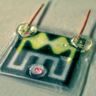
Paper-based electroanalytical devices for accessible diagnostic testing
-
- Journal:
- MRS Bulletin / Volume 38 / Issue 4 / April 2013
- Published online by Cambridge University Press:
- 12 April 2013, pp. 309-314
- Print publication:
- April 2013
-
- Article
- Export citation
MRS volume 38 issue 4 Cover and Front matter
-
- Journal:
- MRS Bulletin / Volume 38 / Issue 4 / April 2013
- Published online by Cambridge University Press:
- 12 April 2013, pp. f1-f7
- Print publication:
- April 2013
-
- Article
-
- You have access
- Export citation
CAREER CENTRAL
-
- Journal:
- MRS Bulletin / Volume 38 / Issue 4 / April 2013
- Published online by Cambridge University Press:
- 12 April 2013, pp. 348-351
- Print publication:
- April 2013
-
- Article
-
- You have access
- Export citation
Nano Focus: Langmuir–Schaefer assembled carbon nanotube arrays show superior electronic properties
-
- Journal:
- MRS Bulletin / Volume 38 / Issue 4 / April 2013
- Published online by Cambridge University Press:
- 12 April 2013, p. 294
- Print publication:
- April 2013
-
- Article
-
- You have access
- HTML
- Export citation
Nano Focus: Superdiffusive electron transport mediates laser-induced demagnetization
-
- Journal:
- MRS Bulletin / Volume 38 / Issue 4 / April 2013
- Published online by Cambridge University Press:
- 12 April 2013, p. 296
- Print publication:
- April 2013
-
- Article
-
- You have access
- HTML
- Export citation
Rare-earth oxide ceramics found to be robustly hydrophobic
-
- Journal:
- MRS Bulletin / Volume 38 / Issue 4 / April 2013
- Published online by Cambridge University Press:
- 12 April 2013, p. 295
- Print publication:
- April 2013
-
- Article
-
- You have access
- HTML
- Export citation
LOOK AGAIN…
-
- Journal:
- MRS Bulletin / Volume 38 / Issue 4 / April 2013
- Published online by Cambridge University Press:
- 12 April 2013, p. 352
- Print publication:
- April 2013
-
- Article
-
- You have access
- HTML
- Export citation
Energy Focus: Charge-density waves may be competing with superconductivity
-
- Journal:
- MRS Bulletin / Volume 38 / Issue 4 / April 2013
- Published online by Cambridge University Press:
- 12 April 2013, pp. 295-296
- Print publication:
- April 2013
-
- Article
-
- You have access
- HTML
- Export citation
Addendum
-
- Journal:
- MRS Bulletin / Volume 38 / Issue 4 / April 2013
- Published online by Cambridge University Press:
- 12 April 2013, p. 298
- Print publication:
- April 2013
-
- Article
-
- You have access
- HTML
- Export citation
MRS volume 38 issue 4 Cover and Back matter
-
- Journal:
- MRS Bulletin / Volume 38 / Issue 4 / April 2013
- Published online by Cambridge University Press:
- 12 April 2013, pp. b1-b2
- Print publication:
- April 2013
-
- Article
-
- You have access
- Export citation
