Refine search
Actions for selected content:
106129 results in Materials Science
Li and Mao to receive MRS Postdoctoral Awards
-
- Journal:
- MRS Bulletin / Volume 45 / Issue 4 / April 2020
- Published online by Cambridge University Press:
- 09 April 2020, p. 310
- Print publication:
- April 2020
-
- Article
-
- You have access
- HTML
- Export citation
Spoof Surface Plasmon Metamaterials
-
- Journal:
- MRS Bulletin / Volume 45 / Issue 4 / April 2020
- Published online by Cambridge University Press:
- 09 April 2020, p. 318
- Print publication:
- April 2020
-
- Article
-
- You have access
- HTML
- Export citation
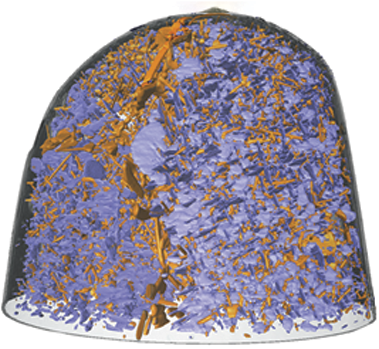
Nanoscale x-ray and electron tomography
-
- Journal:
- MRS Bulletin / Volume 45 / Issue 4 / April 2020
- Published online by Cambridge University Press:
- 09 April 2020, pp. 264-271
- Print publication:
- April 2020
-
- Article
-
- You have access
- HTML
- Export citation
South African science ministries respond favorably to State of the Nation address by president
-
- Journal:
- MRS Bulletin / Volume 45 / Issue 4 / April 2020
- Published online by Cambridge University Press:
- 09 April 2020, p. 260
- Print publication:
- April 2020
-
- Article
-
- You have access
- HTML
- Export citation
The ultimate microscopy—enabling nanotechnology
-
- Journal:
- MRS Bulletin / Volume 45 / Issue 4 / April 2020
- Published online by Cambridge University Press:
- 09 April 2020, p. 261
- Print publication:
- April 2020
-
- Article
-
- You have access
- HTML
- Export citation

High-dimensional and high-resolution x-ray tomography for energy materials science
-
- Journal:
- MRS Bulletin / Volume 45 / Issue 4 / April 2020
- Published online by Cambridge University Press:
- 09 April 2020, pp. 283-289
- Print publication:
- April 2020
-
- Article
- Export citation
Fall detection for elderly-people monitoring using learned features and recurrent neural networks
-
- Journal:
- Experimental Results / Volume 1 / 2020
- Published online by Cambridge University Press:
- 01 April 2020, e7
-
- Article
-
- You have access
- Open access
- HTML
- Export citation
The Alchemy of Us
-
- Journal:
- MRS Bulletin / Volume 45 / Issue 4 / April 2020
- Published online by Cambridge University Press:
- 09 April 2020, pp. 317-318
- Print publication:
- April 2020
-
- Article
-
- You have access
- HTML
- Export citation
Guo to receive MRS Innovation in Materials Characterization Award
-
- Journal:
- MRS Bulletin / Volume 45 / Issue 4 / April 2020
- Published online by Cambridge University Press:
- 09 April 2020, p. 308
- Print publication:
- April 2020
-
- Article
-
- You have access
- HTML
- Export citation
MRS Communications Abstracts
-
- Journal:
- MRS Bulletin / Volume 45 / Issue 4 / April 2020
- Published online by Cambridge University Press:
- 09 April 2020, pp. 314-315
- Print publication:
- April 2020
-
- Article
-
- You have access
- HTML
- Export citation
LOOK AGAIN
-
- Journal:
- MRS Bulletin / Volume 45 / Issue 4 / April 2020
- Published online by Cambridge University Press:
- 09 April 2020, p. 320
- Print publication:
- April 2020
-
- Article
-
- You have access
- HTML
- Export citation
RESEARCH HIGHLIGHTS: Perovskites
-
- Journal:
- MRS Bulletin / Volume 45 / Issue 4 / April 2020
- Published online by Cambridge University Press:
- 09 April 2020, pp. 253-254
- Print publication:
- April 2020
-
- Article
-
- You have access
- HTML
- Export citation
Career progression through professional engagement: The impact of MRS student-led activities
-
- Journal:
- MRS Bulletin / Volume 45 / Issue 4 / April 2020
- Published online by Cambridge University Press:
- 09 April 2020, pp. 306-307
- Print publication:
- April 2020
-
- Article
-
- You have access
- HTML
- Export citation

Atomic electron tomography in three and four dimensions
-
- Journal:
- MRS Bulletin / Volume 45 / Issue 4 / April 2020
- Published online by Cambridge University Press:
- 09 April 2020, pp. 290-297
- Print publication:
- April 2020
-
- Article
- Export citation
Rivnay to receive Outstanding Early-Career Investigator Award
-
- Journal:
- MRS Bulletin / Volume 45 / Issue 4 / April 2020
- Published online by Cambridge University Press:
- 09 April 2020, p. 309
- Print publication:
- April 2020
-
- Article
-
- You have access
- HTML
- Export citation
Duan to receive Mid-Career Researcher Award
-
- Journal:
- MRS Bulletin / Volume 45 / Issue 4 / April 2020
- Published online by Cambridge University Press:
- 09 April 2020, p. 308
- Print publication:
- April 2020
-
- Article
-
- You have access
- HTML
- Export citation
Aselage to receive MRS Woody White Service Award
-
- Journal:
- MRS Bulletin / Volume 45 / Issue 4 / April 2020
- Published online by Cambridge University Press:
- 09 April 2020, p. 310
- Print publication:
- April 2020
-
- Article
-
- You have access
- HTML
- Export citation
MRS volume 45 issue 4 Cover and Front matter
-
- Journal:
- MRS Bulletin / Volume 45 / Issue 4 / April 2020
- Published online by Cambridge University Press:
- 09 April 2020, pp. f1-f6
- Print publication:
- April 2020
-
- Article
-
- You have access
- Export citation
Defect engineering increases polarization retention in ferroelectric thin films
-
- Journal:
- MRS Bulletin / Volume 45 / Issue 4 / April 2020
- Published online by Cambridge University Press:
- 09 April 2020, pp. 254-255
- Print publication:
- April 2020
-
- Article
-
- You have access
- HTML
- Export citation
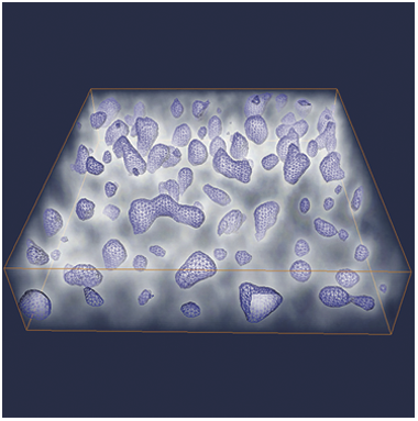
Electron tomography for functional nanomaterials
-
- Journal:
- MRS Bulletin / Volume 45 / Issue 4 / April 2020
- Published online by Cambridge University Press:
- 09 April 2020, pp. 298-304
- Print publication:
- April 2020
-
- Article
- Export citation
