Refine search
Actions for selected content:
238502 results in Physics and Astronomy
10 - Quantification of Quantum Resources
- from Part III - The General Framework of Resource Theories
-
- Book:
- Quantum Resource Theories
- Published online:
- 03 May 2025
- Print publication:
- 10 April 2025, pp 426-466
-
- Chapter
- Export citation
Frontmatter
-
- Book:
- Quantum Resource Theories
- Published online:
- 03 May 2025
- Print publication:
- 10 April 2025, pp i-iv
-
- Chapter
- Export citation

Marangoni-driven patterns, ridges and hills in surfactant-covered parametric surface waves
-
- Journal:
- Journal of Fluid Mechanics / Volume 1008 / 10 April 2025
- Published online by Cambridge University Press:
- 10 April 2025, R4
-
- Article
-
- You have access
- Open access
- HTML
- Export citation
-
Parametric oscillations of an interface separating two fluid phases create nonlinear surface waves, called Faraday waves, which organise into simple patterns, such as squares and hexagons, as well as complex structures, such as double hexagonal and superlattice patterns. In this work, we study the influence of surfactant-induced Marangoni stresses on the formation and transition of Faraday-wave patterns. We use a control parameter,
 $B$, that assesses the relative importance of Marangoni stresses as compared with the surface-wave dynamics. Our results show that the threshold acceleration required to destabilise a surfactant-covered interface through vibration increases with increasing
  $B$. For a surfactant-free interface, a square-wave pattern is observed. As
  $B$ is incremented, we report transitions from squares to asymmetric squares, weakly wavy stripes and ultimately to ridges and hills. These hills are a consequence of the bidirectional Marangoni stresses at the neck of the ridges. The mechanisms underlying the pattern transitions and the formation of exotic ridges and hills are discussed.


7 - Conditional Entropy
- from Part II - Tools and Methods
-
- Book:
- Quantum Resource Theories
- Published online:
- 03 May 2025
- Print publication:
- 10 April 2025, pp 308-351
-
- Chapter
- Export citation
8 - The Asymptotic Regime
- from Part II - Tools and Methods
-
- Book:
- Quantum Resource Theories
- Published online:
- 03 May 2025
- Print publication:
- 10 April 2025, pp 352-400
-
- Chapter
- Export citation
16 - The Resource Theory of Nonuniformity
- from Part V - Additional Examples of Static Resource Theories
-
- Book:
- Quantum Resource Theories
- Published online:
- 03 May 2025
- Print publication:
- 10 April 2025, pp 736-748
-
- Chapter
- Export citation

Inertial focusing of spherical capsule in pulsatile channel flows
-
- Journal:
- Journal of Fluid Mechanics / Volume 1008 / 10 April 2025
- Published online by Cambridge University Press:
- 09 April 2025, A46
-
- Article
-
- You have access
- Open access
- HTML
- Export citation
-
We present numerical analysis of the lateral movement of a spherical capsule in the steady and pulsatile channel flow of a Newtonian fluid for a wide range of oscillatory frequencies. Each capsule membrane satisfying strain-hardening characteristics is simulated for different Reynolds numbers
 $Re$ and capillary numbers
  $Ca$. Our numerical results showed that capsules with high
  $Ca$ exhibit axial focusing at finite
  $Re$ similarly to the inertialess case. We observe that the speed of the axial focusing can be substantially accelerated by making the driving pressure gradient oscillate in time. We also confirm the existence of an optimal frequency that maximises the speed of axial focusing, which remains the same found in the absence of inertia. For relatively low
  $Ca$, however, the capsule exhibits off-centre focusing, resulting in various equilibrium radial positions depending on
  $Re$. Our numerical results further clarify the existence of a specific
  $Re$ for which the effect of the flow pulsation to the equilibrium radial position is maximum. The roles of channel size on the lateral movements of the capsule are also addressed. Throughout our analyses, we have quantified the radial position of the capsule in a tube based on an empirical expression. Given that the speed of inertial focusing can be controlled by the oscillatory frequency, the results obtained here can be used for label-free cell alignment/sorting/separation techniques, e.g. for circulating tumour cells in cancer patients or precious haematopoietic cells such as colony-forming cells.


Numerical simulations of three-dimensional ion crystal dynamics in a Penning trap using the fast multipole method
- Part of
-
- Journal:
- Journal of Plasma Physics / Volume 91 / Issue 2 / April 2025
- Published online by Cambridge University Press:
- 08 April 2025, E53
-
- Article
-
- You have access
- Open access
- HTML
- Export citation
Simultaneous realization of time and carrier-envelope phase synchronization for an ultra-intense few-cycle laser pulse coherent combining system
-
- Journal:
- High Power Laser Science and Engineering / Volume 13 / 2025
- Published online by Cambridge University Press:
- 07 April 2025, e47
-
- Article
-
- You have access
- Open access
- HTML
- Export citation
Large-scale timing synchronization based on linear-optics timing detectors
-
- Journal:
- High Power Laser Science and Engineering / Volume 13 / 2025
- Published online by Cambridge University Press:
- 07 April 2025, e48
-
- Article
-
- You have access
- Open access
- HTML
- Export citation
A main sequence CH-star in the globular cluster M55 (NGC 6809)
-
- Journal:
- Publications of the Astronomical Society of Australia / Volume 42 / 2025
- Published online by Cambridge University Press:
- 07 April 2025, e044
-
- Article
-
- You have access
- Open access
- HTML
- Export citation
-
Spectra have been obtained with the multi-fibre instrument 2dF on the Anglo-Australian Telescope of 89 candidate main sequence stars in the globular cluster M55 (NGC 6809). Radial velocities and Gaia proper motions confirm 72 candidates as cluster members. Among these stars one stands out as having a substantially stronger G-band (CH) than the rest of the member sample. The star is a dwarf carbon star that most likely acquired the high carbon abundance ([C/Fe]
 $\approx$ 1.2
  $\pm$ 0.2) via mass transfer from a
  $\sim$1
 $-$3 M
 $_{\odot}$ binary companion (now a white dwarf) during its AGB phase of evolution. Interestingly, M55 also contains a CH-star that lies on the cluster red giant branch – the low central concentration/low density of this cluster presumably allows the survival of binaries that would otherwise be disrupted in denser systems. The existence of carbon stars in six other globular clusters is consistent with this hypothesis, while the origin of the carbon-enhanced star in M15 (NGC 7078) is attributed to a merger process similar to that proposed for the origin of the carbon-rich R stars.


Brown dwarf number density in the JWST COSMOS-Web field
-
- Journal:
- Publications of the Astronomical Society of Australia / Volume 42 / 2025
- Published online by Cambridge University Press:
- 07 April 2025, e042
-
- Article
-
- You have access
- Open access
- HTML
- Export citation
-
Brown dwarfs are failed stars with very low mass (13–75 Jupiter mass) and an effective temperature lower than 2 500 K. Their mass range is between Jupiter and red dwarfs. Thus, they play a key role in understanding the gap in the mass function between stars and planets. However, due to their faint nature, previous searches are inevitably limited to the solar neighbourhood (20 pc). To improve our knowledge of the low mass part of the initial stellar mass function and the star formation history of the Milky Way, it is crucial to find more distant brown dwarfs. Using James Webb Space Telescope (JWST) COSMOS-Web data, this study seeks to enhance our comprehension of the physical characteristics of brown dwarfs situated at a distance of kpc scale. The exceptional sensitivity of the JWST enables the detection of brown dwarfs that are up to 100 times more distant than those discovered in the earlier all-sky infrared surveys. The large area coverage of the JWST COSMOS-Web survey allows us to find more distant brown dwarfs than earlier JWST studies with smaller area coverages. To capture prominent water absorption features around 2.7
 ${\unicode{x03BC}}$m, we apply two colour criteria,
  $\text{F115W}-\text{F277W}+1\lt\text{F277W}-\text{F444W}$ and
  $\text{F277W}-\text{F444W}\gt\,0.9$. We then select point sources by CLASS_STAR, FLUX_RADIUS, and SPREAD_MODEL criteria. Faint sources are visually checked to exclude possibly extended sources. We conduct SED fitting and MCMC simulations to determine their physical properties and associated uncertainties. Our search reveals 25 T-dwarf candidates and 2 Y-dwarf candidates, more than any previous JWST brown dwarf searches. They are located from 0.3 to 4 kpc away from the Earth. The spatial number density of 900–1 050 K dwarf is
 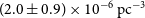 $(2.0\pm0.9) \times10^{-6}\text{ pc}^{-3}$, 1 050–1 200 K dwarf is
 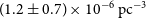 $(1.2\pm0.7) \times10^{-6}\text{ pc}^{-3}$, and 1 200–1 350 K dwarf is
 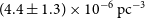 $(4.4\pm1.3) \times10^{-6}\text{ pc}^{-3}$. The cumulative number count of our brown dwarf candidates is consistent with the prediction from a standard double exponential model. Three of our brown dwarf candidates were detected by HST, with transverse velocities
  $12\pm5$,
  $12\pm4$, and
  $17\pm6$ km s
 $^{-1}$. Along with earlier studies, the JWST has opened a new window of brown dwarf research in the Milky Way thick disk and halo.


Dynamics of a film bounded by a pinned contact line
-
- Journal:
- Journal of Fluid Mechanics / Volume 1008 / 10 April 2025
- Published online by Cambridge University Press:
- 07 April 2025, A47
-
- Article
- Export citation
-
We consider the dynamics of a liquid film with a pinned contact line (for example, a drop), as described by the one-dimensional, surface-tension-driven thin-film equation
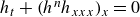 $h_t + (h^n h_{xxx})_x = 0$, where
  $h(x,t)$ is the thickness of the film. The case
  $n=3$ corresponds to a film on a solid substrate. We derive an evolution equation for the contact angle
  $\theta (t)$, which couples to the shape of the film. Starting from a regular initial condition
  $h_0(x)$, we investigate the dynamics of the drop both analytically and numerically, focusing on the contact angle. For short times
  $t\ll 1$, and if
  $n\ne 3$, the contact angle changes according to a power law
  $\displaystyle t^{\frac {n-2}{4-n}}$. In the critical case
  $n=3$, the dynamics become non-local, and
  $\dot {\theta }$ is now of order
  $\displaystyle {\rm{e}}^{-3/(2t^{1/3})}$. This implies that, for
  $n=3$, the standard contact line problem with prescribed contact angle is ill posed. In the long time limit, the solution relaxes exponentially towards equilibrium.


Investigating high-energy Hermite–Gaussian and vortex laser generation in alexandrite
-
- Journal:
- High Power Laser Science and Engineering / Volume 13 / 2025
- Published online by Cambridge University Press:
- 07 April 2025, e43
-
- Article
-
- You have access
- Open access
- HTML
- Export citation

Resonant triad interactions of two acoustic modes and a gravity wave
-
- Journal:
- Journal of Fluid Mechanics / Volume 1008 / 10 April 2025
- Published online by Cambridge University Press:
- 07 April 2025, A15
-
- Article
-
- You have access
- Open access
- HTML
- Export citation

An analytical study on oblique wave scattering involving flexible porous structures in a two-layer fluid
-
- Journal:
- Journal of Fluid Mechanics / Volume 1008 / 10 April 2025
- Published online by Cambridge University Press:
- 07 April 2025, A49
-
- Article
- Export citation

The onset of filamentation on vorticity interfaces in two-dimensional Euler flows
-
- Journal:
- Journal of Fluid Mechanics / Volume 1008 / 10 April 2025
- Published online by Cambridge University Press:
- 07 April 2025, A48
-
- Article
-
- You have access
- Open access
- HTML
- Export citation
Ten-megawatt-level peak power Mamyshev oscillator enabled by anti-resonant hollow-core fiber
-
- Journal:
- High Power Laser Science and Engineering / Volume 13 / 2025
- Published online by Cambridge University Press:
- 07 April 2025, e46
-
- Article
-
- You have access
- Open access
- HTML
- Export citation

On the wave kinetic equation in the presence of forcing and dissipation
-
- Journal:
- Journal of Fluid Mechanics / Volume 1008 / 10 April 2025
- Published online by Cambridge University Press:
- 07 April 2025, A44
-
- Article
-
- You have access
- Open access
- HTML
- Export citation

A viscous drop in a planar linear flow: the role of deformation on streamline topology
-
- Journal:
- Journal of Fluid Mechanics / Volume 1008 / 10 April 2025
- Published online by Cambridge University Press:
- 07 April 2025, A45
-
- Article
- Export citation
-
Planar linear flows are a one-parameter family, with the parameter
 $\hat {\alpha }\in [-1,1]$ being a measure of the relative magnitudes of extension and vorticity;
  $\hat {\alpha } = -1$,
  $0$ and
  $1$ correspond to solid-body rotation, simple shear flow and planar extension, respectively. For a neutrally buoyant spherical drop in a hyperbolic planar linear flow with
  $\hat {\alpha }\in (0,1]$, the near-field streamlines are closed for
  $\lambda \gt \lambda _c = 2 \hat {\alpha } / (1 - \hat {\alpha })$,
  $\lambda$ being the drop-to-medium viscosity ratio; all streamlines are closed for an ambient elliptic linear flow with
  $\hat {\alpha }\in [-1,0)$. We use both analytical and numerical tools to show that drop deformation, as characterized by a non-zero capillary number (
 $Ca$), destroys the aforementioned closed-streamline topology. While inertia has previously been shown to transform closed Stokesian streamlines into open spiralling ones that run from upstream to downstream infinity, the streamline topology around a deformed drop, for small but finite
  $Ca$, is more complicated. Only a subset of the original closed streamlines transforms to open spiralling ones, while the remaining ones densely wind around a configuration of nested invariant tori. Our results contradict previous efforts pointing to the persistence of the closed streamline topology exterior to a deformed drop, and have important implications for transport and mixing.


