Refine search
Actions for selected content:
1868 results in Physiology and biological physics
Unraveling the nature of physicochemical and biological processes underlying vesicular exocytotic release events through modeling of amperometric current spikes
- Part of
-
- Journal:
- QRB Discovery / Volume 6 / 2025
- Published online by Cambridge University Press:
- 24 July 2025, e21
- Print publication:
- 2025
-
- Article
-
- You have access
- Open access
- HTML
- Export citation
On the possibility of carbon-free heteropolymers on Venus: a computational astrobiology study
-
- Journal:
- QRB Discovery / Volume 6 / 2025
- Published online by Cambridge University Press:
- 25 September 2025, e23
- Print publication:
- 2025
-
- Article
-
- You have access
- Open access
- HTML
- Export citation
High-throughput single-molecule nanofluidic studies on B. subtilis Rok protein interaction with DNA
- Part of
-
- Journal:
- QRB Discovery / Volume 6 / 2025
- Published online by Cambridge University Press:
- 19 May 2025, e17
- Print publication:
- 2025
-
- Article
-
- You have access
- Open access
- HTML
- Export citation
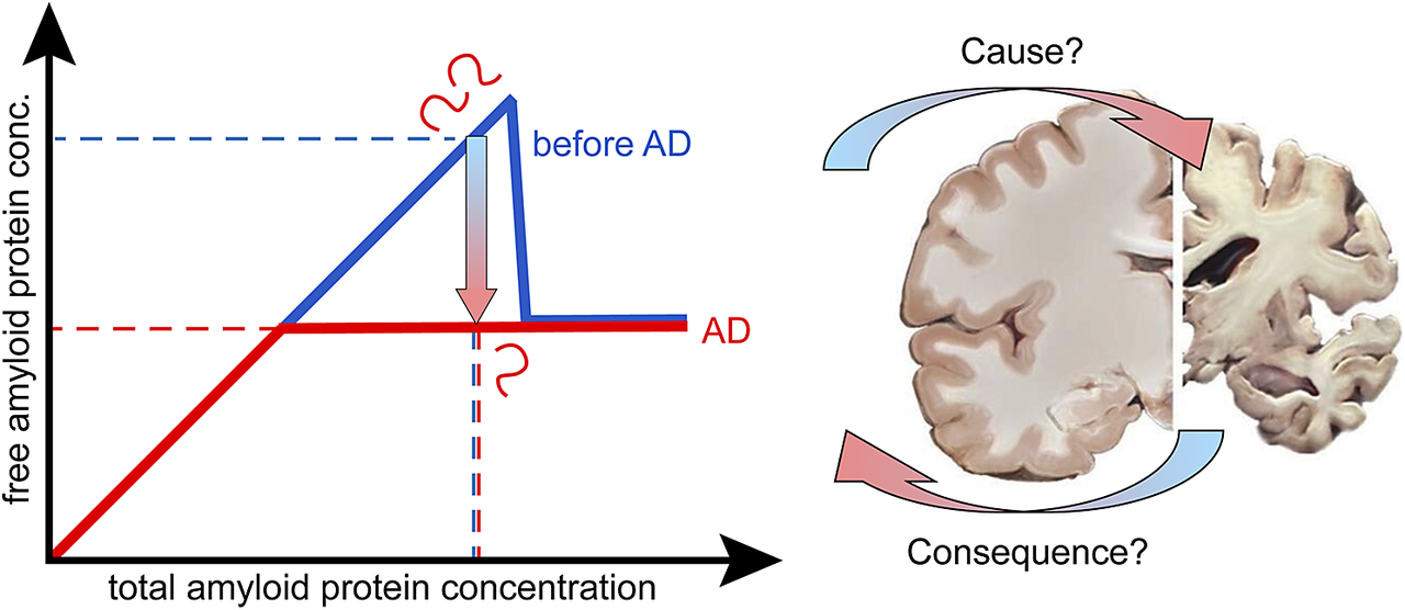
Reduced protein solubility – cause or consequence in amyloid disease?
-
- Journal:
- QRB Discovery / Volume 6 / 2025
- Published online by Cambridge University Press:
- 17 February 2025, e8
- Print publication:
- 2025
-
- Article
-
- You have access
- Open access
- HTML
- Export citation
Protein secondary structure determined from independent and integrated infra-red absorbance and circular dichroism data using the algorithm SELCON
- Part of
-
- Journal:
- QRB Discovery / Volume 6 / 2025
- Published online by Cambridge University Press:
- 03 February 2025, e10
- Print publication:
- 2025
-
- Article
-
- You have access
- Open access
- HTML
- Export citation
A structural and functional bioinformatics study of QTY-designed retinylidene proteins
-
- Journal:
- QRB Discovery / Volume 6 / 2025
- Published online by Cambridge University Press:
- 14 July 2025, e20
- Print publication:
- 2025
-
- Article
-
- You have access
- Open access
- HTML
- Export citation
Unzipping of knotted DNA via nanopore translocation
- Part of
-
- Journal:
- QRB Discovery / Volume 6 / 2025
- Published online by Cambridge University Press:
- 09 January 2025, e4
- Print publication:
- 2025
-
- Article
-
- You have access
- Open access
- HTML
- Export citation
Structural bioinformatic study of human mitochondrial respiratory integral membrane megacomplex and its AlphaFold3 predicted water-soluble QTY megacomplex analog
-
- Journal:
- QRB Discovery / Volume 6 / 2025
- Published online by Cambridge University Press:
- 05 February 2025, e12
- Print publication:
- 2025
-
- Article
-
- You have access
- Open access
- HTML
- Export citation
Probing DNA melting behaviour under vibrational strong coupling
-
- Journal:
- QRB Discovery / Volume 6 / 2025
- Published online by Cambridge University Press:
- 10 March 2025, e13
- Print publication:
- 2025
-
- Article
-
- You have access
- Open access
- HTML
- Export citation
Continuous-time random walk model for the diffusive motion of helicases
-
- Journal:
- QRB Discovery / Volume 6 / 2025
- Published online by Cambridge University Press:
- 09 October 2025, e26
- Print publication:
- 2025
-
- Article
-
- You have access
- Open access
- HTML
- Export citation
An integrated structural and biophysical approach to study carbon metabolism in Mycobacterium tuberculosis
- Part of
-
- Journal:
- QRB Discovery / Volume 6 / 2025
- Published online by Cambridge University Press:
- 12 March 2025, e15
- Print publication:
- 2025
-
- Article
-
- You have access
- Open access
- HTML
- Export citation
Modeling for understanding and engineering metabolism
-
- Journal:
- QRB Discovery / Volume 6 / 2025
- Published online by Cambridge University Press:
- 18 February 2025, e11
- Print publication:
- 2025
-
- Article
-
- You have access
- Open access
- HTML
- Export citation
Filling in missing puzzle pieces in protein structural biology with cross-linking mass spectrometry
- Part of
-
- Journal:
- QRB Discovery / Volume 6 / 2025
- Published online by Cambridge University Press:
- 27 December 2024, e6
- Print publication:
- 2025
-
- Article
-
- You have access
- Open access
- HTML
- Export citation
28 - Communication between Bacteria
- from Part III - Interacting Bacteria and Biofilms
-
- Book:
- The Physics of Bacteria
- Published online:
- 12 December 2024
- Print publication:
- 19 December 2024, pp 321-324
-
- Chapter
- Export citation
3 - The Electrochemical Potential of a Cell
- from Part I - Physical Tools
-
- Book:
- The Physics of Bacteria
- Published online:
- 12 December 2024
- Print publication:
- 19 December 2024, pp 32-38
-
- Chapter
- Export citation
11 - Systems Biology
- from Part I - Physical Tools
-
- Book:
- The Physics of Bacteria
- Published online:
- 12 December 2024
- Print publication:
- 19 December 2024, pp 95-98
-
- Chapter
- Export citation
31 - Other Applications
- from Part III - Interacting Bacteria and Biofilms
-
- Book:
- The Physics of Bacteria
- Published online:
- 12 December 2024
- Print publication:
- 19 December 2024, pp 348-353
-
- Chapter
- Export citation
17 - Motility
- from Part II - Single Bacteria
-
- Book:
- The Physics of Bacteria
- Published online:
- 12 December 2024
- Print publication:
- 19 December 2024, pp 176-189
-
- Chapter
- Export citation
4 - Mesoscopic Forces and Adhesion
- from Part I - Physical Tools
-
- Book:
- The Physics of Bacteria
- Published online:
- 12 December 2024
- Print publication:
- 19 December 2024, pp 39-51
-
- Chapter
- Export citation
Index
-
- Book:
- The Physics of Bacteria
- Published online:
- 12 December 2024
- Print publication:
- 19 December 2024, pp 354-362
-
- Chapter
- Export citation
