Refine search
Actions for selected content:
106117 results in Materials Science
Enhanced phase transformation and variant selection by electric current pulses in a Cu–Zn alloy
-
- Journal:
- Journal of Materials Research / Volume 29 / Issue 8 / 28 April 2014
- Published online by Cambridge University Press:
- 15 April 2014, pp. 975-980
- Print publication:
- 28 April 2014
-
- Article
- Export citation
Resolution of structural transformation of intermediates in Al–Cu alloys during non-isothermal precipitation
-
- Journal:
- Journal of Materials Research / Volume 29 / Issue 7 / 14 April 2014
- Published online by Cambridge University Press:
- 06 October 2020, pp. 874-879
- Print publication:
- 14 April 2014
-
- Article
- Export citation
Impact of interface thermodynamics on Al-induced crystallization of amorphous SixGe1–x alloys – CORRIGENDUM
-
- Journal:
- Journal of Materials Research / Volume 29 / Issue 8 / 28 April 2014
- Published online by Cambridge University Press:
- 09 April 2014, p. 1026
- Print publication:
- 28 April 2014
-
- Article
-
- You have access
- HTML
- Export citation
Phase transformation and microstructural development of zirconia/stainless steel bonded with a Ti/Ni/Ti interlayer for the potential application in solid oxide fuel cells
-
- Journal:
- Journal of Materials Research / Volume 29 / Issue 8 / 28 April 2014
- Published online by Cambridge University Press:
- 09 April 2014, pp. 923-934
- Print publication:
- 28 April 2014
-
- Article
- Export citation
-
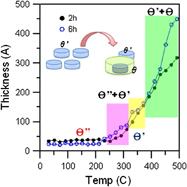
The 8 mol% yttria-stabilized zirconia (8Y-ZrO2) was bonded to stainless steel 316L at 900 °C for 1 h in a protective Ar atmosphere using an interlayer of Ti/Ni/Ti. Interfacial microstructures were characterized using both secondary electron microscope (SEM) and transmission electron microscope (TEM), each with an attached energy dispersive spectroscope (EDS). A layer sequence of σ-phase/TiFe2/TiFe + β-Ti/Ti2Fe was observed at the stainless steel 316L/Ti interface, whereas a layer sequence of Ti2Ni/Ti2Ni + TiNi/TiNi3 was found at the Ti/Ni interface. Furthermore, TiO and c-ZrO2−x formed at the Ti/8Y-ZrO2 interface. An acicular α-Ti and a fine ω-phase existed along with β-Ti in the residual Ti foil adjacent to the stainless steel 316L, but α-Ti and Ti2Ni were observed within β-Ti in the other residual Ti foil adjacent to the 8Y-ZrO2. The orientation relationships of the ω-phase and β-Ti were ${\left[ {1\bar 10} \right]_{{\rm{ \beta {\hbox-} Ti}}}}//{\left[ {1\bar 210} \right]_{\rm{\omega }}}$ and ${\left( {111} \right)_{{\rm{\beta {\hbox-} Ti}}}}//{\left( {0001} \right)_{\rm{\omega }}}$, respectively. The microstructural development was elucidated with the aid of Fe–Ti and Ni–Ti binary phase diagrams.


Regulation of Cu precipitation by intercritical tempering in a HSLA steel
-
- Journal:
- Journal of Materials Research / Volume 29 / Issue 8 / 28 April 2014
- Published online by Cambridge University Press:
- 09 April 2014, pp. 950-958
- Print publication:
- 28 April 2014
-
- Article
- Export citation
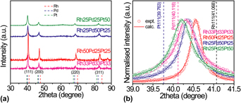
Composition-controlled ternary Rh–Pd–Pt solid-solution alloy nanoparticles by laser irradiation of mixed solution of metallic ions
-
- Journal:
- Journal of Materials Research / Volume 29 / Issue 7 / 14 April 2014
- Published online by Cambridge University Press:
- 09 April 2014, pp. 856-864
- Print publication:
- 14 April 2014
-
- Article
- Export citation
JMR volume 29 issue 7 Cover and Front matter
-
- Journal:
- Journal of Materials Research / Volume 29 / Issue 7 / 14 April 2014
- Published online by Cambridge University Press:
- 09 April 2014, pp. f1-f5
- Print publication:
- 14 April 2014
-
- Article
-
- You have access
- Export citation
X-ray diffraction characterization of a distorted Debye–Scherrer film strip – the effect of deacetylation on cellulose triacetate and an improved structural model for cellulose II
-
- Journal:
- Powder Diffraction / Volume 29 / Issue 2 / June 2014
- Published online by Cambridge University Press:
- 09 April 2014, pp. 108-112
-
- Article
- Export citation
JMR volume 29 issue 7 Cover and Back matter
-
- Journal:
- Journal of Materials Research / Volume 29 / Issue 7 / 14 April 2014
- Published online by Cambridge University Press:
- 09 April 2014, pp. b1-b5
- Print publication:
- 14 April 2014
-
- Article
-
- You have access
- Export citation
Synthesis and X-ray diffraction data of 1-N-(3-pyridylmethyl)aminonaphthalene hydrochloride
-
- Journal:
- Powder Diffraction / Volume 29 / Issue 2 / June 2014
- Published online by Cambridge University Press:
- 09 April 2014, pp. 186-189
-
- Article
- Export citation
X-ray powder diffraction data for meloxicam, C14H13N3O4S2
-
- Journal:
- Powder Diffraction / Volume 29 / Issue 2 / June 2014
- Published online by Cambridge University Press:
- 09 April 2014, pp. 196-198
-
- Article
- Export citation
A new “chain” of events: polymers in the Powder Diffraction FileTM (PDF®)
-
- Journal:
- Powder Diffraction / Volume 29 / Issue 2 / June 2014
- Published online by Cambridge University Press:
- 09 April 2014, pp. 102-107
-
- Article
- Export citation
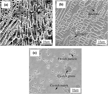
Influence of Al addition on microstructure and properties of Cu–Fe-based coatings by laser induction hybrid rapid cladding
-
- Journal:
- Journal of Materials Research / Volume 29 / Issue 7 / 14 April 2014
- Published online by Cambridge University Press:
- 09 April 2014, pp. 865-873
- Print publication:
- 14 April 2014
-
- Article
- Export citation
Tuning the monoclinic-to-orthorhombic phase transition temperature of Fe2Mo3O12 by substitutional co-incorporation of Zr4+ and Mg2+
-
- Journal:
- Journal of Materials Research / Volume 29 / Issue 7 / 14 April 2014
- Published online by Cambridge University Press:
- 09 April 2014, pp. 849-855
- Print publication:
- 14 April 2014
-
- Article
- Export citation
Mechanical properties of Bombyx mori silkworm silk subjected to microwave radiation
-
- Journal:
- Journal of Materials Research / Volume 29 / Issue 7 / 14 April 2014
- Published online by Cambridge University Press:
- 09 April 2014, pp. 833-842
- Print publication:
- 14 April 2014
-
- Article
- Export citation

Ion-modulated transistors on paper using phase-separated semiconductor/insulator blends
-
- Journal:
- MRS Communications / Volume 4 / Issue 2 / June 2014
- Published online by Cambridge University Press:
- 07 April 2014, pp. 51-55
- Print publication:
- June 2014
-
- Article
- Export citation
Yury Gogotsi to present Kavli Lecture on nanoscience at 2014 MRS Spring Meeting
-
- Journal:
- MRS Bulletin / Volume 39 / Issue 4 / April 2014
- Published online by Cambridge University Press:
- 09 April 2014, p. 375
- Print publication:
- April 2014
-
- Article
-
- You have access
- HTML
- Export citation

Focused ion beam and scanning electron microscopy for 3D materials characterization
-
- Journal:
- MRS Bulletin / Volume 39 / Issue 4 / April 2014
- Published online by Cambridge University Press:
- 09 April 2014, pp. 361-365
- Print publication:
- April 2014
-
- Article
- Export citation
Light converts monomers into large single-crystal linear polymers
-
- Journal:
- MRS Bulletin / Volume 39 / Issue 4 / April 2014
- Published online by Cambridge University Press:
- 09 April 2014, pp. 310-311
- Print publication:
- April 2014
-
- Article
-
- You have access
- HTML
- Export citation
Bio Focus: Turkey skin inspires biomimetic sensor
-
- Journal:
- MRS Bulletin / Volume 39 / Issue 4 / April 2014
- Published online by Cambridge University Press:
- 09 April 2014, pp. 309-310
- Print publication:
- April 2014
-
- Article
-
- You have access
- HTML
- Export citation
