Refine search
Actions for selected content:
106117 results in Materials Science

A topological twist on materials science
-
- Journal:
- MRS Bulletin / Volume 39 / Issue 3 / March 2014
- Published online by Cambridge University Press:
- 13 March 2014, pp. 265-279
- Print publication:
- March 2014
-
- Article
- Export citation
MRS expands materials publications landscape: www.mrs.org/mrs-publishing
-
- Journal:
- MRS Bulletin / Volume 39 / Issue 3 / March 2014
- Published online by Cambridge University Press:
- 13 March 2014, p. 287
- Print publication:
- March 2014
-
- Article
-
- You have access
- HTML
- Export citation
Remembering the legacy of Fred Kavli
-
- Journal:
- MRS Bulletin / Volume 39 / Issue 3 / March 2014
- Published online by Cambridge University Press:
- 13 March 2014, p. 285
- Print publication:
- March 2014
-
- Article
-
- You have access
- HTML
- Export citation
MRC volume 4 issue 1 Cover and Back matter
-
- Journal:
- MRS Communications / Volume 4 / Issue 1 / March 2014
- Published online by Cambridge University Press:
- 27 March 2014, pp. b1-b3
- Print publication:
- March 2014
-
- Article
-
- You have access
- Export citation
Bio Focus: Contact lens elutes glaucoma medication sustainably for one month
-
- Journal:
- MRS Bulletin / Volume 39 / Issue 3 / March 2014
- Published online by Cambridge University Press:
- 13 March 2014, pp. 204-206
- Print publication:
- March 2014
-
- Article
-
- You have access
- HTML
- Export citation

Understanding nano-bio interactions to improve nanocarriers for drug delivery
-
- Journal:
- MRS Bulletin / Volume 39 / Issue 3 / March 2014
- Published online by Cambridge University Press:
- 13 March 2014, pp. 227-237
- Print publication:
- March 2014
-
- Article
- Export citation
US Congress proposes alternative to COMPETES Act
-
- Journal:
- MRS Bulletin / Volume 39 / Issue 3 / March 2014
- Published online by Cambridge University Press:
- 13 March 2014, pp. 209-210
- Print publication:
- March 2014
-
- Article
-
- You have access
- HTML
- Export citation
Famous last words
-
- Journal:
- MRS Bulletin / Volume 39 / Issue 3 / March 2014
- Published online by Cambridge University Press:
- 13 March 2014, pp. 303-304
- Print publication:
- March 2014
-
- Article
-
- You have access
- HTML
- Export citation
MRS volume 39 issue 3 Cover and Front matter
-
- Journal:
- MRS Bulletin / Volume 39 / Issue 3 / March 2014
- Published online by Cambridge University Press:
- 13 March 2014, pp. f1-f7
- Print publication:
- March 2014
-
- Article
-
- You have access
- Export citation
John J. Tracy of Boeing to give plenary address at 2014 MRS Spring Meeting: www.mrs.org/spring2014
-
- Journal:
- MRS Bulletin / Volume 39 / Issue 3 / March 2014
- Published online by Cambridge University Press:
- 13 March 2014, p. 286
- Print publication:
- March 2014
-
- Article
-
- You have access
- HTML
- Export citation

Clinical translational challenges in nanomedicine
-
- Journal:
- MRS Bulletin / Volume 39 / Issue 3 / March 2014
- Published online by Cambridge University Press:
- 13 March 2014, pp. 259-264
- Print publication:
- March 2014
-
- Article
- Export citation
The MRS mission and industry
-
- Journal:
- MRS Bulletin / Volume 39 / Issue 3 / March 2014
- Published online by Cambridge University Press:
- 13 March 2014, pp. 198-199
- Print publication:
- March 2014
-
- Article
-
- You have access
- HTML
- Export citation
MRC volume 4 issue 1 Cover and Front matter
-
- Journal:
- MRS Communications / Volume 4 / Issue 1 / March 2014
- Published online by Cambridge University Press:
- 27 March 2014, pp. f1-f4
- Print publication:
- March 2014
-
- Article
-
- You have access
- Export citation

Organic nanoparticles for drug delivery and imaging
-
- Journal:
- MRS Bulletin / Volume 39 / Issue 3 / March 2014
- Published online by Cambridge University Press:
- 13 March 2014, pp. 219-223
- Print publication:
- March 2014
-
- Article
-
- You have access
- HTML
- Export citation
European Commission launches pilot to open publicly funded research data: http://ec.europa.eu
-
- Journal:
- MRS Bulletin / Volume 39 / Issue 3 / March 2014
- Published online by Cambridge University Press:
- 13 March 2014, p. 211
- Print publication:
- March 2014
-
- Article
-
- You have access
- HTML
- Export citation
Quantitative STEM technique extracts 3D atom stacking information from 2D image
-
- Journal:
- MRS Bulletin / Volume 39 / Issue 3 / March 2014
- Published online by Cambridge University Press:
- 13 March 2014, p. 208
- Print publication:
- March 2014
-
- Article
-
- You have access
- HTML
- Export citation

Study of quasi-amorphous to nanocrystalline phase transition in thermally evaporated CuInS2 thin films
-
- Journal:
- Journal of Materials Research / Volume 29 / Issue 4 / 28 February 2014
- Published online by Cambridge University Press:
- 03 March 2014, pp. 542-555
- Print publication:
- 28 February 2014
-
- Article
- Export citation

Measuring the volume charge in dielectric films using single frequency electro-acoustic waves
-
- Journal:
- Journal of Materials Research / Volume 29 / Issue 4 / 28 February 2014
- Published online by Cambridge University Press:
- 03 March 2014, pp. 501-508
- Print publication:
- 28 February 2014
-
- Article
- Export citation

Microstructural evolution and bonding mechanisms of the brazed Ti/ZrO2 joint using an Ag68.8Cu26.7Ti4.5 interlayer at 900 °C
-
- Journal:
- Journal of Materials Research / Volume 29 / Issue 5 / 14 March 2014
- Published online by Cambridge University Press:
- 28 February 2014, pp. 684-694
- Print publication:
- 14 March 2014
-
- Article
- Export citation
-
In this study, 3 mol% Y2O3-stabilized zirconia (3Y–ZrO2) and commercially pure titanium (cp-Ti) joints were fabricated with an Ag68.8Cu26.7Ti4.5 interlayer (Ticusil) at 900 °C for various brazing periods. After brazing at 900 °C/0.1 h, Ti2Cu, TiCu, Ti3Cu4, and TiCu4 layers were present at the Ti/Ticusil interface, while TiCu and TiO layers were observed at the Ticusil/3Y–ZrO2 interface. In the residual interlayer, clumpy TiCu4 was formed along with the Ag solid phase. After brazing at 900 °C/1 h, Ti3Cu3O and Ti2O layers were formed at the interlayer/ZrO2 interface, while Cu2O was precipitated in the residual interlayer with
 $\left[ {111} \right]_{{\rm{Cu}}_{\rm{2}} {\rm{O}}} //\left[ {111} \right]_{{\rm{Ag}}}$ and
 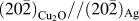 $\left( {20\bar 2} \right)_{{\rm{Cu}}_{\rm{2}} {\rm{O}}} //\left( {20\bar 2} \right){}_{{\rm{Ag}}}$ . After brazing at 900 °C/6 h, a two-phase (α-Ti + Ti2Cu) region was observed on the Ti side with
 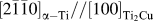 $\left[ {2\bar 1\bar 10} \right]_{{\rm{\alpha - Ti}}} //\left[ {100} \right]_{{\rm{Ti}}_{\rm{2}} {\rm{Cu}}}$ and
 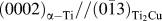 $\left( {0002} \right)_{{\rm{\alpha - Ti}}} //\left( {0\bar 13} \right)_{{\rm{Ti}}_{\rm{2}} {\rm{Cu}}}$ , while the TiCu layer grew at the expense of Ti3Cu4 and TiCu4. The bonding mechanisms and diffusion paths were explored with the aid of Ag–Cu–Ti and Ti–Cu–O ternary phase diagrams.


JMR volume 29 issue 4 Cover and Front matter
-
- Journal:
- Journal of Materials Research / Volume 29 / Issue 4 / 28 February 2014
- Published online by Cambridge University Press:
- 03 March 2014, pp. f1-f5
- Print publication:
- 28 February 2014
-
- Article
-
- You have access
- Export citation
