To save content items to your account,
please confirm that you agree to abide by our usage policies.
If this is the first time you use this feature, you will be asked to authorise Cambridge Core to connect with your account.
Find out more about saving content to .
To save content items to your Kindle, first ensure no-reply@cambridge.org
is added to your Approved Personal Document E-mail List under your Personal Document Settings
on the Manage Your Content and Devices page of your Amazon account. Then enter the ‘name’ part
of your Kindle email address below.
Find out more about saving to your Kindle.
Note you can select to save to either the @free.kindle.com or @kindle.com variations.
‘@free.kindle.com’ emails are free but can only be saved to your device when it is connected to wi-fi.
‘@kindle.com’ emails can be delivered even when you are not connected to wi-fi, but note that service fees apply.
A series of TiO2 samples were prepared from the precursor P25 using impregnation method followed by a calcination. The phase compositions of the samples were controlled by addition of various additives, including NaNO3, NaHCO3, Na2SO4, Na3PO4, Na2SiO3, and Na2MoO4. X-ray diffraction patterns demonstrate that the phase transformation from anatase to rutile can be suppressed to a different extent by these additives. Photocatalytic H2 production reactions were performed in methanol aqueous solution. The catalysts, with the addition of NaNO3, NaHCO3, and Na2SO4, show a great improvement in the H2 production activity, while others show a negative effect. This indicates that both the phase structure and the properties of the additives are important for the photocatalytic activity. Infrared spectra of pyridine adsorption on the catalysts reveal that the decreased acidity of the samples may be responsible for the suppressed CO production in the photocatalytic methanol reforming.
The hydrophobic rutile titania aerogels were successfully prepared by nonalkoxide ambient pressure drying through a modification process. The resulted materials were characterized by x-ray diffraction, scanning electronic microscope, transmission electron microscope, contact angle analyzer, Brunauer–Emmett–Teller specific surface area, and ultraviolet (UV)–visible diffuse reflection spectrum. The experimental results demonstrated that the as-prepared samples nanoparticles with rutile crystalline structure were uniformly distributed. The UV-resistant hydrophobic samples having high surface area were used as photocatalysts for dye degradation reaction.
TiO2@C core–shell nanostructures with various crystal structures of TiO2-B, anatase, and rutile were successfully synthesized by a simple hydrothermal process and postheat treatments. As-synthesized precursor hydrogen titanate@carbonaceous nanoribbons transformed into TiO2-B@C nanoribbons at 400 °C and further transformed into anatase and rutile TiO2@C nanoribbons at 700 and 800 °C, respectively. The morphology of nanoribbons can be retained up to 800 °C. The transformation temperature (800 °C) from anatase to rutile phase is lower than that of TiO2 nanofibers without carbon layers and anatase TiO2@C nanoparticles. These results show that the carbon shell plays important roles in promoting the phase transition from anatase to rutile phase and protecting the nanoribbon-like morphology. The formation mechanism of the TiO2@C core–shell nanostructures with various crystal structures was discussed.
We report on the photovoltaic properties of polymer solar cells with an inverted structure wherein the electron-collecting electrode comprises an indium tin oxide (ITO) electrode coated with titanium dioxide (TiO2) nanoparticles dispersed into poly(N-vinylpyrrolidone) (PVP). The optimization of performance of polymer solar cells in which the TiO2 concentration in PVP was varied is presented. Pristine solar cells with the TiO2:PVP-coated ITO electrodes showed S-shape current-voltage characteristics. The S-shaped feature disappeared after the continuous exposure of the solar cells to light from an AM 1.5G solar simulator, leading to an improved device performance compared with solar cells that use an ITO or ITO/TiO2 electron-collecting electrodes.
In this article, we review our recent research efforts on the synthesis, crystallinity control, and photocatalysis of nanostructured titanium dioxide (TiO2) shells. First, we introduce several synthetic methods for preparing TiO2 shell structures using either template-free or template-assisted approaches. Several methods to change the structures from amorphous to crystalline and subsequently ways to enhance the crystallinity are then discussed, including those involving the “silica-protected calcination” and “partial etching and recalcination” strategies. We also discuss the photocatalytic applications of the TiO2 nanoshells and the methods for improving their catalytic activities. Finally, we conclude with a summary and our perspective on the further development of the nanostructured TiO2 shells. It is believed that more rational design and modification strategies such as well-controlled nonmetal doping, plasmonic metal decoration and the hybridization with other semiconducting materials will significantly enhance the photocatalytic efficiency of TiO2-based catalyst materials.
Use of nanostructured TiO2 for photocatalysis is a cost-effective and sustainable technology. However, to make this an attractive viable technology will require the design of TiO2 photocatalyst capable of harnessing the energy of visible light. One possible solution is the doping of TiO2 to reduce its band gap. In this paper, the effect of Cr-doping by ion implantation on the in situ crystallization and phase stability of anodic TiO2 nanotubes at elevated temperature is described. Cr-doping has dramatically reduced the fraction of anatase-to-rutile transformation and lowered the crystallization temperature of anatase from 600 to 400 °C and rutile from 600 to 500 °C. Ion beam analysis by Rutherford backscattering spectrometry has confirmed the existence of Cr ions composition gradation in doped TiO2 nanotubes. The real doping of Ti lattices with Cr ions was evidenced by the analyses of surface compositions and chemical states of the nanotubes using x-ray photoelectron spectroscopy.
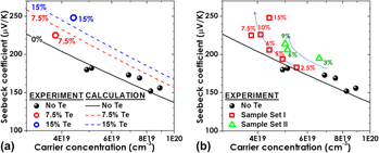
We present experimental evidence of enhancement of thermoelectric properties in tellurium (Te) nanoparticle-embedded bismuth antimony telluride (BiSbTe) alloys. Bi0.5Sb1.5Te3 films with a high density of Te particles of 10–20 nm size were prepared by growth of alternating multilayers of ultrathin Te and Bi0.5Sb1.5Te3. As the amount of Te nanoinclusions increased up to ∼15%, the Seebeck coefficient and thermoelectric power factor were increased. Based on the concept of band bending at heterointerfaces as a carrier energy filter, the energy relaxation calculation was made to confirm that the Te nanoinclusions result in a carrier energy filtering effect in p-type bismuth antimony telluride. In addition, thermal conductivities were reduced in the Te-embedded samples, permitting possible further enhancement of the thermoelectric figure of merit. The advantages of Te nanoinclusions in p-type Bi0.5Sb1.5Te3alloys on thermoelectric performance are experimentally realized by both electron- and phonon scattering.
ZnO/TiO2 heterojunction composite fibers were prepared via a physical route, i.e., first electrospinning titanium dioxide (TiO2) fibers, then pulse plating zinc (Zn) on the fibers, and at last thermal treating the fibers. The morphologies, phase structures, and photocatalytic property of the composite fibers were characterized by using field-emission gun scanning electron microscope, x-ray diffractometer, high-resolution transmission electron microscope, and ultraviolet–visible spectrophotometer. It was found that a full or partial lattice coherent heterojunction was formed between the TiO2 fibers and zinc oxide (ZnO) particles, due to thermal treatment at 400 °C, which simultaneously resulted in the phase transformations including Zn to ZnO and amorphous TiO2 to anatase TiO2. The experimental results demonstrated that the photocatalytic activity of the composite fibers was improved and exhibited a value more than two times higher than that of TiO2 fibers.
Advances in nanotechnology have prompted rapid progress and versatile imaging modalities for diagnostics and treatment of diseases. Molecular imaging is a powerful technique for quantifying physiological changes in vivo using noninvasive imaging probes. These probes are used to image specific cells and tissues within a whole organism. Currently, imaging is an essential part of clinical protocols providing morphological, structural, metabolic and functional information. Using theranostic micro- or nanoparticles, which combine both therapeutic and diagnostic capabilities in one single entity, holds a true promise to propel the biomedical field toward personalized medicine. With this approach, biological processes can be directly and simultaneously monitored with the treatment of the diseases. This mini-review highlights the recent innovative diagnostic imaging aspects of porous silicon (PSi) materials and emphasizes their potential as theranostic platforms and tools for the clinic. Multiple biomedical imaging applications of the PSi materials are also outlined.
A new technique based on the detection of the amplitude of the second harmonic was described in a previous paper. To compute the elastic modulus and the hardness of materials, the technique uses only the derivative of the contact radius with respect to the indentation depth. For this reason, this method is applicable only to homogeneous materials. In this paper, the method is extended to any materials with constant Young modulus. The indentation depth value is not needed at all, thus eliminating uncertainties related to the displacement measurement, which are very influent at small penetration depths. Furthermore, we also explain how to compute the indentation depth from the detection of the amplitude of the second harmonic. This new measurement technique was tested on three samples: fused silica, Poly(methyl methacrylate) (PMMA), and calcite, which is expected to exhibit indentation size effect. The obtained results show that mechanical properties and the indentation depth can be determined with good accuracy for penetration depths between 25 and 100 nm using this method.
In this paper, a novel sintering method is introduced for the forming of microcomponents in which the loose powders were loaded directly into the die, sintered with an external electric field, a thermal field, and an external stress field (called coupled multifields activation), where the fields were generated by a Gleeble thermal simulation instrument. Two kinds of 316L stainless steel powders of different particle sizes (20 and 70 μm) with no binder were sintered with microforming using a multifield coupling method. For particle size of 20 μm, a nearly fully densified microsintered compact (relative density is 99.2%) was manufactured at a relatively low sintering temperature (900 °C) and within a relatively short sintering time (4 min). The fluctuated temperature–time curve reveals that the rapid mass transfer of liquid phase is the dominant densification mechanism in the compacts with a starting particle size of 20 μm.
Backscattered electron microscopy provides gray-level contrast resulting from variations in atomic composition. Through the use of reference materials, quantitative backscattered electron (qBSE) imaging can be used to measure the mineral content of mineralized tissues at submicron resolution. We have developed novel tunable reference materials that can be adjusted for analysis of an individual tissue or a wide range of tissues with variable atomic density. As an alternative to conventional metallic reference materials, these amorphous materials maintain long-term stability and possess no long-range order that may induce channeling contrast. Using these reference materials, we characterized the mineral content of a broad range of mineralized tissues from immature mouse femur to whale bulla. Mineral volume fraction correlated to more traditional measurements of mineral content with microcomputed tomography and ashing techniques. Further, we demonstrate the advantage of location-matched measurements of nanomechanical properties and qBSE mineral content.
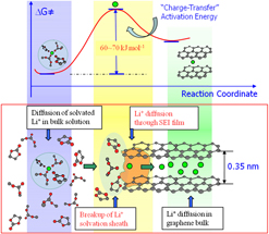
In any electrochemical device, the interface between electrolyte and electrode should be the only “legitimate” location where redox reactions happen. Particularly in Li ion batteries, these interfaces become “interphases” due to the reactivity of the electrode materials used, and they mainly consist of chemical species from the sacrificial decomposition of electrolyte components. Since the emergence of Li ion technology, it has been recognized that interphase on graphitic anodes, usually referred as SEI (solid electrolyte interphase) after its electrolyte attributes, is the key component supporting the reversibility of Li+-intercalation chemistry. Research attention focused on this component during the past two decades has led to substantial understanding about both its chemistry and mechanism. This article summarizes these progresses, and elaborates on the relatively recent insights, including the effect of Li+-solvation sheath structure on the interphasial processes at graphitic anode. A new strategy of forming a more desirable interphase is also discussed.
Nitrogen-doped titanium dioxide (denoted as N-doped TiO2) nanomaterials were prepared through the ion exchange of sodium titanate nanotube (the precursor; denoted as STN) with aqueous NH4Cl and follow-up sintering at different temperatures in air. The morphology, structure, surface component, and optical properties of as-obtained N-doped TiO2 nanomaterials have been analyzed by transmission electron microscopy, x-ray diffraction, x-ray photoelectron spectroscopy, and ultraviolet–visible light diffuse reflectance spectrometry. The formation mechanism and the origin of the visible light absorption for N-doped TiO2 nanomaterials have been discussed. Moreover, the thermogravimetric analysis and differential thermal analysis of N-doped TiO2 nanomaterial calcined at 100 °C are conducted as an example to examine the thermal stability of as-synthesized N-doped TiO2. It has been found that, as the calcination temperature rises, the initial nanotubular morphology of STN is transformed to the final nanoscale granular one, accompanied by a phase transformation from orthorhombic crystalline system to anatase TiO2. The N content in N-doped TiO2 is 7.04%, 6.22%, 3.20%, 1.14%, 0.61%, and 0.40% (atomic percentage), depending on calcination temperature rising from 100 to 600 °C. Moreover, N-doped TiO2 samples experience three stages of weight losses, and that calcinated at 300 °C and above have strong visible light absorption, due to the formation of Ti–O–N bonds thereat.
The Nd1.7Ba0.3Ni0.9Cr0.1O4+δ polycrystalline sample was synthesized by the sol–gel process and a subsequent annealing at 1523 K in 1 atm of flowing argon. X-ray diffraction (XRD) analysis and electrical transport properties have been investigated as well. The oxygen non-stoichiometry was determined by iodometric titration. The sample shows adoption of the K2NiF4-type structure based on a tolerance factor calculation. Rietveld refinement of the crystal structure from X-ray powder diffraction data confirmed that Nd1.7Ba0.3Ni0.9Cr0.1O4+δ adopts the tetragonal structure (space group I4/mmm, Z = 2). The room temperature unit-cell parameters are determined to be a = 3.82515(2) and c = 12.47528(6) Å. The reliability factors are: RB = 0.043, Rwp = 0.012 and χ2 = 3.00. The Nd1.7Ba0.3Ni0.9Cr0.1O4+δ compound exhibits a semi-conductive behaviour. The electrical transport mechanism has been investigated and it agrees with the adiabatic small polaron hopping model in the temperature range 313 K ≤ T ≤ 708 K.