Refine search
Actions for selected content:
106117 results in Materials Science
South Africa and Italy step up S&T cooperation
-
- Journal:
- MRS Bulletin / Volume 39 / Issue 10 / October 2014
- Published online by Cambridge University Press:
- 15 October 2014, p. 841
- Print publication:
- October 2014
-
- Article
-
- You have access
- HTML
- Export citation
Rodney S. Ruoff selected for 2014 David Turnbull Lectureship
-
- Journal:
- MRS Bulletin / Volume 39 / Issue 10 / October 2014
- Published online by Cambridge University Press:
- 15 October 2014, p. 896
- Print publication:
- October 2014
-
- Article
-
- You have access
- HTML
- Export citation
Marvin L. Cohen to receive 2014 Von Hippel Award for modeling of materials and nanoscale structures
-
- Journal:
- MRS Bulletin / Volume 39 / Issue 10 / October 2014
- Published online by Cambridge University Press:
- 15 October 2014, pp. 895-896
- Print publication:
- October 2014
-
- Article
-
- You have access
- HTML
- Export citation
Preview: 2014 Materials Research Society Fall Meeting & Exhibit: www.mrs.org/fall2014
-
- Journal:
- MRS Bulletin / Volume 39 / Issue 10 / October 2014
- Published online by Cambridge University Press:
- 15 October 2014, pp. 891-894
- Print publication:
- October 2014
-
- Article
-
- You have access
- HTML
- Export citation
CAREER CENTRAL
-
- Journal:
- MRS Bulletin / Volume 39 / Issue 10 / October 2014
- Published online by Cambridge University Press:
- 15 October 2014, pp. 911-919
- Print publication:
- October 2014
-
- Article
-
- You have access
- Export citation
Bio Focus: Conducting polymers utilized to overcome electrode limits in ionic transport systems
-
- Journal:
- MRS Bulletin / Volume 39 / Issue 10 / October 2014
- Published online by Cambridge University Press:
- 15 October 2014, pp. 838-839
- Print publication:
- October 2014
-
- Article
-
- You have access
- HTML
- Export citation
Long-Qing Chen receives 2014 Materials Theory Award
-
- Journal:
- MRS Bulletin / Volume 39 / Issue 10 / October 2014
- Published online by Cambridge University Press:
- 15 October 2014, p. 897
- Print publication:
- October 2014
-
- Article
-
- You have access
- HTML
- Export citation
In pursuit of damage tolerance in engineering and biological materials
-
- Journal:
- MRS Bulletin / Volume 39 / Issue 10 / October 2014
- Published online by Cambridge University Press:
- 15 October 2014, pp. 880-890
- Print publication:
- October 2014
-
- Article
- Export citation
Introduction to Nanoscience and Nanomaterials Dinesh C. Agrawal: World Scientific, 2013 556 pages, $89.00 ISBN 978-981-4397-97-1
-
- Journal:
- MRS Bulletin / Volume 39 / Issue 10 / October 2014
- Published online by Cambridge University Press:
- 15 October 2014, p. 909
- Print publication:
- October 2014
-
- Article
-
- You have access
- HTML
- Export citation
Mercouri G. Kanatzidis selected as MRS Medalist for nanostructured thermoelectric materials
-
- Journal:
- MRS Bulletin / Volume 39 / Issue 10 / October 2014
- Published online by Cambridge University Press:
- 15 October 2014, pp. 897-898
- Print publication:
- October 2014
-
- Article
-
- You have access
- HTML
- Export citation
Nano Focus: Precursor molecules enable custom-made CNTs
-
- Journal:
- MRS Bulletin / Volume 39 / Issue 10 / October 2014
- Published online by Cambridge University Press:
- 15 October 2014, p. 837
- Print publication:
- October 2014
-
- Article
-
- You have access
- HTML
- Export citation
Calendar of Short Courses & Workshops Powder Diffraction September 2014
-
- Journal:
- Powder Diffraction / Volume 29 / Issue 3 / September 2014
- Published online by Cambridge University Press:
- 30 September 2014, p. 316
-
- Article
-
- You have access
- HTML
- Export citation
Calendar of Forthcoming Meetings Powder Diffraction September 2014
-
- Journal:
- Powder Diffraction / Volume 29 / Issue 3 / September 2014
- Published online by Cambridge University Press:
- 30 September 2014, p. 315
-
- Article
-
- You have access
- HTML
- Export citation
JMR volume 29 issue 19 Cover and Back matter
-
- Journal:
- Journal of Materials Research / Volume 29 / Issue 19 / 14 October 2014
- Published online by Cambridge University Press:
- 30 September 2014, pp. b1-b3
- Print publication:
- 14 October 2014
-
- Article
-
- You have access
- Export citation
JMR volume 29 issue 19 Cover and Front matter
-
- Journal:
- Journal of Materials Research / Volume 29 / Issue 19 / 14 October 2014
- Published online by Cambridge University Press:
- 30 September 2014, pp. f1-f4
- Print publication:
- 14 October 2014
-
- Article
-
- You have access
- Export citation
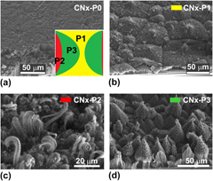
Pine-tree-like morphologies of nitrogen-doped carbon nanotubes: Electron field emission enhancement
-
- Journal:
- Journal of Materials Research / Volume 29 / Issue 20 / 28 October 2014
- Published online by Cambridge University Press:
- 30 September 2014, pp. 2441-2450
- Print publication:
- 28 October 2014
-
- Article
- Export citation
International Report: BCA Spring Meeting 2014
-
- Journal:
- Powder Diffraction / Volume 29 / Issue 3 / September 2014
- Published online by Cambridge University Press:
- 30 September 2014, pp. 311-314
-
- Article
- Export citation
PDJ volume 29 issue 3 Cover and Back matter
-
- Journal:
- Powder Diffraction / Volume 29 / Issue 3 / September 2014
- Published online by Cambridge University Press:
- 30 September 2014, pp. b1-b4
-
- Article
-
- You have access
- Export citation

Plasma spraying of cerium-doped YAG
-
- Journal:
- Journal of Materials Research / Volume 29 / Issue 19 / 14 October 2014
- Published online by Cambridge University Press:
- 30 September 2014, pp. 2344-2351
- Print publication:
- 14 October 2014
-
- Article
- Export citation
Introduction
-
- Journal:
- Journal of Materials Research / Volume 29 / Issue 19 / 14 October 2014
- Published online by Cambridge University Press:
- 30 September 2014, p. 2251
- Print publication:
- 14 October 2014
-
- Article
- Export citation
