Refine search
Actions for selected content:
106129 results in Materials Science
Liquid phase electron microscopy of biological specimens
-
- Journal:
- MRS Bulletin / Volume 45 / Issue 9 / September 2020
- Published online by Cambridge University Press:
- 10 September 2020, pp. 754-760
- Print publication:
- September 2020
-
- Article
- Export citation
Mrs Movers & Shakers: Spread the good news!
-
- Journal:
- MRS Bulletin / Volume 45 / Issue 9 / September 2020
- Published online by Cambridge University Press:
- 10 September 2020, pp. 783-784
- Print publication:
- September 2020
-
- Article
-
- You have access
- HTML
- Export citation
How to define your “field of endeavor” for your EB-1A/NIW petition
-
- Journal:
- MRS Bulletin / Volume 45 / Issue 9 / September 2020
- Published online by Cambridge University Press:
- 10 September 2020, p. 782
- Print publication:
- September 2020
-
- Article
-
- You have access
- HTML
- Export citation

In situ electrochemical scanning/transmission electron microscopy of electrode–electrolyte interfaces
-
- Journal:
- MRS Bulletin / Volume 45 / Issue 9 / September 2020
- Published online by Cambridge University Press:
- 10 September 2020, pp. 738-745
- Print publication:
- September 2020
-
- Article
- Export citation
MRC volume 10 issue 3 Cover and Back matter
-
- Journal:
- MRS Communications / Volume 10 / Issue 3 / September 2020
- Published online by Cambridge University Press:
- 14 September 2020, pp. b1-b2
- Print publication:
- September 2020
-
- Article
-
- You have access
- Export citation

Electron-beam-driven chemical processes during liquid phase transmission electron microscopy
-
- Journal:
- MRS Bulletin / Volume 45 / Issue 9 / September 2020
- Published online by Cambridge University Press:
- 10 September 2020, pp. 746-753
- Print publication:
- September 2020
-
- Article
- Export citation
Materials science, energy transition, and the pandemic
-
- Journal:
- MRS Bulletin / Volume 45 / Issue 9 / September 2020
- Published online by Cambridge University Press:
- 10 September 2020, p. 697
- Print publication:
- September 2020
-
- Article
-
- You have access
- HTML
- Export citation
Takiya Ahmed Foskey Chemist and community outreach champion
-
- Journal:
- MRS Bulletin / Volume 45 / Issue 9 / September 2020
- Published online by Cambridge University Press:
- 10 September 2020, p. 781
- Print publication:
- September 2020
-
- Article
-
- You have access
- HTML
- Export citation
Liquid phase transmission electron microscopy for imaging of nanoscale processes in solution
-
- Journal:
- MRS Bulletin / Volume 45 / Issue 9 / September 2020
- Published online by Cambridge University Press:
- 10 September 2020, pp. 704-712
- Print publication:
- September 2020
-
- Article
-
- You have access
- HTML
- Export citation
MRS plans joint 2022 MRS Spring Meeting events with societies in South Korea, Japan, and Singapore
-
- Journal:
- MRS Bulletin / Volume 45 / Issue 9 / September 2020
- Published online by Cambridge University Press:
- 10 September 2020, pp. 779-780
- Print publication:
- September 2020
-
- Article
-
- You have access
- HTML
- Export citation
Greening the production and utilization of ammonia
-
- Journal:
- MRS Bulletin / Volume 45 / Issue 9 / September 2020
- Published online by Cambridge University Press:
- 10 September 2020, pp. 698-699
- Print publication:
- September 2020
-
- Article
-
- You have access
- HTML
- Export citation
Chemical and bonding analysis of liquids using liquid cell electron microscopy
-
- Journal:
- MRS Bulletin / Volume 45 / Issue 9 / September 2020
- Published online by Cambridge University Press:
- 10 September 2020, pp. 761-768
- Print publication:
- September 2020
-
- Article
- Export citation
MRS Journal Highlights
-
- Journal:
- MRS Bulletin / Volume 45 / Issue 9 / September 2020
- Published online by Cambridge University Press:
- 10 September 2020, p. 703
- Print publication:
- September 2020
-
- Article
-
- You have access
- HTML
- Export citation
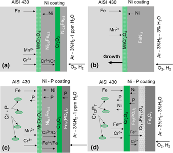
Corrosion of nickel and nickel–phosphorous-coated AISI 430 in dry (Ar–3%H2) and humid hydrogen (Ar–3%H2–3%H2O) atmosphere
-
- Journal:
- Journal of Materials Research , First View
- Published online by Cambridge University Press:
- 01 September 2020, pp. 1-11
-
- Article
- Export citation
MRC volume 10 issue 3 Cover and Front matter
-
- Journal:
- MRS Communications / Volume 10 / Issue 3 / September 2020
- Published online by Cambridge University Press:
- 14 September 2020, pp. f1-f5
- Print publication:
- September 2020
-
- Article
-
- You have access
- Export citation
Smart textiles respond to human emotions
-
- Journal:
- MRS Bulletin / Volume 45 / Issue 9 / September 2020
- Published online by Cambridge University Press:
- 10 September 2020, pp. 694-696
- Print publication:
- September 2020
-
- Article
-
- You have access
- HTML
- Export citation
High-speed video analyzes microcracking during additive manufacturing of tungsten
-
- Journal:
- MRS Bulletin / Volume 45 / Issue 9 / September 2020
- Published online by Cambridge University Press:
- 10 September 2020, p. 686
- Print publication:
- September 2020
-
- Article
-
- You have access
- HTML
- Export citation
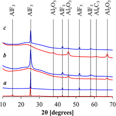
Balancing processing ease with combustion performance in aluminum/PVDF energetic filaments
-
- Journal:
- Journal of Materials Research , First View
- Published online by Cambridge University Press:
- 01 September 2020, pp. 1-8
-
- Article
- Export citation
MRS volume 45 issue 9 Cover and Back matter
-
- Journal:
- MRS Bulletin / Volume 45 / Issue 9 / September 2020
- Published online by Cambridge University Press:
- 10 September 2020, pp. b1-b2
- Print publication:
- September 2020
-
- Article
-
- You have access
- Export citation
MRS volume 45 issue 9 Cover and Front matter
-
- Journal:
- MRS Bulletin / Volume 45 / Issue 9 / September 2020
- Published online by Cambridge University Press:
- 10 September 2020, pp. f1-f6
- Print publication:
- September 2020
-
- Article
-
- You have access
- Export citation
