Refine search
Actions for selected content:
106129 results in Materials Science
MRS volume 44 issue 12 Cover and Back matter
-
- Journal:
- MRS Bulletin / Volume 44 / Issue 12 / December 2019
- Published online by Cambridge University Press:
- 10 December 2019, pp. b1-b2
- Print publication:
- December 2019
-
- Article
-
- You have access
- Export citation
MRC volume 9 issue 4 Cover and Back matter
-
- Journal:
- MRS Communications / Volume 9 / Issue 4 / December 2019
- Published online by Cambridge University Press:
- 16 December 2019, pp. b1-b2
- Print publication:
- December 2019
-
- Article
-
- You have access
- Export citation
US Department of Energy celebrates 10 years at the frontiers of energy research
-
- Journal:
- MRS Bulletin / Volume 44 / Issue 12 / December 2019
- Published online by Cambridge University Press:
- 10 December 2019, pp. 912-913
- Print publication:
- December 2019
-
- Article
-
- You have access
- HTML
- Export citation
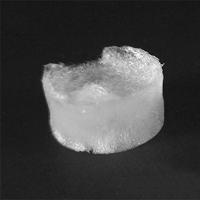
Cryogenic transmission electron microscopy for materials research
-
- Journal:
- MRS Bulletin / Volume 44 / Issue 12 / December 2019
- Published online by Cambridge University Press:
- 10 December 2019, pp. 924-928
- Print publication:
- December 2019
-
- Article
-
- You have access
- HTML
- Export citation
The G-filter: A simple high-tech solution to India’s water pollution
-
- Journal:
- MRS Bulletin / Volume 44 / Issue 12 / December 2019
- Published online by Cambridge University Press:
- 10 December 2019, pp. 914-915
- Print publication:
- December 2019
-
- Article
-
- You have access
- HTML
- Export citation

Soft matter and nanomaterials characterization by cryogenic transmission electron microscopy
-
- Journal:
- MRS Bulletin / Volume 44 / Issue 12 / December 2019
- Published online by Cambridge University Press:
- 10 December 2019, pp. 942-948
- Print publication:
- December 2019
-
- Article
- Export citation
CAREER CENTRAL
-
- Journal:
- MRS Bulletin / Volume 44 / Issue 12 / December 2019
- Published online by Cambridge University Press:
- 10 December 2019, pp. 974-975
- Print publication:
- December 2019
-
- Article
-
- You have access
- Export citation
LOOK AGAIN
-
- Journal:
- MRS Bulletin / Volume 44 / Issue 12 / December 2019
- Published online by Cambridge University Press:
- 10 December 2019, p. 976
- Print publication:
- December 2019
-
- Article
-
- You have access
- HTML
- Export citation

Damage in electron cryomicroscopy: Lessons from biology for materials science
-
- Journal:
- MRS Bulletin / Volume 44 / Issue 12 / December 2019
- Published online by Cambridge University Press:
- 10 December 2019, pp. 935-941
- Print publication:
- December 2019
-
- Article
- Export citation
PDJ volume 34 issue 4 Cover and Back matter
-
- Journal:
- Powder Diffraction / Volume 34 / Issue 4 / December 2019
- Published online by Cambridge University Press:
- 29 November 2019, pp. b1-b10
-
- Article
-
- You have access
- Export citation
Special section: Crystallography and properties of metal–organic framework compounds
-
- Journal:
- Powder Diffraction / Volume 34 / Issue 4 / December 2019
- Published online by Cambridge University Press:
- 29 November 2019, p. 295
-
- Article
-
- You have access
- HTML
- Export citation
2019 American Crystallographic Association Annual Meeting Report
-
- Journal:
- Powder Diffraction / Volume 34 / Issue 4 / December 2019
- Published online by Cambridge University Press:
- 29 November 2019, pp. 396-399
-
- Article
- Export citation
Calendar of Short Courses and Workshops
-
- Journal:
- Powder Diffraction / Volume 34 / Issue 4 / December 2019
- Published online by Cambridge University Press:
- 29 November 2019, p. 402
-
- Article
-
- You have access
- HTML
- Export citation
PDJ volume 34 issue 4 Cover and Front matter
-
- Journal:
- Powder Diffraction / Volume 34 / Issue 4 / December 2019
- Published online by Cambridge University Press:
- 29 November 2019, pp. f1-f6
-
- Article
-
- You have access
- Export citation
Calendar of Forthcoming Meetings
-
- Journal:
- Powder Diffraction / Volume 34 / Issue 4 / December 2019
- Published online by Cambridge University Press:
- 29 November 2019, pp. 400-401
-
- Article
-
- You have access
- HTML
- Export citation
Powder Diffraction Journal is now Open Access – Hybrid
-
- Journal:
- Powder Diffraction / Volume 34 / Issue 4 / December 2019
- Published online by Cambridge University Press:
- 29 November 2019, p. 296
-
- Article
-
- You have access
- HTML
- Export citation
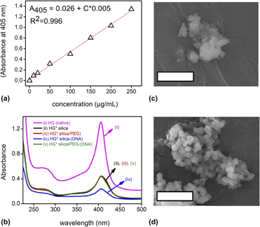
Sol–gel derived silica/polyethylene glycol hybrids as potential oligonucleotide vectors
-
- Journal:
- Journal of Materials Research / Volume 34 / Issue 22 / 28 November 2019
- Published online by Cambridge University Press:
- 28 November 2019, pp. 3787-3797
- Print publication:
- 28 November 2019
-
- Article
- Export citation
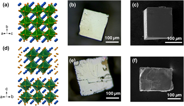
Crystal growth and structural analysis of perovskite chalcogenide BaZrS3 and Ruddlesden–Popper phase Ba3Zr2S7
-
- Journal:
- Journal of Materials Research / Volume 34 / Issue 22 / 28 November 2019
- Published online by Cambridge University Press:
- 28 November 2019, pp. 3819-3826
- Print publication:
- 28 November 2019
-
- Article
- Export citation
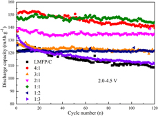
Rheological phase reaction method synthesis and characterizations of xLiMn0.5Fe0.5PO4–yLi3V2(PO4)3/C composites as cathode materials for lithium ion batteries
-
- Journal:
- Journal of Materials Research / Volume 35 / Issue 1 / 14 January 2020
- Published online by Cambridge University Press:
- 28 November 2019, pp. 2-11
- Print publication:
- 14 January 2020
-
- Article
- Export citation
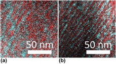
Phase evolution of radio frequency magnetron sputtered Cr-rich (Cr,Zr)2O3 coatings studied by in situ synchrotron X-ray diffraction during annealing in air or vacuum
-
- Journal:
- Journal of Materials Research / Volume 34 / Issue 22 / 28 November 2019
- Published online by Cambridge University Press:
- 28 November 2019, pp. 3735-3746
- Print publication:
- 28 November 2019
-
- Article
-
- You have access
- Open access
- HTML
- Export citation
