Refine search
Actions for selected content:
106129 results in Materials Science
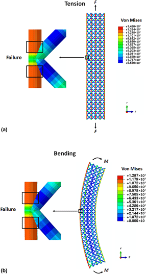
Mechanical and failure behaviors of lattice–plate hybrid structures
-
- Journal:
- MRS Communications / Volume 10 / Issue 1 / March 2020
- Published online by Cambridge University Press:
- 03 December 2019, pp. 42-54
- Print publication:
- March 2020
-
- Article
- Export citation
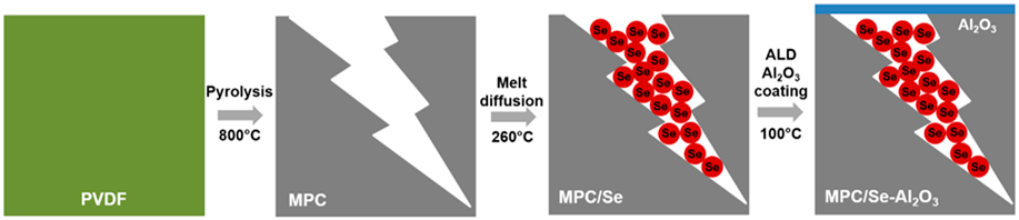
Nanoscale Al2O3 coating to stabilize selenium cathode for sodium–selenium batteries
-
- Journal:
- Journal of Materials Research / Volume 35 / Issue 7 / 14 April 2020
- Published online by Cambridge University Press:
- 02 December 2019, pp. 747-755
- Print publication:
- 14 April 2020
-
- Article
-
- You have access
- HTML
- Export citation
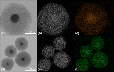
General strategy for doping rare earth metals into Au–ZnO core–shell nanospheres
-
- Journal:
- Journal of Materials Research / Volume 34 / Issue 23 / 16 December 2019
- Published online by Cambridge University Press:
- 02 December 2019, pp. 3877-3886
- Print publication:
- 16 December 2019
-
- Article
- Export citation
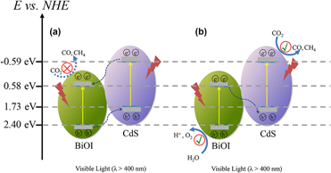
Construction of visible light–responsive Z-scheme CdS/BiOI photocatalyst with enhanced photocatalytic CO2 reduction activity
-
- Journal:
- Journal of Materials Research / Volume 34 / Issue 23 / 16 December 2019
- Published online by Cambridge University Press:
- 02 December 2019, pp. 3907-3917
- Print publication:
- 16 December 2019
-
- Article
- Export citation
MRS Journal Highlights
-
- Journal:
- MRS Bulletin / Volume 44 / Issue 12 / December 2019
- Published online by Cambridge University Press:
- 10 December 2019, p. 916
- Print publication:
- December 2019
-
- Article
-
- You have access
- HTML
- Export citation
Recommended reading
-
- Journal:
- MRS Bulletin / Volume 44 / Issue 12 / December 2019
- Published online by Cambridge University Press:
- 10 December 2019, p. 967
- Print publication:
- December 2019
-
- Article
-
- You have access
- HTML
- Export citation
Fuel efficiency drives the auto industry to reduce vehicle weight
-
- Journal:
- MRS Bulletin / Volume 44 / Issue 12 / December 2019
- Published online by Cambridge University Press:
- 10 December 2019, pp. 920-923
- Print publication:
- December 2019
-
- Article
-
- You have access
- HTML
- Export citation

Cryo-electron microscopy instrumentation and techniques for life sciences and materials science
-
- Journal:
- MRS Bulletin / Volume 44 / Issue 12 / December 2019
- Published online by Cambridge University Press:
- 10 December 2019, pp. 929-934
- Print publication:
- December 2019
-
- Article
- Export citation
Surface tension determines tissue shape during growth
-
- Journal:
- MRS Bulletin / Volume 44 / Issue 12 / December 2019
- Published online by Cambridge University Press:
- 10 December 2019, p. 909
- Print publication:
- December 2019
-
- Article
-
- You have access
- HTML
- Export citation

Cryogenic electron microscopy for quantum science
-
- Journal:
- MRS Bulletin / Volume 44 / Issue 12 / December 2019
- Published online by Cambridge University Press:
- 10 December 2019, pp. 961-966
- Print publication:
- December 2019
-
- Article
- Export citation
The allure of the bezoar endures
-
- Journal:
- MRS Bulletin / Volume 44 / Issue 12 / December 2019
- Published online by Cambridge University Press:
- 10 December 2019, p. 968
- Print publication:
- December 2019
-
- Article
-
- You have access
- HTML
- Export citation
Little precious lithium?
-
- Journal:
- MRS Bulletin / Volume 44 / Issue 12 / December 2019
- Published online by Cambridge University Press:
- 10 December 2019, p. 917
- Print publication:
- December 2019
-
- Article
-
- You have access
- HTML
- Export citation
There’s a lithium battery in your future
-
- Journal:
- MRS Bulletin / Volume 44 / Issue 12 / December 2019
- Published online by Cambridge University Press:
- 10 December 2019, pp. 918-919
- Print publication:
- December 2019
-
- Article
-
- You have access
- HTML
- Export citation
3D carbon nanotubes and graphene sheets grown simultaneously on copper
-
- Journal:
- MRS Bulletin / Volume 44 / Issue 12 / December 2019
- Published online by Cambridge University Press:
- 10 December 2019, pp. 910-911
- Print publication:
- December 2019
-
- Article
-
- You have access
- HTML
- Export citation

Microcrystal electron diffraction methodology and applications
-
- Journal:
- MRS Bulletin / Volume 44 / Issue 12 / December 2019
- Published online by Cambridge University Press:
- 10 December 2019, pp. 956-960
- Print publication:
- December 2019
-
- Article
- Export citation
Thermal oxidation toughens carbon fiber/polysulfone composites
-
- Journal:
- MRS Bulletin / Volume 44 / Issue 12 / December 2019
- Published online by Cambridge University Press:
- 10 December 2019, p. 910
- Print publication:
- December 2019
-
- Article
-
- You have access
- HTML
- Export citation

Cryogenic specimens for nanoscale characterization of solid–liquid interfaces
-
- Journal:
- MRS Bulletin / Volume 44 / Issue 12 / December 2019
- Published online by Cambridge University Press:
- 10 December 2019, pp. 949-955
- Print publication:
- December 2019
-
- Article
- Export citation
MRS volume 44 issue 12 Cover and Front matter
-
- Journal:
- MRS Bulletin / Volume 44 / Issue 12 / December 2019
- Published online by Cambridge University Press:
- 10 December 2019, pp. f1-f6
- Print publication:
- December 2019
-
- Article
-
- You have access
- Export citation
MRC volume 9 issue 4 Cover and Front matter
-
- Journal:
- MRS Communications / Volume 9 / Issue 4 / December 2019
- Published online by Cambridge University Press:
- 16 December 2019, pp. f1-f6
- Print publication:
- December 2019
-
- Article
-
- You have access
- Export citation
Zakya Kafafi: An international leader in materials research
-
- Journal:
- MRS Bulletin / Volume 44 / Issue 12 / December 2019
- Published online by Cambridge University Press:
- 10 December 2019, pp. 969-973
- Print publication:
- December 2019
-
- Article
-
- You have access
- HTML
- Export citation
