Refine search
Actions for selected content:
106117 results in Materials Science
2 - Electrical Transport in Graphene: Carrier Scattering by Impurities and Phonons
- from Part I
-
-
- Book:
- 2D Materials
- Published online:
- 22 June 2017
- Print publication:
- 29 June 2017, pp 25-37
-
- Chapter
- Export citation
Contributors
-
- Book:
- 2D Materials
- Published online:
- 22 June 2017
- Print publication:
- 29 June 2017, pp xi-xvi
-
- Chapter
- Export citation
22 - Anisotropic Properties of Black Phosphorus
- from Part III
-
-
- Book:
- 2D Materials
- Published online:
- 22 June 2017
- Print publication:
- 29 June 2017, pp 413-434
-
- Chapter
- Export citation
1 - Graphene: Basic Properties
- from Part I
-
-
- Book:
- 2D Materials
- Published online:
- 22 June 2017
- Print publication:
- 29 June 2017, pp 7-24
-
- Chapter
- Export citation
Part II
-
- Book:
- 2D Materials
- Published online:
- 22 June 2017
- Print publication:
- 29 June 2017, pp 257-378
-
- Chapter
- Export citation
10 - Graphene: Optoelectronic Devices
- from Part I
-
-
- Book:
- 2D Materials
- Published online:
- 22 June 2017
- Print publication:
- 29 June 2017, pp 180-196
-
- Chapter
- Export citation
18 - TMDs – Optoelectronic Devices
- from Part II
-
-
- Book:
- 2D Materials
- Published online:
- 22 June 2017
- Print publication:
- 29 June 2017, pp 329-343
-
- Chapter
- Export citation

Manufacturing of porous mullite fiber compacts by uniaxial hot pressing of semicrystalline MAFTEC® MLS-2 organic bound mats
-
- Journal:
- Journal of Materials Research / Volume 32 / Issue 17 / 14 September 2017
- Published online by Cambridge University Press:
- 29 June 2017, pp. 3294-3301
- Print publication:
- 14 September 2017
-
- Article
- Export citation

Synergy of palladium species and hydrogenation for enhanced photocatalytic activity of {001} facets dominant TiO2 nanosheets
-
- Journal:
- Journal of Materials Research / Volume 32 / Issue 14 / 28 July 2017
- Published online by Cambridge University Press:
- 29 June 2017, pp. 2781-2789
- Print publication:
- 28 July 2017
-
- Article
- Export citation
4 - Graphene Mechanical Properties
- from Part I
-
-
- Book:
- 2D Materials
- Published online:
- 22 June 2017
- Print publication:
- 29 June 2017, pp 52-70
-
- Chapter
- Export citation
Introduction
-
-
- Book:
- 2D Materials
- Published online:
- 22 June 2017
- Print publication:
- 29 June 2017, pp 1-4
-
- Chapter
- Export citation
8 - Electron Optics with Graphene p–n Junctions
- from Part I
-
-
- Book:
- 2D Materials
- Published online:
- 22 June 2017
- Print publication:
- 29 June 2017, pp 141-158
-
- Chapter
- Export citation
6 - Thermal Properties of Graphene: From Physics to Applications
- from Part I
-
-
- Book:
- 2D Materials
- Published online:
- 22 June 2017
- Print publication:
- 29 June 2017, pp 90-103
-
- Chapter
- Export citation
25 - Predictions of Single-Layer Honeycomb Structures from First Principles
- from Part III
-
-
- Book:
- 2D Materials
- Published online:
- 22 June 2017
- Print publication:
- 29 June 2017, pp 472-484
-
- Chapter
- Export citation
X-ray powder diffraction data for three red azo pigments: sodium, barium, and ammonium lithol salts
-
- Journal:
- Powder Diffraction / Volume 32 / Issue 3 / September 2017
- Published online by Cambridge University Press:
- 29 June 2017, pp. 187-192
-
- Article
- Export citation

Dopant-controlled photoluminescence of Ag-doped Zn–In–S nanocrystals
-
- Journal:
- Journal of Materials Research / Volume 32 / Issue 18 / 28 September 2017
- Published online by Cambridge University Press:
- 28 June 2017, pp. 3585-3592
- Print publication:
- 28 September 2017
-
- Article
- Export citation

Microstructure evolution and enhanced mechanical properties of hot rolled Mg–3Al–Zn alloy with the addition of Al and Si as a eutectic alloy
-
- Journal:
- Journal of Materials Research / Volume 32 / Issue 18 / 28 September 2017
- Published online by Cambridge University Press:
- 27 June 2017, pp. 3564-3573
- Print publication:
- 28 September 2017
-
- Article
- Export citation
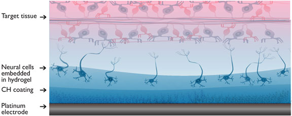
A living electrode construct for incorporation of cells into bionic devices
-
- Journal:
- MRS Communications / Volume 7 / Issue 3 / September 2017
- Published online by Cambridge University Press:
- 27 June 2017, pp. 487-495
- Print publication:
- September 2017
-
- Article
- Export citation
JMR volume 32 issue 12 Cover and Back matter
-
- Journal:
- Journal of Materials Research / Volume 32 / Issue 12 / 28 June 2017
- Published online by Cambridge University Press:
- 27 June 2017, pp. b1-b4
- Print publication:
- 28 June 2017
-
- Article
-
- You have access
- Export citation
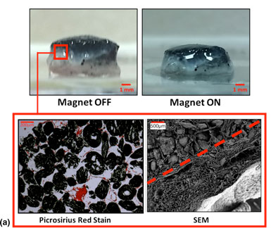
Development of magnetically active scaffolds as intrinsically-deformable bioreactors
-
- Journal:
- MRS Communications / Volume 7 / Issue 3 / September 2017
- Published online by Cambridge University Press:
- 27 June 2017, pp. 367-374
- Print publication:
- September 2017
-
- Article
- Export citation
