Refine search
Actions for selected content:
106117 results in Materials Science
Calendar of Forthcoming Meetings Powder Diffraction March 2015
-
- Journal:
- Powder Diffraction / Volume 30 / Issue 1 / March 2015
- Published online by Cambridge University Press:
- 17 March 2015, pp. 85-86
-
- Article
-
- You have access
- HTML
- Export citation
Recent changes to the Author Notes for Powder Diffraction
-
- Journal:
- Powder Diffraction / Volume 30 / Issue 1 / March 2015
- Published online by Cambridge University Press:
- 17 March 2015, p. 1
-
- Article
-
- You have access
- HTML
- Export citation
Calendar of Short Courses and Workshops Powder Diffraction March 2015
-
- Journal:
- Powder Diffraction / Volume 30 / Issue 1 / March 2015
- Published online by Cambridge University Press:
- 17 March 2015, p. 87
-
- Article
-
- You have access
- HTML
- Export citation
PDJ volume 30 issue 1 Cover and Front matter
-
- Journal:
- Powder Diffraction / Volume 30 / Issue 1 / March 2015
- Published online by Cambridge University Press:
- 17 March 2015, pp. f1-f6
-
- Article
-
- You have access
- Export citation
PDJ volume 30 issue 1 Cover and Back matter
-
- Journal:
- Powder Diffraction / Volume 30 / Issue 1 / March 2015
- Published online by Cambridge University Press:
- 17 March 2015, pp. b1-b7
-
- Article
-
- You have access
- Export citation

Effect of a high axial magnetic field on the structure of directionally solidified Al–Si alloys
-
- Journal:
- Journal of Materials Research / Volume 30 / Issue 8 / 28 April 2015
- Published online by Cambridge University Press:
- 17 March 2015, pp. 1043-1055
- Print publication:
- 28 April 2015
-
- Article
- Export citation
2014 Denver X-ray Conference – Student Reflection
-
- Journal:
- Powder Diffraction / Volume 30 / Issue 1 / March 2015
- Published online by Cambridge University Press:
- 17 March 2015, pp. 82-84
-
- Article
- Export citation

Microstructure and texture evolution in Mg–Al–Zn–(AgIn) alloy during single and multiple pass warm rolling
-
- Journal:
- Journal of Materials Research / Volume 30 / Issue 7 / 14 April 2015
- Published online by Cambridge University Press:
- 17 March 2015, pp. 1011-1018
- Print publication:
- 14 April 2015
-
- Article
- Export citation
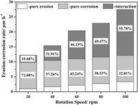
Effect of erosion speed on the interaction between erosion and corrosion of the Fe–3.5 wt% B alloy in a flowing zinc bath
-
- Journal:
- Journal of Materials Research / Volume 30 / Issue 6 / 28 March 2015
- Published online by Cambridge University Press:
- 17 March 2015, pp. 852-859
- Print publication:
- 28 March 2015
-
- Article
- Export citation

Oxidation of Zircaloy-4 during in situ proton irradiation and corrosion in PWR primary water
-
- Journal:
- Journal of Materials Research / Volume 30 / Issue 9 / 14 May 2015
- Published online by Cambridge University Press:
- 16 March 2015, pp. 1335-1348
- Print publication:
- 14 May 2015
-
- Article
- Export citation
JMR volume 30 issue 5 Cover and Back matter
-
- Journal:
- Journal of Materials Research / Volume 30 / Issue 5 / 14 March 2015
- Published online by Cambridge University Press:
- 13 March 2015, pp. b1-b4
- Print publication:
- 14 March 2015
-
- Article
-
- You have access
- Export citation

Preparation and rheo-squeeze casting of semi-solid AZ91–2 wt% Ca magnesium alloy by gas bubbling process
-
- Journal:
- Journal of Materials Research / Volume 30 / Issue 6 / 28 March 2015
- Published online by Cambridge University Press:
- 13 March 2015, pp. 825-832
- Print publication:
- 28 March 2015
-
- Article
- Export citation

Optical properties of Si nanowires: Dependence on substrate crystallographic orientation and light polarization
-
- Journal:
- Journal of Materials Research / Volume 30 / Issue 6 / 28 March 2015
- Published online by Cambridge University Press:
- 13 March 2015, pp. 753-760
- Print publication:
- 28 March 2015
-
- Article
- Export citation
JMR volume 30 issue 5 Cover and Front matter
-
- Journal:
- Journal of Materials Research / Volume 30 / Issue 5 / 14 March 2015
- Published online by Cambridge University Press:
- 13 March 2015, pp. f1-f5
- Print publication:
- 14 March 2015
-
- Article
-
- You have access
- Export citation
The water retention mechanism of a Cs+ and Na+ exchanged montmorillonite: effect of relative humidity and ionic radius on the interlayer
-
- Journal:
- Powder Diffraction / Volume 30 / Issue S1 / June 2015
- Published online by Cambridge University Press:
- 11 March 2015, pp. S70-S75
-
- Article
- Export citation
Face-sharing octahedra in Cs3Al2F9 and Cs2AlF5
-
- Journal:
- Powder Diffraction / Volume 30 / Issue 2 / June 2015
- Published online by Cambridge University Press:
- 11 March 2015, pp. 130-138
-
- Article
- Export citation
Powder diffraction of yellow and red natural earths from Lessini Mountains in NE Italy
-
- Journal:
- Powder Diffraction / Volume 30 / Issue 2 / June 2015
- Published online by Cambridge University Press:
- 11 March 2015, pp. 122-129
-
- Article
-
- You have access
- HTML
- Export citation
X-ray powder diffraction studies of (Bax Sr1− x )2Co2Fe12O22 and (Bax Sr1− x )Co2Fe16O27
-
- Journal:
- Powder Diffraction / Volume 30 / Issue 2 / June 2015
- Published online by Cambridge University Press:
- 11 March 2015, pp. 139-148
-
- Article
- Export citation
-
X-ray structural characterization and X-ray reference powder patterns have been determined for two series of iron- and cobalt-containing layered compounds (Bax Sr1− x )2Co2Fe12O22 (x = 0.2, 0.4, 0.6, 0.8) and (Bax Sr1− x )Co2Fe16O27 (x = 0.2, 0.4, 0.6, 0.8). The (Bax Sr1− x )2Co2Fe12O22 series of compounds crystallized in the space group R $\bar 3$ m (No. 166), with Z = 3. The structure is essentially that of the Y-type hexagonal ferrite, BaM 2+Fe6 3+O11. The lattice parameters range from a = 5.859 15(8) to 5.843 72(8) Å, and c = 43.4975(9) to 43.3516(9) Å for x = 0.2 to 0.8, respectively. The (Bax Sr1− x )Co2Fe16O27 series (W-type hexagonal ferrite) crystallized in the space group P63/mmc (No. 194) and Z = 2. The lattice parameters range from a = 5.902 05(12) to 5.8979(2) Å and c = 32.9002(10) to 32.8110(13) Å for x = 0.2 to 0.8. Results of measurements of the Seebeck coefficient and resistivity of these two sets of samples indicated that they are insulators. Powder X-ray diffraction patterns of these two series of compounds have been submitted to be included in the Powder Diffraction File.

Cystamindi-ium tetrachlorocuprate [NH3(CH2)2SS(CH2)2NH3][CuCl4]: synthesis, crystal structure, and thermal decomposition
-
- Journal:
- Powder Diffraction / Volume 30 / Issue 2 / June 2015
- Published online by Cambridge University Press:
- 11 March 2015, pp. 163-169
-
- Article
- Export citation

Importance of dislocation pile-ups on the mechanical properties and the Bauschinger effect in microcantilevers
-
- Journal:
- Journal of Materials Research / Volume 30 / Issue 6 / 28 March 2015
- Published online by Cambridge University Press:
- 10 March 2015, pp. 791-797
- Print publication:
- 28 March 2015
-
- Article
- Export citation
