Refine search
Actions for selected content:
106129 results in Materials Science
MRC volume 2 issue 2 Cover and Back matter
-
- Journal:
- MRS Communications / Volume 2 / Issue 2 / June 2012
- Published online by Cambridge University Press:
- 15 June 2012, pp. b1-b2
- Print publication:
- June 2012
-
- Article
-
- You have access
- Export citation
Survey reveals interdisciplinarity of MSE faculty
-
- Journal:
- MRS Bulletin / Volume 37 / Issue 6 / June 2012
- Published online by Cambridge University Press:
- 08 June 2012, pp. 541-542
- Print publication:
- June 2012
-
- Article
-
- You have access
- HTML
- Export citation

Thin films and microelectrode arrays for neuroprosthetics
-
- Journal:
- MRS Bulletin / Volume 37 / Issue 6 / June 2012
- Published online by Cambridge University Press:
- 08 June 2012, pp. 590-598
- Print publication:
- June 2012
-
- Article
- Export citation
Nano Focus: Nanomechanical mass sensor boasts yoctogram resolution
-
- Journal:
- MRS Bulletin / Volume 37 / Issue 6 / June 2012
- Published online by Cambridge University Press:
- 08 June 2012, p. 543
- Print publication:
- June 2012
-
- Article
-
- You have access
- HTML
- Export citation

Extracellular matrix-based materials for neural interfacing
-
- Journal:
- MRS Bulletin / Volume 37 / Issue 6 / June 2012
- Published online by Cambridge University Press:
- 08 June 2012, pp. 606-613
- Print publication:
- June 2012
-
- Article
- Export citation
Energy efficiency with organic electronics: Ching W. Tang revisits his days at Kodak
-
- Journal:
- MRS Bulletin / Volume 37 / Issue 6 / June 2012
- Published online by Cambridge University Press:
- 08 June 2012, pp. 552-553
- Print publication:
- June 2012
-
- Article
-
- You have access
- HTML
- Export citation
MRS volume 37 issue 6 Cover and Back matter
-
- Journal:
- MRS Bulletin / Volume 37 / Issue 6 / June 2012
- Published online by Cambridge University Press:
- 08 June 2012, pp. b1-b2
- Print publication:
- June 2012
-
- Article
-
- You have access
- Export citation
High-temperature materials increase efficiency of gas power plants
-
- Journal:
- MRS Bulletin / Volume 37 / Issue 6 / June 2012
- Published online by Cambridge University Press:
- 08 June 2012, pp. 550-551
- Print publication:
- June 2012
-
- Article
-
- You have access
- HTML
- Export citation

Carrier-induced nonlinearities in InGaN/GaN quantum wells with V-pits
-
- Journal:
- MRS Communications / Volume 2 / Issue 2 / June 2012
- Published online by Cambridge University Press:
- 15 June 2012, pp. 55-60
- Print publication:
- June 2012
-
- Article
- Export citation
MRC volume 2 issue 2 Cover and Front matter
-
- Journal:
- MRS Communications / Volume 2 / Issue 2 / June 2012
- Published online by Cambridge University Press:
- 15 June 2012, pp. f1-f4
- Print publication:
- June 2012
-
- Article
-
- You have access
- Export citation
China’s nuclear resolve
-
- Journal:
- MRS Bulletin / Volume 37 / Issue 6 / June 2012
- Published online by Cambridge University Press:
- 08 June 2012, pp. 554-555
- Print publication:
- June 2012
-
- Article
-
- You have access
- HTML
- Export citation
LOOK AGAIN...
-
- Journal:
- MRS Bulletin / Volume 37 / Issue 6 / June 2012
- Published online by Cambridge University Press:
- 08 June 2012, p. 616
- Print publication:
- June 2012
-
- Article
-
- You have access
- HTML
- Export citation
MRS volume 37 issue 6 Cover and Front matter
-
- Journal:
- MRS Bulletin / Volume 37 / Issue 6 / June 2012
- Published online by Cambridge University Press:
- 08 June 2012, pp. f1-f6
- Print publication:
- June 2012
-
- Article
-
- You have access
- Export citation
Meet our Authors
-
- Journal:
- MRS Bulletin / Volume 37 / Issue 6 / June 2012
- Published online by Cambridge University Press:
- 08 June 2012, pp. 562-565
- Print publication:
- June 2012
-
- Article
-
- You have access
- HTML
- Export citation

Mechanically adaptive nanocomposites for neural interfacing
-
- Journal:
- MRS Bulletin / Volume 37 / Issue 6 / June 2012
- Published online by Cambridge University Press:
- 08 June 2012, pp. 581-589
- Print publication:
- June 2012
-
- Article
- Export citation
CAREER CENTRAL
-
- Journal:
- MRS Bulletin / Volume 37 / Issue 6 / June 2012
- Published online by Cambridge University Press:
- 08 June 2012, pp. 614-615
- Print publication:
- June 2012
-
- Article
-
- You have access
- Export citation
Canadian composites industry receives C$9.8 million federal boost: www.composites.ubc.ca
-
- Journal:
- MRS Bulletin / Volume 37 / Issue 6 / June 2012
- Published online by Cambridge University Press:
- 08 June 2012, p. 548
- Print publication:
- June 2012
-
- Article
-
- You have access
- HTML
- Export citation

Material considerations for in vitro neural interface technology
-
- Journal:
- MRS Bulletin / Volume 37 / Issue 6 / June 2012
- Published online by Cambridge University Press:
- 08 June 2012, pp. 566-572
- Print publication:
- June 2012
-
- Article
- Export citation
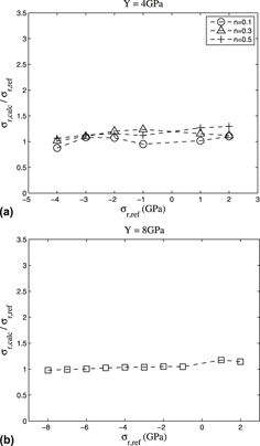
Numerical analysis of different methods to calculate residual stresses in thin films based on instrumented indentation data
-
- Journal:
- Journal of Materials Research / Volume 27 / Issue 13 / 14 July 2012
- Published online by Cambridge University Press:
- 01 June 2012, pp. 1732-1741
- Print publication:
- 14 July 2012
-
- Article
- Export citation
Energy Focus
-
- Journal:
- MRS Bulletin / Volume 37 / Issue 6 / June 2012
- Published online by Cambridge University Press:
- 08 June 2012, p. 556
- Print publication:
- June 2012
-
- Article
-
- You have access
- HTML
- Export citation
