Refine search
Actions for selected content:
106107 results in Materials Science
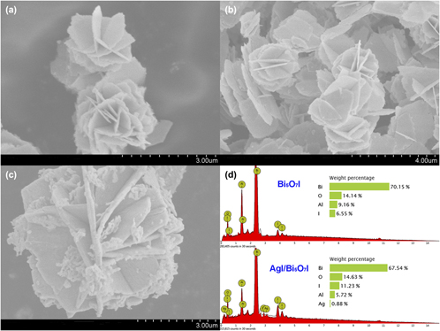
Synthesis of flower-like AgI/Bi5O7I hybrid photocatalysts with enhanced photocatalytic activity in rhodamine B degradation
-
- Journal:
- Journal of Materials Research / Volume 33 / Issue 16 / 28 August 2018
- Published online by Cambridge University Press:
- 28 June 2018, pp. 2385-2395
- Print publication:
- 28 August 2018
-
- Article
- Export citation
JMR volume 33 issue 12 Cover and Back matter
-
- Journal:
- Journal of Materials Research / Volume 33 / Issue 12 / 28 June 2018
- Published online by Cambridge University Press:
- 28 June 2018, pp. b1-b2
- Print publication:
- 28 June 2018
-
- Article
-
- You have access
- Export citation
Scandium on the formation of in situ TiB2 particulates in an aluminum matrix
-
- Journal:
- Journal of Materials Research / Volume 33 / Issue 18 / 28 September 2018
- Published online by Cambridge University Press:
- 28 June 2018, pp. 2721-2727
- Print publication:
- 28 September 2018
-
- Article
- Export citation
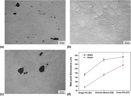
Effect of laser hatch style on densification behavior, microstructure, and tribological performance of aluminum alloys by selective laser melting
-
- Journal:
- Journal of Materials Research / Volume 33 / Issue 12 / 28 June 2018
- Published online by Cambridge University Press:
- 28 June 2018, pp. 1713-1722
- Print publication:
- 28 June 2018
-
- Article
- Export citation
Alginate-honey bioinks with improved cell responses for applications as bioprinted tissue engineered constructs
-
- Journal:
- Journal of Materials Research / Volume 33 / Issue 14 / 27 July 2018
- Published online by Cambridge University Press:
- 28 June 2018, pp. 2029-2039
- Print publication:
- 27 July 2018
-
- Article
- Export citation
JMR volume 33 issue 12 Cover and Front matter
-
- Journal:
- Journal of Materials Research / Volume 33 / Issue 12 / 28 June 2018
- Published online by Cambridge University Press:
- 28 June 2018, pp. f1-f4
- Print publication:
- 28 June 2018
-
- Article
-
- You have access
- Export citation
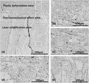
Laser solid forming assisted by friction stir processing for preparation of Ni–16Cr–8Fe alloys: Crack repairing and grain refinement
-
- Journal:
- Journal of Materials Research / Volume 33 / Issue 20 / 29 October 2018
- Published online by Cambridge University Press:
- 27 June 2018, pp. 3521-3529
- Print publication:
- 29 October 2018
-
- Article
- Export citation
3D printing of poly(ε-caprolactone)/poly(D,L-lactide-co-glycolide)/hydroxyapatite composite constructs for bone tissue engineering
-
- Journal:
- Journal of Materials Research / Volume 33 / Issue 14 / 27 July 2018
- Published online by Cambridge University Press:
- 27 June 2018, pp. 1972-1986
- Print publication:
- 27 July 2018
-
- Article
- Export citation
Using differential scanning calorimetry to characterize the precipitation and dissolution of V(CN) and VC particles during continuous casting and reheating process
-
- Journal:
- Journal of Materials Research / Volume 33 / Issue 18 / 28 September 2018
- Published online by Cambridge University Press:
- 26 June 2018, pp. 2784-2795
- Print publication:
- 28 September 2018
-
- Article
- Export citation
Improved strengthening efficiency of nanoreinforcements realized by a novel melt spinning process
-
- Journal:
- Journal of Materials Research / Volume 33 / Issue 18 / 28 September 2018
- Published online by Cambridge University Press:
- 26 June 2018, pp. 2711-2720
- Print publication:
- 28 September 2018
-
- Article
- Export citation
A superfine eutectic microstructure and the mechanical properties of CoCrFeNiMox high-entropy alloys
-
- Journal:
- Journal of Materials Research / Volume 33 / Issue 19 / 14 October 2018
- Published online by Cambridge University Press:
- 26 June 2018, pp. 3258-3265
- Print publication:
- 14 October 2018
-
- Article
- Export citation
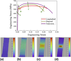
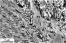
Anisotropy and microstructural evolutions of X70 pipeline steel during tensile deformation
-
- Journal:
- Journal of Materials Research / Volume 33 / Issue 20 / 29 October 2018
- Published online by Cambridge University Press:
- 26 June 2018, pp. 3512-3520
- Print publication:
- 29 October 2018
-
- Article
- Export citation
-
Tensile properties of different directions of X70 pipeline steel plate were tested, and microstructural evolutions of different zones along the transverse direction (TD) were also investigated using electron backscatter diffraction. The highest strength values (yield strength and ultimate strength) appear at TD, and the diagonal direction shows the largest uniform elongation. The elongations of the polygonal ferrite and quasi polygonal ferrite grains increase with the decrease in the distance to the fracture zone. The ratio between high-angle grain boundaries and low-angle grain boundaries in the as-received steel is about 7/3 and starts to decrease from the fillet zone to the fracture zone. The refinement of grains occurs adjacent to the fracture section with the formation of subgrains. With the increase in tensile strain, the intensities of cube and γ-fiber textures increase sharply, and the reinforcement of the (111) $\left[ {\bar{1}\bar{1}2} \right]$ component was obviously larger than the (111) $\left[ {1\bar{2}1} \right]$ component in the γ-fiber texture during tensile deformation.


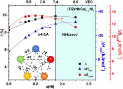
Structure property relationship in (TiZrNbCu)1−xNix metallic glasses
-
- Journal:
- Journal of Materials Research / Volume 33 / Issue 19 / 14 October 2018
- Published online by Cambridge University Press:
- 26 June 2018, pp. 3170-3183
- Print publication:
- 14 October 2018
-
- Article
- Export citation

Europium(III)-induced water-soluble nano-aggregates of hyaluronic acid and chitosan: structure and fluorescence
-
- Journal:
- MRS Communications / Volume 8 / Issue 3 / September 2018
- Published online by Cambridge University Press:
- 25 June 2018, pp. 1224-1229
- Print publication:
- September 2018
-
- Article
- Export citation
Correlation between the atomic configurations and the amorphous-to-icosahedral phase transition in metallic glasses
-
- Journal:
- Journal of Materials Research / Volume 33 / Issue 18 / 28 September 2018
- Published online by Cambridge University Press:
- 22 June 2018, pp. 2775-2783
- Print publication:
- 28 September 2018
-
- Article
- Export citation
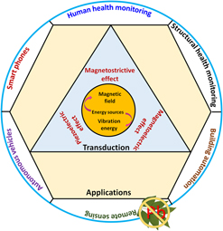
Lead-free piezoelectric materials and composites for high power density energy harvesting
-
- Journal:
- Journal of Materials Research / Volume 33 / Issue 16 / 28 August 2018
- Published online by Cambridge University Press:
- 22 June 2018, pp. 2235-2263
- Print publication:
- 28 August 2018
-
- Article
- Export citation
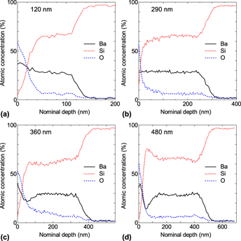
Diffusion process in BaSi2 film formation by thermal evaporation and its relation to electrical properties
-
- Journal:
- Journal of Materials Research / Volume 33 / Issue 16 / 28 August 2018
- Published online by Cambridge University Press:
- 22 June 2018, pp. 2297-2305
- Print publication:
- 28 August 2018
-
- Article
- Export citation

In situ neutron diffraction study on tensile deformation behavior of carbon-strengthened CoCrFeMnNi high-entropy alloys at room and elevated temperatures
-
- Journal:
- Journal of Materials Research / Volume 33 / Issue 19 / 14 October 2018
- Published online by Cambridge University Press:
- 22 June 2018, pp. 3192-3203
- Print publication:
- 14 October 2018
-
- Article
- Export citation
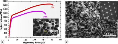
L12-strengthened high-entropy alloys for advanced structural applications
-
- Journal:
- Journal of Materials Research / Volume 33 / Issue 19 / 14 October 2018
- Published online by Cambridge University Press:
- 22 June 2018, pp. 2983-2997
- Print publication:
- 14 October 2018
-
- Article
- Export citation
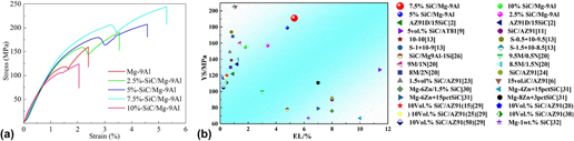
Dependence of microstructure characteristics and mechanical properties on nanosize SiCp contents in Mg–9Al matrix composites fabricated by ultrasonic-assisted semisolid powder hot pressing
-
- Journal:
- Journal of Materials Research / Volume 33 / Issue 18 / 28 September 2018
- Published online by Cambridge University Press:
- 21 June 2018, pp. 2689-2699
- Print publication:
- 28 September 2018
-
- Article
- Export citation
