Refine search
Actions for selected content:
106117 results in Materials Science
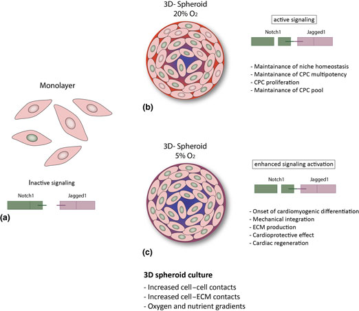
Spheroid three-dimensional culture enhances Notch signaling in cardiac progenitor cells
-
- Journal:
- MRS Communications / Volume 7 / Issue 3 / September 2017
- Published online by Cambridge University Press:
- 11 September 2017, pp. 496-501
- Print publication:
- September 2017
-
- Article
-
- You have access
- Open access
- HTML
- Export citation
Cultivating curves
-
- Journal:
- MRS Bulletin / Volume 42 / Issue 9 / September 2017
- Published online by Cambridge University Press:
- 08 September 2017, p. 688
- Print publication:
- September 2017
-
- Article
-
- You have access
- HTML
- Export citation

Eco-friendly synthesis of egg-white capped silver nanoparticles for rapid, selective, and sensitive detection of Hg(II)
-
- Journal:
- MRS Communications / Volume 7 / Issue 3 / September 2017
- Published online by Cambridge University Press:
- 04 September 2017, pp. 695-700
- Print publication:
- September 2017
-
- Article
- Export citation
CAREER CENTRAL
-
- Journal:
- MRS Bulletin / Volume 42 / Issue 9 / September 2017
- Published online by Cambridge University Press:
- 08 September 2017, p. 687
- Print publication:
- September 2017
-
- Article
-
- You have access
- Export citation
MRS volume 42 issue 9 Cover and Front matter
-
- Journal:
- MRS Bulletin / Volume 42 / Issue 9 / September 2017
- Published online by Cambridge University Press:
- 08 September 2017, pp. f1-f6
- Print publication:
- September 2017
-
- Article
-
- You have access
- Export citation
MRC volume 7 issue 3 Cover and Front matter
-
- Journal:
- MRS Communications / Volume 7 / Issue 3 / September 2017
- Published online by Cambridge University Press:
- 03 October 2017, pp. f1-f7
- Print publication:
- September 2017
-
- Article
-
- You have access
- Export citation
STEM professionals utilize established routes of science advocacy in United States
-
- Journal:
- MRS Bulletin / Volume 42 / Issue 9 / September 2017
- Published online by Cambridge University Press:
- 08 September 2017, pp. 626-627
- Print publication:
- September 2017
-
- Article
-
- You have access
- HTML
- Export citation
Nano Focus: Laser design emits multicolor light
-
- Journal:
- MRS Bulletin / Volume 42 / Issue 9 / September 2017
- Published online by Cambridge University Press:
- 08 September 2017, p. 623
- Print publication:
- September 2017
-
- Article
-
- You have access
- HTML
- Export citation

Spectroscopic studies of dopant-induced conformational changes in poly(3-hexylthiophene) thin films
-
- Journal:
- MRS Communications / Volume 7 / Issue 3 / September 2017
- Published online by Cambridge University Press:
- 07 September 2017, pp. 728-734
- Print publication:
- September 2017
-
- Article
- Export citation
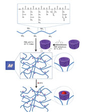
Gelatin-based hydrogels for biomedical applications
-
- Journal:
- MRS Communications / Volume 7 / Issue 3 / September 2017
- Published online by Cambridge University Press:
- 03 October 2017, pp. 416-426
- Print publication:
- September 2017
-
- Article
- Export citation
Third NSF decadal report presents challenges for polymer field
-
- Journal:
- MRS Bulletin / Volume 42 / Issue 9 / September 2017
- Published online by Cambridge University Press:
- 08 September 2017, pp. 621-622
- Print publication:
- September 2017
-
- Article
-
- You have access
- HTML
- Export citation
A visitor’s review of Manchester’s graphene exhibition
-
- Journal:
- MRS Bulletin / Volume 42 / Issue 9 / September 2017
- Published online by Cambridge University Press:
- 08 September 2017, pp. 634-636
- Print publication:
- September 2017
-
- Article
-
- You have access
- HTML
- Export citation
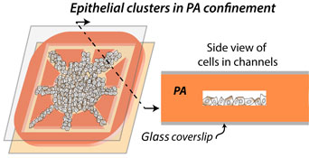
Hydrogel-based microchannels to measure confinement- and stiffness-sensitive Yes-associated-protein activity in epithelial clusters
-
- Journal:
- MRS Communications / Volume 7 / Issue 3 / September 2017
- Published online by Cambridge University Press:
- 07 September 2017, pp. 450-457
- Print publication:
- September 2017
-
- Article
- Export citation
Deep decarbonization faces deep challenges
-
- Journal:
- MRS Bulletin / Volume 42 / Issue 9 / September 2017
- Published online by Cambridge University Press:
- 08 September 2017, pp. 632-633
- Print publication:
- September 2017
-
- Article
-
- You have access
- HTML
- Export citation
MRC volume 7 issue 3 Cover and Back matter
-
- Journal:
- MRS Communications / Volume 7 / Issue 3 / September 2017
- Published online by Cambridge University Press:
- 03 October 2017, pp. b1-b2
- Print publication:
- September 2017
-
- Article
-
- You have access
- Export citation

Designer matter: Fascinating interactions of light and sound with metamaterials
-
- Journal:
- MRS Bulletin / Volume 42 / Issue 9 / September 2017
- Published online by Cambridge University Press:
- 08 September 2017, pp. 677-682
- Print publication:
- September 2017
-
- Article
- Export citation

Strongly scale-dependent charge transport from interconnections of silicon quantum dots and nanowires
-
- Journal:
- MRS Communications / Volume 7 / Issue 3 / September 2017
- Published online by Cambridge University Press:
- 07 September 2017, pp. 621-625
- Print publication:
- September 2017
-
- Article
-
- You have access
- HTML
- Export citation

Single-atom dynamics in scanning transmission electron microscopy
-
- Journal:
- MRS Bulletin / Volume 42 / Issue 9 / September 2017
- Published online by Cambridge University Press:
- 08 September 2017, pp. 644-652
- Print publication:
- September 2017
-
- Article
- Export citation
Kinetics in Materials Science and Engineering Dennis W. Readey: CRC Press, 2017 636 pages, $127.96 (e-book $111.97) ISBN: 9781138732469
-
- Journal:
- MRS Bulletin / Volume 42 / Issue 9 / September 2017
- Published online by Cambridge University Press:
- 08 September 2017, pp. 684-685
- Print publication:
- September 2017
-
- Article
-
- You have access
- HTML
- Export citation
Born in the lab: Hydrocarbon fuels ditch their fossil origins
-
- Journal:
- MRS Bulletin / Volume 42 / Issue 9 / September 2017
- Published online by Cambridge University Press:
- 08 September 2017, pp. 630-631
- Print publication:
- September 2017
-
- Article
-
- You have access
- HTML
- Export citation
