Refine search
Actions for selected content:
106117 results in Materials Science
A (bio) materials approach to three-dimensional cellbiology
-
- Journal:
- MRS Communications / Volume 7 / Issue 3 / September 2017
- Published online by Cambridge University Press:
- 03 October 2017, pp. 287-288
- Print publication:
- September 2017
-
- Article
- Export citation
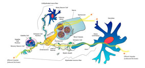
Three-dimensional neuronal cell culture: in pursuit of novel treatments for neurodegenerative disease
-
- Journal:
- MRS Communications / Volume 7 / Issue 3 / September 2017
- Published online by Cambridge University Press:
- 03 October 2017, pp. 320-331
- Print publication:
- September 2017
-
- Article
- Export citation
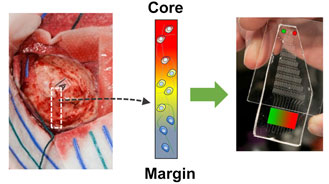
Spatially graded hydrogels for preclinical testing of glioblastoma anticancer therapeutics
-
- Journal:
- MRS Communications / Volume 7 / Issue 3 / September 2017
- Published online by Cambridge University Press:
- 12 September 2017, pp. 442-449
- Print publication:
- September 2017
-
- Article
- Export citation
Realizing true energy impact
-
- Journal:
- MRS Bulletin / Volume 42 / Issue 9 / September 2017
- Published online by Cambridge University Press:
- 08 September 2017, p. 629
- Print publication:
- September 2017
-
- Article
-
- You have access
- HTML
- Export citation
MRS volume 42 issue 9 Cover and Back matter
-
- Journal:
- MRS Bulletin / Volume 42 / Issue 9 / September 2017
- Published online by Cambridge University Press:
- 08 September 2017, pp. b1-b2
- Print publication:
- September 2017
-
- Article
-
- You have access
- Export citation

Densification of thoria through flash sintering
-
- Journal:
- MRS Communications / Volume 7 / Issue 3 / September 2017
- Published online by Cambridge University Press:
- 11 September 2017, pp. 677-682
- Print publication:
- September 2017
-
- Article
- Export citation

Single-atom fabrication with electron and ion beams: From surfaces and two-dimensional materials toward three-dimensional atom-by-atom assembly
-
- Journal:
- MRS Bulletin / Volume 42 / Issue 9 / September 2017
- Published online by Cambridge University Press:
- 08 September 2017, pp. 637-643
- Print publication:
- September 2017
-
- Article
-
- You have access
- HTML
- Export citation
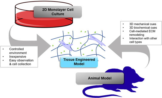
Shooting for the moon: using tissue-mimetic hydrogels to gain new insight on cancer biology and screen therapeutics
-
- Journal:
- MRS Communications / Volume 7 / Issue 3 / September 2017
- Published online by Cambridge University Press:
- 07 September 2017, pp. 427-441
- Print publication:
- September 2017
-
- Article
- Export citation

Structural and luminescence properties of yellow phosphors prepared by a modified sol–gel method
-
- Journal:
- MRS Communications / Volume 7 / Issue 3 / September 2017
- Published online by Cambridge University Press:
- 12 September 2017, pp. 721-727
- Print publication:
- September 2017
-
- Article
- Export citation
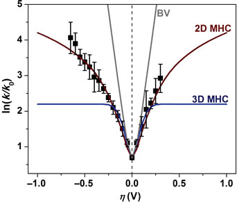
Electrochemical kinetics and dimensional considerations at the nanoscale: the influence of the density of states
-
- Journal:
- MRS Communications / Volume 7 / Issue 3 / September 2017
- Published online by Cambridge University Press:
- 14 September 2017, pp. 651-657
- Print publication:
- September 2017
-
- Article
- Export citation
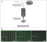
Polymeric scaffolds for three-dimensional culture of nerve cells: a model of peripheral nerve regeneration
-
- Journal:
- MRS Communications / Volume 7 / Issue 3 / September 2017
- Published online by Cambridge University Press:
- 03 October 2017, pp. 391-415
- Print publication:
- September 2017
-
- Article
- Export citation

TaWSi amorphous metal thin films: composition tuning to improve thermal stability
-
- Journal:
- MRS Communications / Volume 7 / Issue 3 / September 2017
- Published online by Cambridge University Press:
- 04 September 2017, pp. 715-720
- Print publication:
- September 2017
-
- Article
- Export citation
Successful Women Ceramic and Glass Scientists and Engineers: 100 Inspirational Profiles Lynnette D. Madsen: Wiley, 2016 640 pages, $65.00 (e-book $52.99) ISBN 978-1-118-73360-8
-
- Journal:
- MRS Bulletin / Volume 42 / Issue 9 / September 2017
- Published online by Cambridge University Press:
- 08 September 2017, p. 684
- Print publication:
- September 2017
-
- Article
-
- You have access
- HTML
- Export citation
Bio Focus: Hierarchical structure of spider dragline silk prevents spinning
-
- Journal:
- MRS Bulletin / Volume 42 / Issue 9 / September 2017
- Published online by Cambridge University Press:
- 08 September 2017, p. 625
- Print publication:
- September 2017
-
- Article
-
- You have access
- HTML
- Export citation
Bio Focus: Graphene-based composites achieve microstructural order at atomic scale
-
- Journal:
- MRS Bulletin / Volume 42 / Issue 9 / September 2017
- Published online by Cambridge University Press:
- 08 September 2017, pp. 624-625
- Print publication:
- September 2017
-
- Article
-
- You have access
- HTML
- Export citation
FUTURE Act to advance clean energy through Carbon Capture Utilization and Storage
-
- Journal:
- MRS Bulletin / Volume 42 / Issue 9 / September 2017
- Published online by Cambridge University Press:
- 08 September 2017, p. 628
- Print publication:
- September 2017
-
- Article
-
- You have access
- HTML
- Export citation

Engineering and modifying two-dimensional materials by electron beams
-
- Journal:
- MRS Bulletin / Volume 42 / Issue 9 / September 2017
- Published online by Cambridge University Press:
- 08 September 2017, pp. 667-676
- Print publication:
- September 2017
-
- Article
- Export citation
Nanomaterials for Wastewater Remediation Ravindra Kumar Gautam and Mahesh Chandra Chattopadhyaya: Butterworth-Heinemann, 2016 366 pages, $170.00 (e-book $170.00) ISBN 9780128046098
-
- Journal:
- MRS Bulletin / Volume 42 / Issue 9 / September 2017
- Published online by Cambridge University Press:
- 08 September 2017, pp. 685-686
- Print publication:
- September 2017
-
- Article
-
- You have access
- HTML
- Export citation
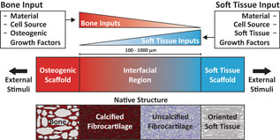
Next generation tissue engineering of orthopedic soft tissue-to-bone interfaces
-
- Journal:
- MRS Communications / Volume 7 / Issue 3 / September 2017
- Published online by Cambridge University Press:
- 03 October 2017, pp. 289-308
- Print publication:
- September 2017
-
- Article
-
- You have access
- HTML
- Export citation
Frank W. Clinard Jr., authority on radiation damage, remembered
-
- Journal:
- MRS Bulletin / Volume 42 / Issue 9 / September 2017
- Published online by Cambridge University Press:
- 08 September 2017, p. 683
- Print publication:
- September 2017
-
- Article
-
- You have access
- HTML
- Export citation
